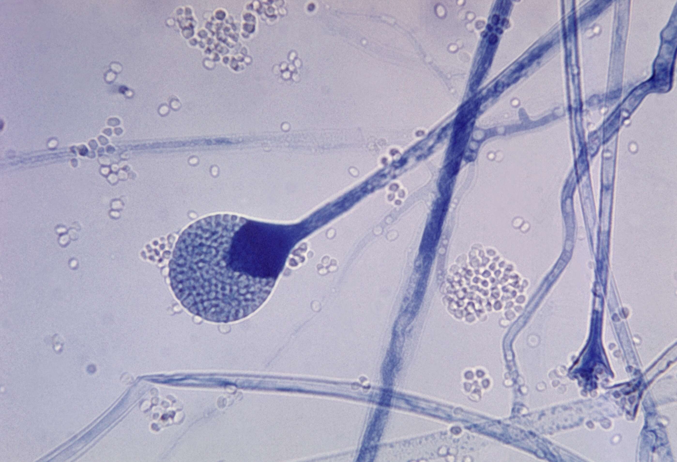
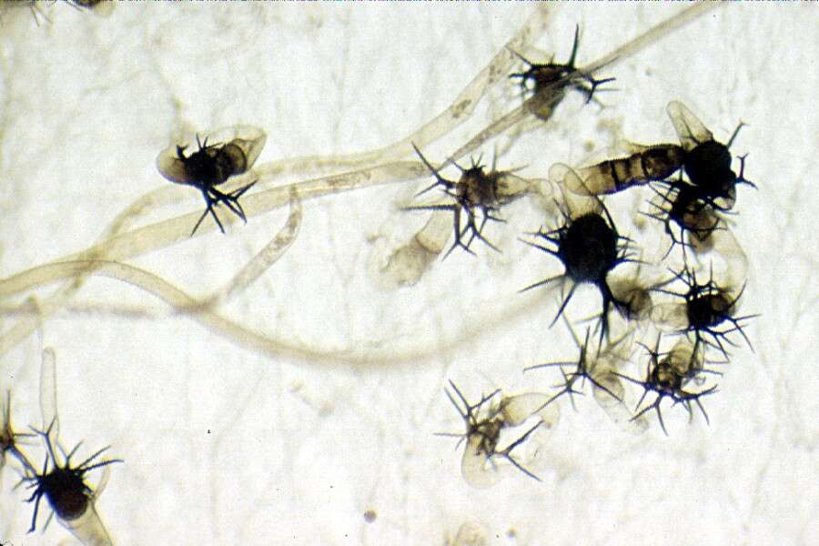
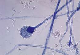
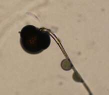
Description: English: This photomicrograph reveals a mature sporangium of a Absidia sp. fungus. Mucor is a common indoor mold, and is among the fungi that cause the group of infections known as zygomycosis. The infection typically involves the rhino-facial-cranial area, lungs, GI tract, skin, or less commonly other organ systems. Français : Sporocyste de la moisissure Absidia. Nederlands: Rijp sporangium van een Absidia schimmelsoort. Русский: Спорангий плесневого гриба рода Absidia со зрелыми спорами. 中文:弓毛黴的成熟孢子囊. 日本語: ユミケカビの胞子嚢. Date: 1955. Source: : This media comes from the Centers for Disease Control and Prevention's Public Health Image Library (PHIL), with identification number #3960. Note: Not all PHIL images are public domain; be sure to check copyright status and credit authors and content providers. العربية | Deutsch | English | македонски | slovenščina | +/−. Author: Photo Credit: Content Providers: CDC/Dr. Lucille K. Georg. Permission(Reusing this file): PD-USGov-HHS-CDC English: None - This image is in the public domain and thus free of any copyright restrictions. As a matter of courtesy we request that the content provider be credited and notified in any public or private usage of this image. Other versions: Low resolution: Image:Mature sporangium of a Mucor sp. fungus PHIL 3960 lores.jpg.
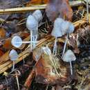
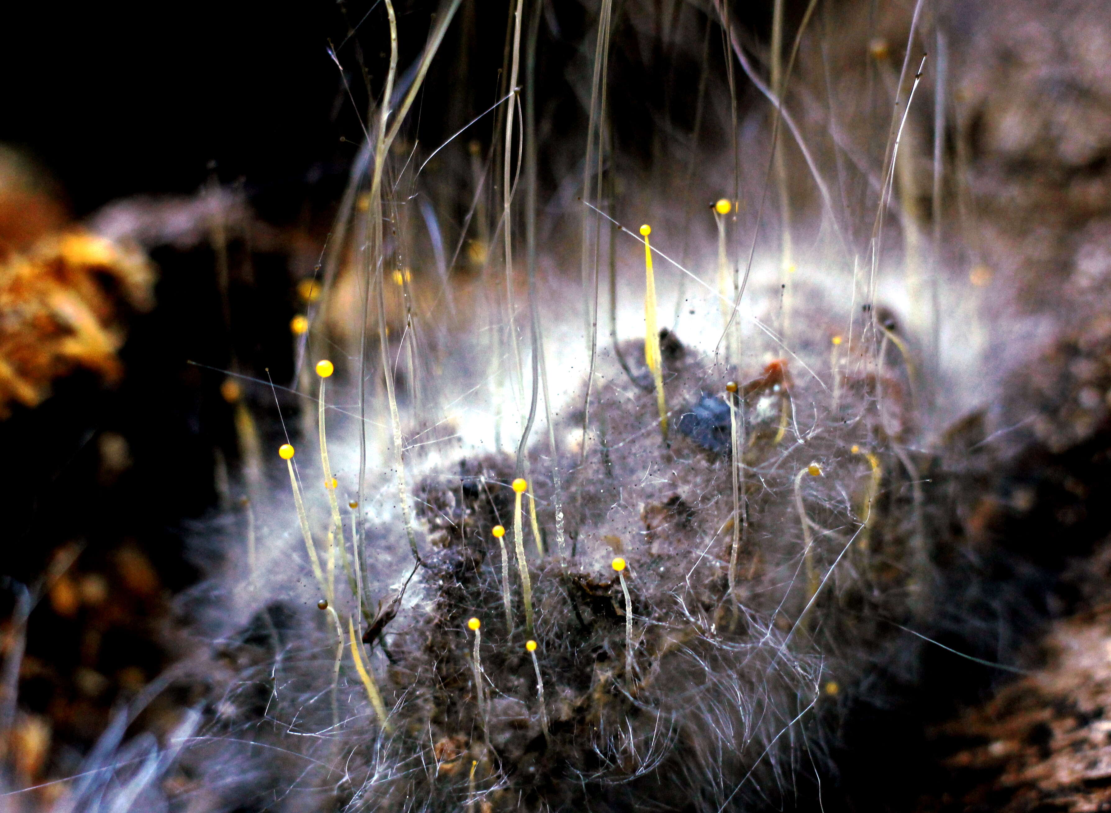
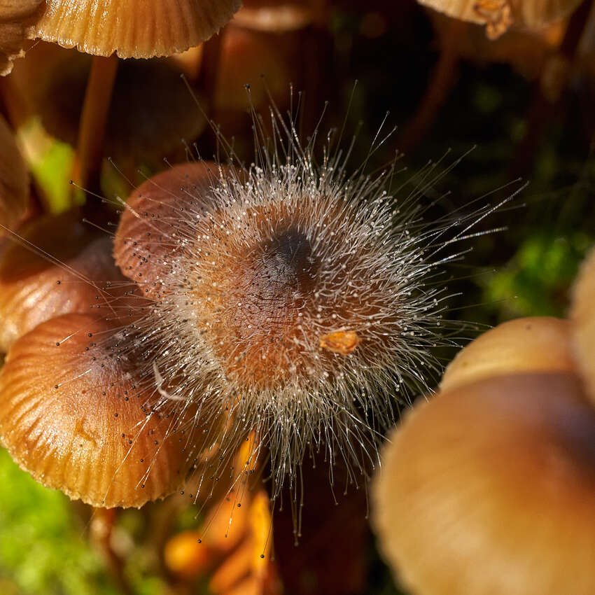
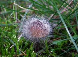
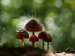
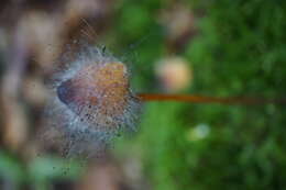
This image was created by user Richard Kneal (bloodworm) at Mushroom Observer, a source for mycological images.You can contact this user here. English | español | français | italiano | македонски | português | +/−
Wikimedia Commons
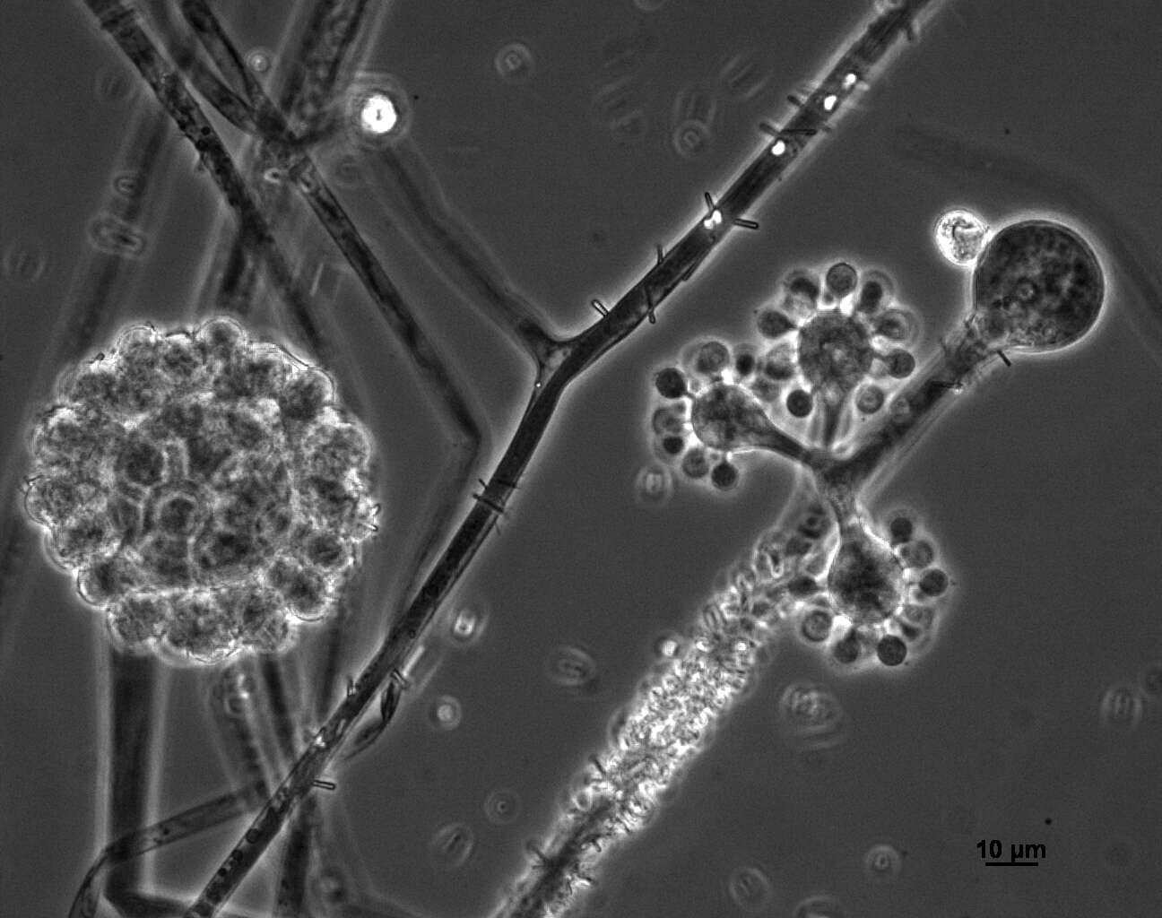
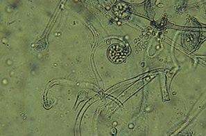
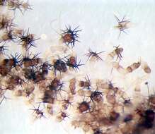
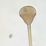
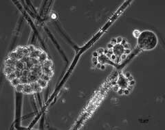
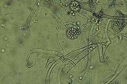
Description: English: Photomicrograph of sporangiophore of Apophysomyces variabilis in bright field microscopy. Date: 16 August 2013. Source: Photomicrograph from culture Previously published: n/a. Author: Medmyco.
No machine-readable author provided. Keisotyo assumed (based on copyright claims).
Wikimedia Commons
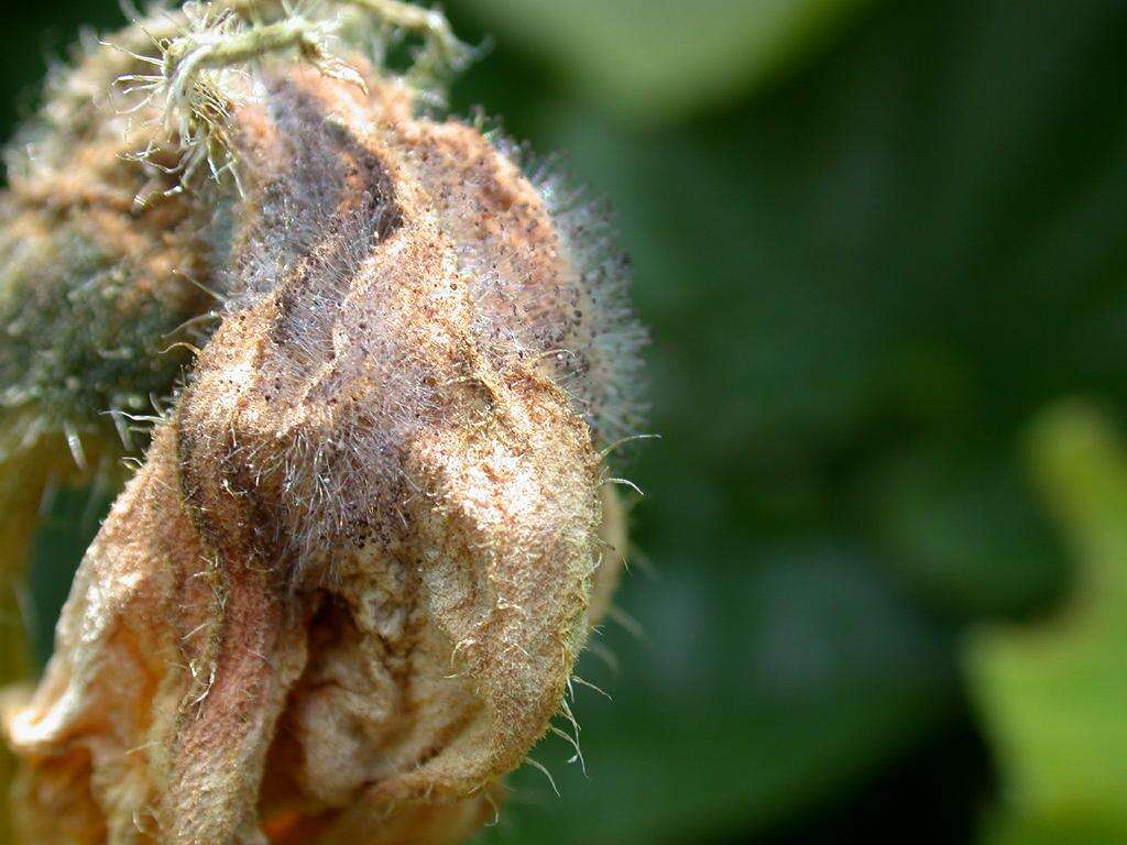
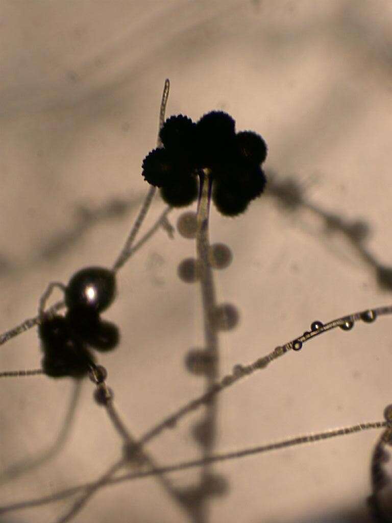
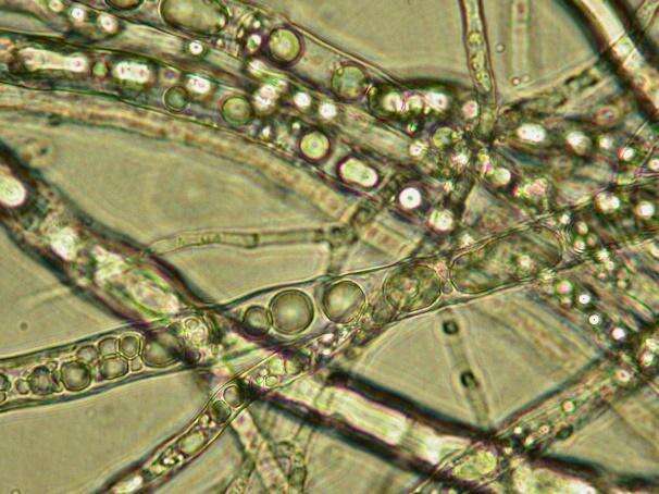
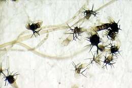
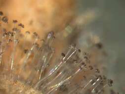
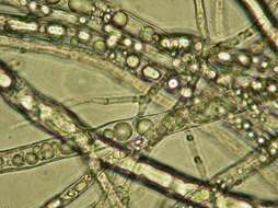
Description: Choanephora cucurbitarum (Choanephoraceae Mucorales) on Pumpkin flower Japanese name;Kougaikekabi Date;2004,07,22;Susami town, Wakayama prefecture, Japan Author;Keisotyo. Date: 23 November 2007 (original upload date). Source: No machine-readable source provided. Own work assumed (based on copyright claims). Author: No machine-readable author provided. Keisotyo assumed (based on copyright claims).
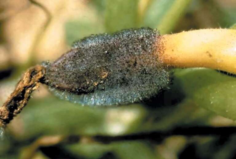
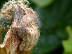
Description: English: Choanephora fruit rot caused by Choanephora cucurbitarum. Date: 6 November 2006. Source: : This image is Image Number 5077010 at Forestry Images, a source for forest health, natural resources and silviculture images operated by The Bugwood Network at the University of Georgia and the USDA Forest Service.. Author: David B. Langston, University of Georgia. Permission(Reusing this file): Cite: David B. Langston, University of Georgia, Bugwood.org.
No machine-readable author provided. Keisotyo assumed (based on copyright claims).
Wikimedia Commons
Description: Choanephora cucurbitarum (Choanephoraceae Mucorales)on Pumpkin flower Japanese name;Kougaikekabi Date;2004,07,22;Susami town, Wakayama prefecture, Japan Author;Keisotyo. Date: 23 November 2007 (original upload date). Source: No machine-readable source provided. Own work assumed (based on copyright claims). Author: No machine-readable author provided. Keisotyo assumed (based on copyright claims).
No machine-readable author provided. Keisotyo assumed (based on copyright claims).
Wikimedia Commons
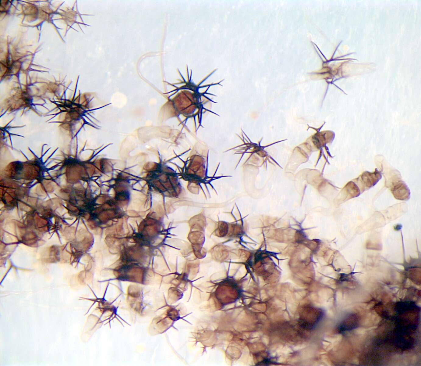
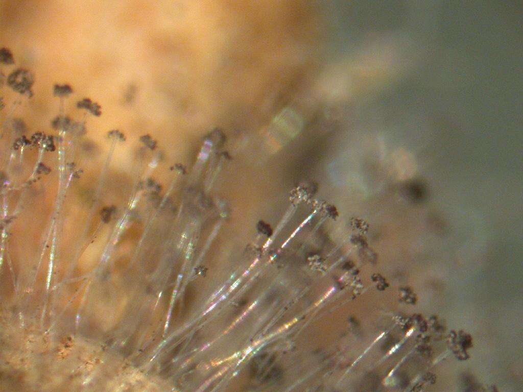
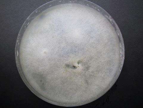
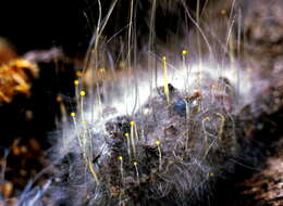
Description: Choanephora cucurbitarum (Choanephoraceae Mucorales) incubated in wet chamber Japanese name;Kougaikekabi Date;2004,07,23;Susami town, Wakayama prefecture, Japan Author;Keisotyo. Date: 23 November 2007 (original upload date). Source: No machine-readable source provided. Own work assumed (based on copyright claims). Author: No machine-readable author provided. Keisotyo assumed (based on copyright claims).
No machine-readable author provided. Keisotyo assumed (based on copyright claims).
Wikimedia Commons
Description: Choanephora cucurbitarum (Choanephoraceae Mucorales) incubated in wet chamber Japanese name;Kougaikekabi Date;2004,07,23;Susami town, Wakayama prefecture, Japan Author;Keisotyo. Date: 23 November 2007 (original upload date). Source: No machine-readable source provided. Own work assumed (based on copyright claims). Author: No machine-readable author provided. Keisotyo assumed (based on copyright claims).
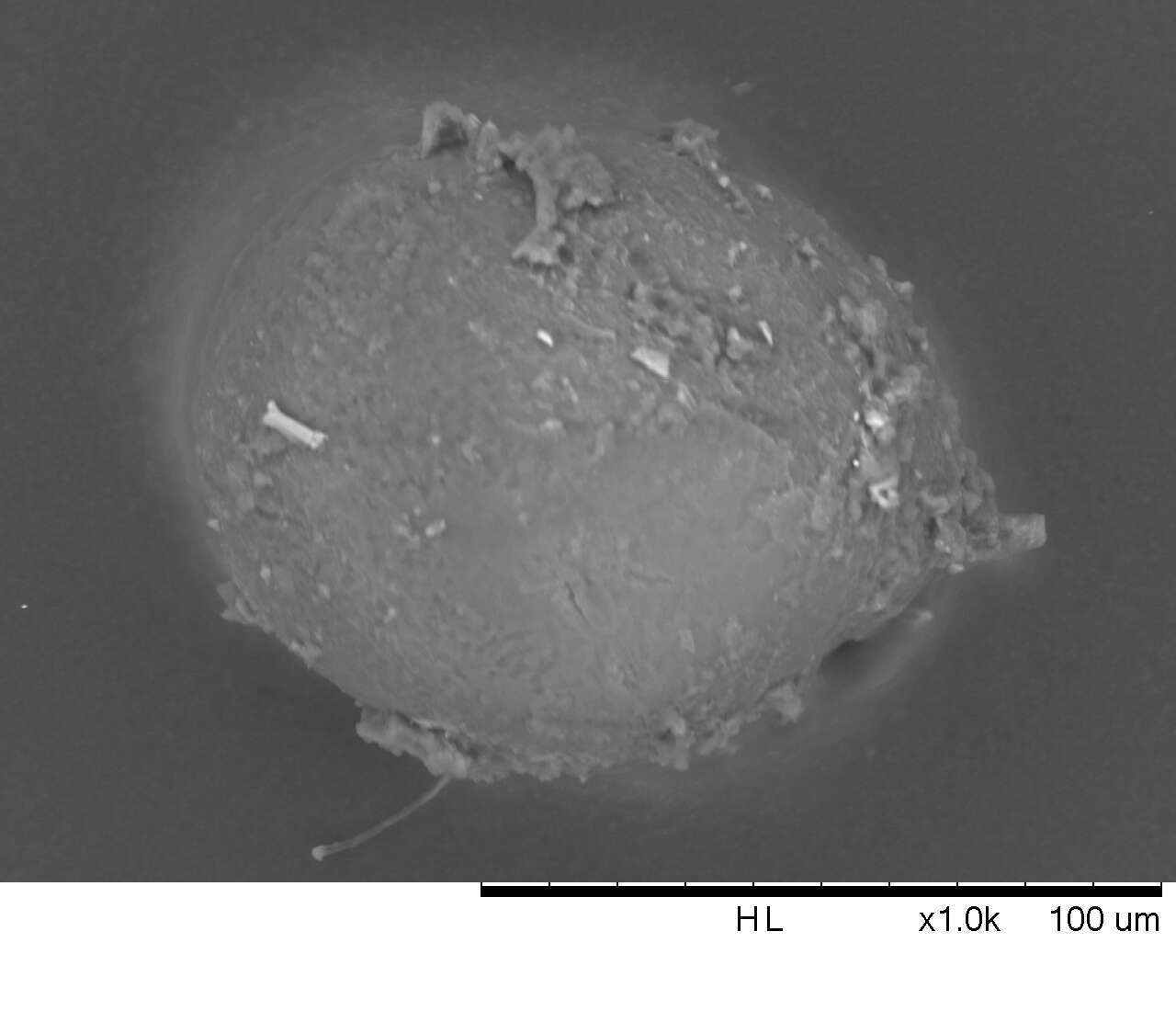
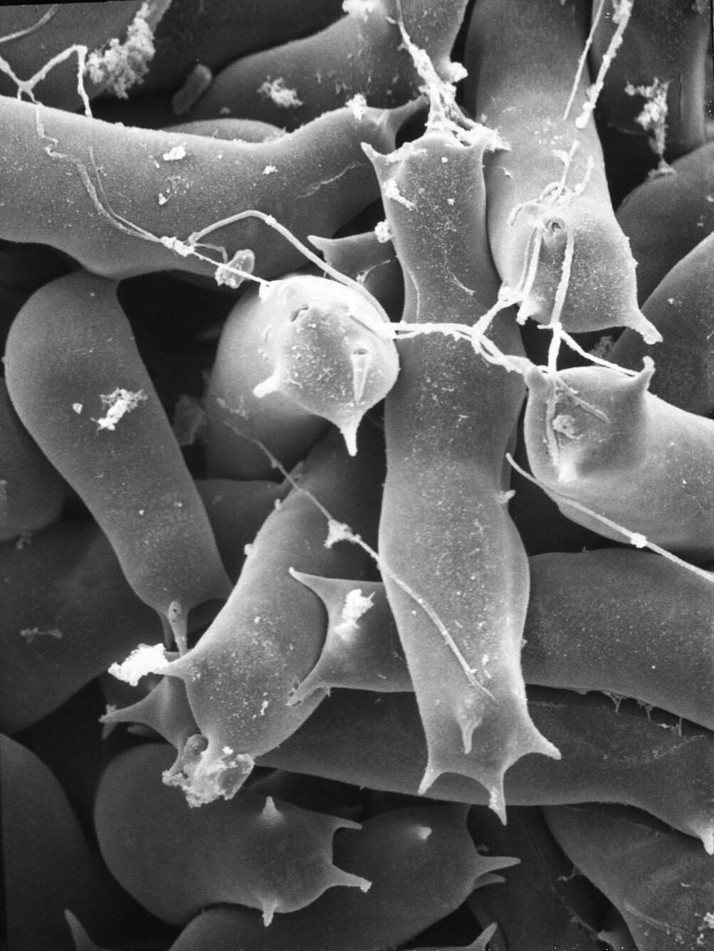
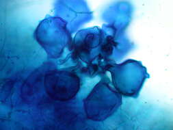
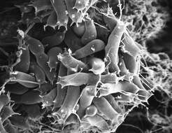
Description: English: 1000x electon microscopy of a mycorrhiza spore, probably Funneliformis mosseae (former 'Glomus mosseae'), wetsieved out of soil. Spore is coated with bacteria, soil and other fungi. Kindly provided by: Dr. rer. nat. tech. Tobias Sieberer, Matthias Salomon. Department 'Biotechnologie gartenbaulicher Kulturen', WZW, Technical University Munich. Date: 23 May 2014, 21:06:23. Source: Own work. Author: Samson90.


