-
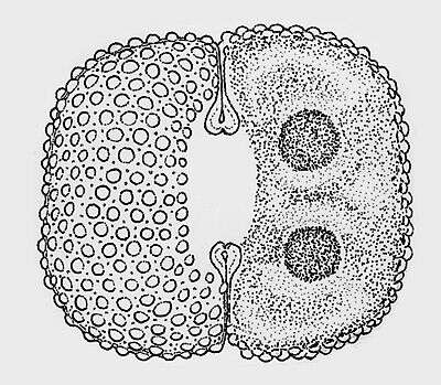
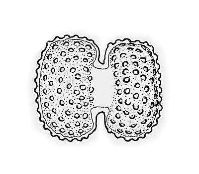
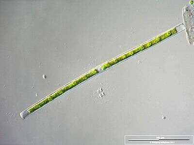
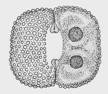
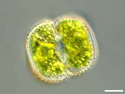
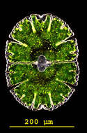
Cosmarium quadrum P. LUNDELL The cells are a little longer than wide, rounded off rectangular in shape. The sides are straight or flat convex. The central cuts are deep, linear, and extended outwards. The cell wall is covered with warts. They run in crossing rows (under an angle of appr. 45°). Around each of the warts are pores, which build symmetrical hexagon in their arrangement. Length 55 - 85 µm, width 50 - 80 µm. Occurrence: Common in littoral region and quaking bogs of moor ponds and in moderate acidic moorlands in Central Europe.
-
Scale bar indicates 25 µm.Sample from the pond Hegne Moor situated in the vicinity of Lake Constance. The image was built up using several photomicrographic frames with manual stacking technique. Images were taken using Zeiss Universal with Olympus C7070 CCD camera.Image under Creative Commons License V 3.0 (CC BY-NC-SA).
-
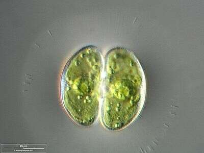
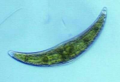
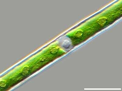
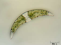
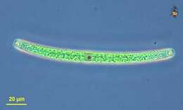
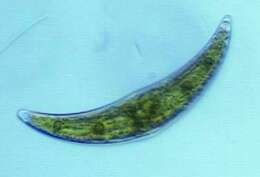
Closterium (claws-tear-ee-um), a common and widespread desmid, a type of green alga. Organisms usually comprised of two mirror image parts. Typically from slightly acidic environments. With a cellulosic cell wall and chlorophyll B containing plastids. Phase contrast.
-
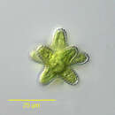
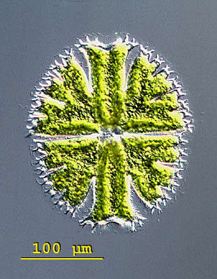
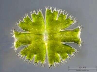
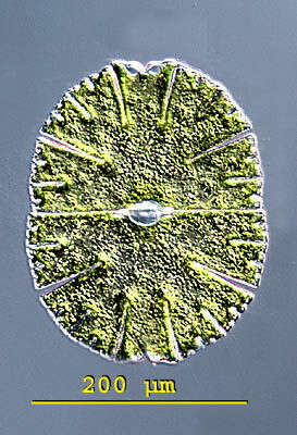
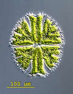
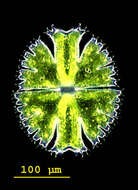
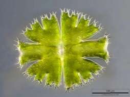
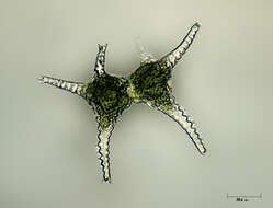
Micrasterias(mike-raz-tear-ee-ass) is a genus of unicellular algae in the family Desmidiaceae. The cells are flattened and disc-like. The cells of the genus Micrasterias are organized in two semi-cells that are mirror images of each other. The semicells have a distinctive shape with an intricate lobes and indentation. At the end of the lobes the cell wall may sometimes form notches or short spines. The nucleus is located in the centre between the semicells. Each semicell has a chloroplast with some pyrenoids. Usually found in oligotrophic, acid waters. This specimen of Micrasterias apiculata was collected in the Salzburger Land, Austria. Differential interference contrast.
-
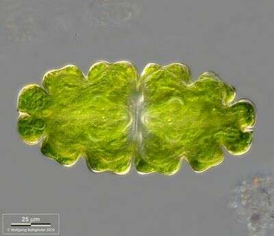
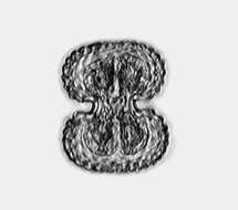
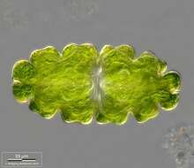
Cosmarium reniforme (RALFS) ARCHER var. alaskanum CROASDALE The cells and the cell halves are rectangular, the cell ends are weakly rounded off. The central cuts are opened widely along the entire length. The cell wall is covered with numbers of spherical warts, between these small pores. Length 50 µm, width 40 µm. Occurrence: The habitat is apparently limited to northern latitudes.
-
Scale bar indicates 10 µm. Sample from a wetland at the Pillersee (Tyrol, Austria). The image was built up using several photomicrographic frames with manual stacking technique. Images were taken using Zeiss Universal with Olympus C7070 CCD camera.Image under Creative Commons License V 3.0 (CC BY-NC-SA).
-
-
-
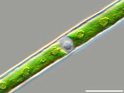
Closterium (claws-tear-ee-um), a common and widespread desmid, a type of green alga. Organisms usually comprised of two mirror image parts. Typically from slightly acidic environments. With a cellulosic cell wall and chlorophyll B containing plastids. Phase contrast.
-
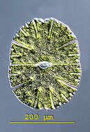
Micrasterias(mike-raz-tear-ee-ass) is a genus of unicellular algae in the family Desmidiaceae. The cells are flattened and disc-like. The cells of the genus Micrasterias are organized in two semi-cells that are mirror images of each other. The semicells have a distinctive shape with an intricate lobes and indentation. At the end of the lobes the cell wall may sometimes form notches or short spines. The nucleus is located in the centre between the semicells. Each semicell has a chloroplast with some pyrenoids. Usually found in oligotrophic, acid waters. This specimen of Micrasterias apiculata was collected in the Salzburger Land, Austria. Dark ground illumination.
-
Cosmarium reniforme (RALFS) ARCHER var. alaskanum CROASDALE The cells and the cell halves are rectangular, the cell ends are weakly rounded off. The central cuts are opened widely along the entire length. The cell wall is covered with numbers of spherical warts, between these small pores. Length 50 µm, width 40 µm. Occurrence: The habitat is apparently limited to northern latitudes.
-
Scale bar indicates 100 µm.Sample from the pond Hegne Moor situated in the vicinity of Lake Constance. The image was built up using several photomicrographic frames with manual stacking technique. Images were taken using Zeiss Universal with Olympus C7070 CCD camera.Image under Creative Commons License V 3.0 (CC BY-NC-SA).
-
Bright field.
-
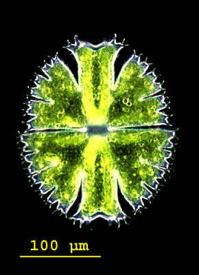
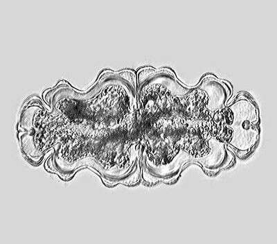
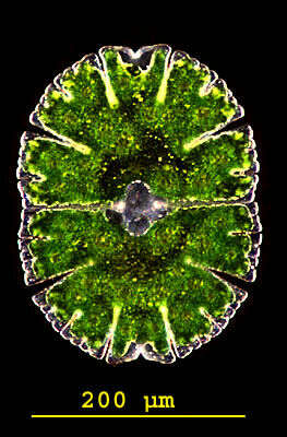
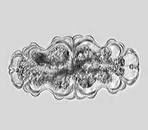
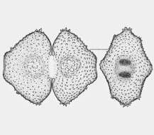
The picture shows the outline and the texture of the cell wall with the numerous spines on the cell wall and was built up using 38 high resolution DIC frames with manual stacking technique using Corel Photopaint. The scale bar indicates 100 µm. Sample from sphagnum pond situated in the northern alpine region of Austria near Salzburg. Images were taken using Zeiss Universal with Olympus C7070 CCD camera.
-
Desmids have the ability to move slowly on the surface excreting mucilage out of special pores. The picture shows this pores of Euastrum oblongum. High resolution DOF picture assembled of over 40 shots (manually stacked) taken with Planapo 63/1.4 showing the multilevel surface structur. See zip archive for details. Sample from sphagnum pond situated in the northern alpine region of Austria near Salzburg. Images were taken using Zeiss Universal with Olympus C7070 CCD camera.
-
Zoom-in to the center of the cell with nucleus, chloroplasts with pyrenoids. Scale bar indicates 25 µm.Sample from the pond Hegne Moor situated in the vicinity of Lake Constance. The image was built up using several photomicrographic frames with manual stacking technique. Images were taken using Zeiss Universal with Olympus C7070 CCD camera.Image under Creative Commons License V 3.0 (CC BY-NC-SA).
-
-
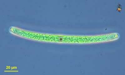
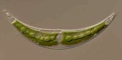
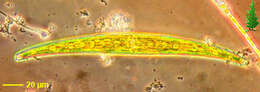
Closterium, a common desmid, is very distinctive because of its smile shape. The nucleus is in the central region and the bright green chloroplasts extend into the two sides of the cell. The small circular pyrenoids are located in the chloroplasts. This desmid was collected from Obsidian Creek.
-
Micrasterias(mike-raz-tear-ee-ass) is a genus of unicellular algae in the family Desmidiaceae. The cells are flattened and disc-like. The cells of the genus Micrasterias are organized in two semi-cells that are mirror images of each other. The semicells have a distinctive shape with an intricate lobes and indentation. At the end of the lobes the cell wall may sometimes form notches or short spines. The nucleus is located in the centre between the semicells. Each semicell has a chloroplast with some pyrenoids. Usually found in oligotrophic, acid waters. This specimen of Micrasterias denticulata collected in the Salzburger Land, Austria. Differential interference contrast.
-
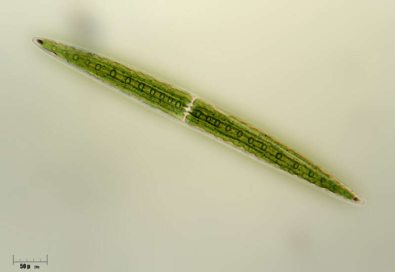
Euastrum oblongum (GREV.) RALFS ex RALFS The cells are appr. 2 times longer than wide and slenderly elliptical in shape. The cell halves consists of two clearly distinguishable sets of lobes which are weakly concave in the center. The vertex lobes are clearly contrasted, widened wedge-shaped with a cut in the center. The cuts in the middle of the cell are not peripherally widened. There exists a big pore in the middle of the cell halves between the two humps. Further humps are visible at the lateral lobes. The cell wall is covered with densely packed pores. Length 150 - 170 µm, width 70 - 85 µm. Occurrence: Adaptable alga, ubiquitous
-
-
Collected from Cumloden Swamp on October 7, 2002.
-
Micrasterias(mike-raz-tear-ee-ass) is a genus of unicellular algae in the family Desmidiaceae. The cells are flattened and disc-like. The cells of the genus Micrasterias are organized in two semi-cells that are mirror images of each other. The semicells have a distinctive shape with an intricate lobes and indentation. At the end of the lobes the cell wall may sometimes form notches or short spines. The nucleus is located in the centre between the semicells. Each semicell has a chloroplast with some pyrenoids. Usually found in oligotrophic, acid waters. This specimen of Micrasterias denticulata collected in the Salzburger Land, Austria. Dark ground illumination.
-
Euastrum verrucosum EHRENB. var. groenlandicum (LARSEN) WILLI KRIEG So far only two quite inaccurate illustrations of this alga exist: The original illustration of LARSEN (1904) and an illustration of GROENBLAD (1952). The cell halves are rounded off trapezoidal, both the lateral lobes and the vertex lobe drawn weakly into the center turn into one another, separated only by a shallow emargination. At the ends of the lateral lobes there are several short pricks. The central cuts are far opened. The hump near the isthmus range of the cell halves are covered with large warts, the remaining cell wall with concentric rows of small warts. Length 100 - 105 µm, width 80 - 85 µm. Occurrence: So far only known from Greenland