-
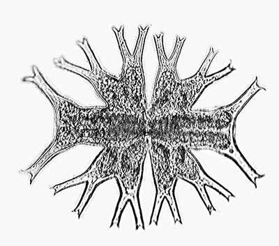
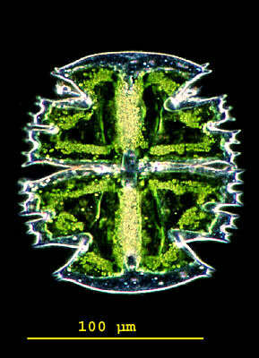
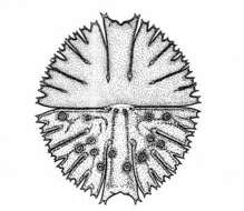
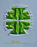
Euastrum germanicum (SCHMIDLE) WILLI KRIEG. he cells are only a little longer then wide. The cell halves constist of five broadly rounded lobes. The vertex lobes which are outward somewhat widened are separated from the lobes by extended, inside rounded recessings. Each center of the cell halves showes a hemispheric bump. The cellwall is covered with parallel rows of spines. The central cuts are slender and broadly opened towards the periphery. Dimension: Length 50 â 60 µm, width 40 â 50 µm Ecology: Especially in medium acidic to weakly alcaline waters of ponds and alluvial areas (Danube meadows) Occurrence: Mainly in Europe, but probably ubiquitous
-
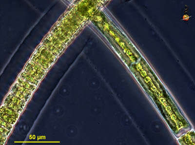
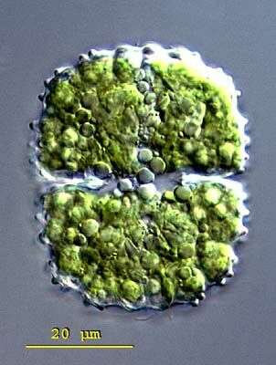
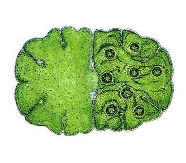
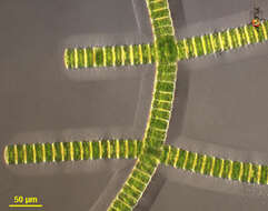
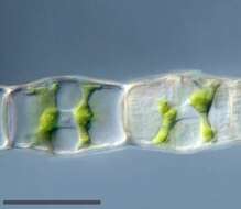
Filemaoentous desmids, cells located within a thick mucus sheath. A green alga, with cellulosic cell walls and bright green chloroplasts. Two forms are shown here. Phase contrast micrograph.
-
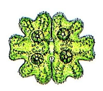
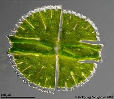
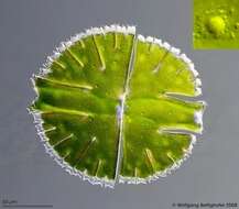
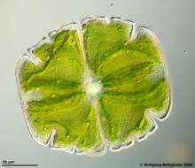
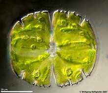
Micrasterias rotata (GREV.) RALFS Length: 200 â 300 µm, width: 200 â 270 µm. This specie is very tolerant concerning living conditions. Therefore the species is widely spread in all altitudes, in forestal ditches and lowland fens sometimes abundant. The cells are 1.08 to 1.15 times longer than wide, the shape seems almost circular or wide elliptical. The cell is devided into lobes due to deep cuts, the terminations of lobes are denticulated. The central lobe is broadened evenly at the end. The termination is formed concavely and is lightly arched upwards at both sides. The lateral angles of the central lobe are little denticulated. The cut in the middle of the cell (sinus) is very deep and peripherally a little widened. The cellwall is densly punctuated by tiny pores. The Chromatophores have several scattered pyrenoids with varying sizes.
-
Euastrum germanicum (SCHMIDLE) WILLI KRIEG. The cells are only a little longer then wide. The cell halves constist of five broadly rounded lobes. The vertex lobes which are outward somewhat widened are separated from the lobes by extended, inside rounded recessings. Each center of the cell halves showes a hemispheric bump. The cellwall is covered with parallel rows of spines. The central cuts are slender and broadly opened towards the periphery. Dimension: Length 50 â 60 µm, width 40 â 50 µm Ecology: Especially in medium acidic to weakly alcaline waters of ponds and alluvial areas (Danube meadows) Occurrence: Mainly in Europe, but probably ubiquitous
-
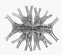
FIlaments (unhappy) observed in freshwater sediments in the vicinity of Broome, Western Australia in September 2003. This image was taken using differential interference contrast optics. Â Â This work was supported by the Australian Biological Resources Study.
-
The picture shows the outline, the texture of the cell wall and some dictyosomes producing mucilage for motion (see also inserted image). This multi layer image was built up using 40 high resolution DIC frames with manual stacking technique using Corel Photopaint. The scale bar indicates 50 µm. Sample from sphagnum pond situated in the northern alpine region of Austria near Salzburg. Images were taken using Zeiss Universal with Olympus C7070 CCD camera.
-
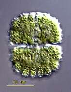
Euastrum intermedium CLEVE The celles are almost two times longer than wide. The cell sides are loboid broadly rounded. The vertex lobes are protruding clearly, broadened on the ends and with a cut in the middle. In the center of the cell halves there are pairs of bumps which are covered with small warts. The central cuts are far opened towards the periphery. Dimension: Length 70 â 80 µm, width 35 â 45 µm Ecology: Acidophilic alga, rather rare in shallow waters between sphagnum. Occurrence: Ubiquitous, prefers boreal areas (mountains), rather rare in Central Europe.
-
This image of filamentous desmids shows the mucus layer in which the cells are embedded.
-
This optical medial section of the desmid cell shows the outline, the texture of the chloroplast with many pyrenoids and the nucleus at the center of the cell. This multi layer image was built up using 9 high resolution DIC frames with manual stacking technique using Corel Photopaint. Sample from sphagnum pond situated in the northern alpine region of Austria near Salzburg. Images were taken using Zeiss Universal with Olympus C7070 CCD camera.
-
Euastrum intermedium CLEVE The celles are almost two times longer than wide. The cell sides are loboid broadly rounded. The vertex lobes are protruding clearly, broadened on the ends and with a cut in the middle. In the center of the cell halves there are pairs of bumps which are covered with small warts. The central cuts are far opened towards the periphery. Dimension: Length 70 â 80 µm, width 35 â 45 µm Ecology: Acidophilic alga, rather rare in shallow waters between sphagnum. Occurrence: Ubiquitous, prefers boreal areas (mountains), rather rare in Central Europe.
-
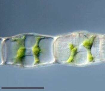
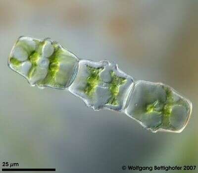
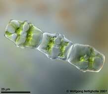
This filamentous desmid with its charcteristic outline has a delicate textured cell wall. Each cell holds two chloroplasts with nucleus between them. Multi layer image using 7 high resolution DIC frames with manual stacking technique using Corel Photopaint. The scale bar indicates 25 µm. Sample from sphagnum pond Dosenmoor near Neumuenster (Schleswig-Holstein, Germany). Images were taken using Zeiss Universal with Olympus C7070 CCD camera.
-
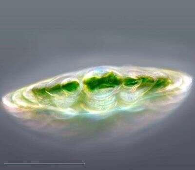
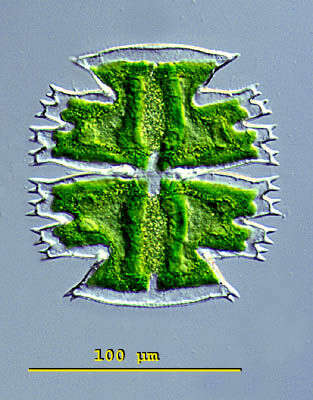
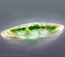
A very special view of Micrasterias rotata using an inverted microscope. The cell ist standing perpenticular upon its apical lobe. Like all the desmids Micrasterias can move using mucilage for backstroke. They move towards the light. When it's dark they erect with help of mucilage ejection. The scale bar indicates 100 µm. Sample from sphagnum pond situated in the northern alpine region of Austria near Salzburg. Images were taken using Zeiss IM35 with Olympus C7070 CCD camera.
-
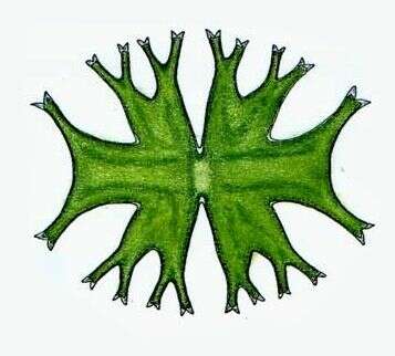
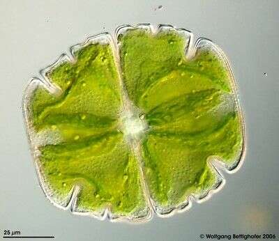
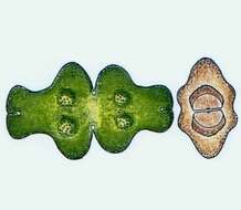
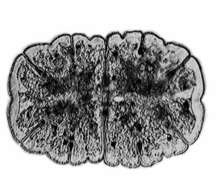
Micrasterias furcata RALFS The cells are about as long as wide and have a circular shape. The cell halves consist of three lobes. The lateral lobes are clearly separated. At the ends of the lateral lobes there are two outwards standing projections. Between the projections there is a concave area. The central cut is deep and peripherally strongly widened. Dimension: Length 150 - 180 µm, width 150 â 160 µm Ecology: In acidic to moderate acidic fens and bogs. Occurrence: Ubiquitous, rather rare in Central Europe.
-
A developing filament with only 3 cells is shown. Mulit layer image using 3 high resolution DIC frames with manual stacking technique using Corel Photopaint. Sample from sphagnum pond Dosenmoor near Neumuenster (Schleswig-Holstein, Germany). Images were taken using Zeiss Universal with Olympus C7070 CCD camera.
-
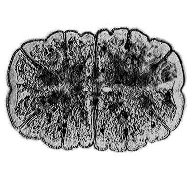
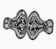
Micrasterias(mike-raz-tear-ee-ass) is a genus of unicellular algae in the family Desmidiaceae. The cells are flattened and disc-like. The cells of the genus Micrasterias are organized in two semi-cells that are mirror images of each other. The semicells have a distinctive shape with an intricate lobes and indentation. At the end of the lobes the cell wall may sometimes form notches or short spines. The nucleus is located in the centrfe between the semicells. Each semicell has a chloroplast with some pyrenoids. Usually found in oligotrophic, acid waters. This is a specimen of Micrasterias truncata collected in a moor pond located in the vicinity of Konstanz, Germany. Differential interference contrast.
-
Micrasterias furcata RALFS The cells are about as long as wide and have a circular shape. The cell halves consist of three lobes. The lateral lobes are clearly separated. At the ends of the lateral lobes there are two outwards standing projections. Between the projections there is a concave area. The central cut is deep and peripherally strongly widened. Dimension: Length 150 - 180 µm, width 150 â 160 µm Ecology: In acidic to moderate acidic fens and bogs. Occurrence: Ubiquitous, rather rare in Central Europe.
-
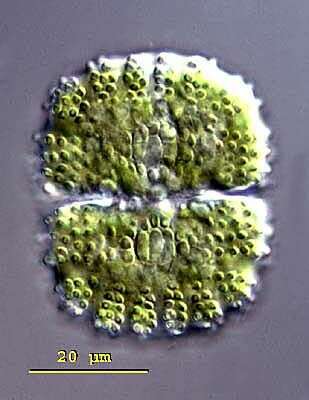
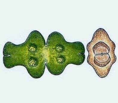
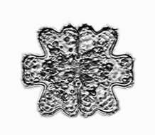
Cosmarium (cos-may-ree-um) caelatum. Cosmarium is a very common and large genus of alga found usually in oligotrophic, acid waters. The cells of this genus are composed of two semi-cells, constricted in the middle. This region is termed the isthmus and is where the nucleus is found. The outer portions of each semi-cell contain a single, large chloroplast. The outer cell wall of each semi-cell is covered with pores and can be very ornate with the pattern being useful in distinguishing among species. The cells move slowly using mucilage secretion to create the force for movement. Both asexual and sexual reproduction occurs. The asexual reproduction is by cell division and the sexual reproduction involves the formation of zygospores. The gametes migrate from the parental cells, passing through pores to fuse in a region midway between the parental walls. The zygote can form a very ornate wall. This specimen was collected in a moor located in the Salzburger Land, Austria. This image emphasizes the ornate cell wall of Cosmarium ornatum. This specimen measures 49 microns long and 38 microns wide.
-
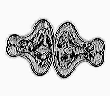
Micrasterias(mike-raz-tear-ee-ass) is a genus of unicellular algae in the family Desmidiaceae. The cells are flattened and disc-like. The cells of the genus Micrasterias are organized in two semi-cells that are mirror images of each other. The semicells have a distinctive shape with an intricate lobes and indentation. At the end of the lobes the cell wall may sometimes form notches or short spines. The nucleus is located in the centrfe between the semicells. Each semicell has a chloroplast with some pyrenoids. Usually found in oligotrophic, acid waters. This is a specimen of Micrasterias truncata collected in a moor pond located in the vicinity of Konstanz, Germany. Dark ground illumination.
-
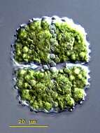
Micrasterias jenneri RALFS The cells are appr. 1.5 times longer than wide, rounded off rectangular in shape. The cells have five lobes, the lateral lobes are broadened towards the periphery and emaginated in the center, the cuts between them are closed and short. The lateral lobes arenât spread apart, they are strongly widened peripherally and rounded with shallow emarginations in the center. The central cut is deep, closed, and not peripherally widened. Dimension: Length 150 - 180 µm, width 100 â 130 µm Ecology: Acidophilic alga, lives in sphagnum ponds together with other algae which prefer upland moor waters. Occurrence: Probably ubiquitous, mainly in colder zones.
-
Cosmarium (cos-may-ree-um) caelatum. Cosmarium is a very common and large genus of alga found usually in oligotrophic, acid waters. The cells of this genus are composed of two semi-cells, constricted in the middle. This region is termed the isthmus and is where the nucleus is found. The outer portions of each semi-cell contain a single, large chloroplast. The outer cell wall of each semi-cell is covered with pores and can be very ornate with the pattern being useful in distinguishing among species. The cells move slowly using mucilage secretion to create the force for movement. Both asexual and sexual reproduction occurs. The asexual reproduction is by cell division and the sexual reproduction involves the formation of zygospores. The gametes migrate from the parental cells, passing through pores to fuse in a region midway between the parental walls. The zygote can form a very ornate wall. This specimen is 49 microns long and 38 microns wide.
-
Non-filamentous desmids have the ability to move slowly by means of directed mucilage secretion. Depht of focus approach can show cell surface together with folded chloroplasts, cell contour and nucleus. In zip archive there are more DOF pictures of this object. Picture generated from 9 shots using CombineZ by Alan Hadley, MicroPicS by Bernhard Wiedemann and Photoshop. Sample from spagnum pond Dosenmoor near Neumuenster (Schleswig-Holstein, Germany). Images were taken using Zeiss Universal with Olympus C7070 CCD camera.
-
Micrasterias jenneri RALFS The cells are appr. 1.5 times longer than wide, rounded off rectangular in shape. The cells have five lobes, the lateral lobes are broadened towards the periphery and emaginated in the center, the cuts between them are closed and short. The lateral lobes arenât spread apart, they are strongly widened peripherally and rounded with shallow emarginations in the center. The central cut is deep, closed, and not peripherally widened. Dimension: Length 150 - 180 µm, width 100 â 130 µm Ecology: Acidophilic alga, lives in sphagnum ponds together with other algae which prefer upland moor waters. Occurrence: Probably ubiquitous, mainly in colder zones.
-
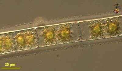
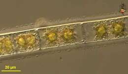
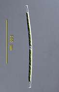
Gonatozygon (go-nat-owes-eye-gone) brebissonii. Cells are mostly elongated and cylindrical and slightly bent. The apices are swollen and rounded. The nucleus is located centrally. The cell wall may be covered with dots, granules or short spines. Each semi-cell contains a single, ribbon-like chloroplast with many aligned pyrenoids. The chloroplasts may be slightly spiralled and are centrally placed. Cells can be loosely attached together to form a filament. This genus is found in moderate to weakly alkaline waters (pH 6-6.8) in bog ponds or moors. This specimen was collected in freshwater ponds near Konstanz, Germany. The two separated chloroplasts are visible as is the central located nucleus. 200 microns. Differential interference contrast.
-
In photosynthesis the enzyme RuBisCO (Ribulose-1,5-Biphosphat- Carboxylase/Oxygenase) plays a very important role. Many algae concentrate RuBisCO in special grains named pyrenoids. The depth of focus picture shows the structured chloroplasts wth their pyrenoids. Micrasterias truncata has a mucous envelope. The genus have two types of pores, small ones situated all over the cell surface for building their mucous envelope and special ones with bigger lumen to excrete mucilage for movement. A high resolution taking in zip archive showes the numerous small ones. Deph of focus technique was used to gather details from over 40 shots. See zip archive for more. Sample from spagnum pond situated in the northern alpine region of Austria near Salzburg. Images were taken using Zeiss Universal with Olympus C7070 CCD camera.