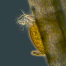
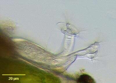
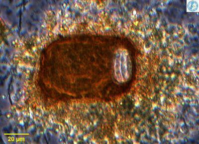
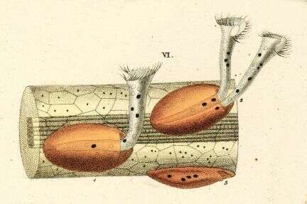
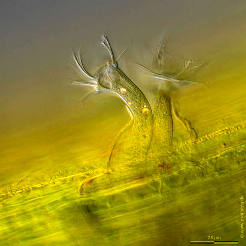
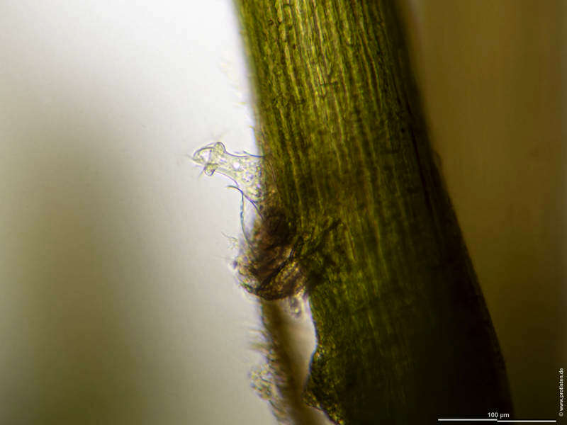
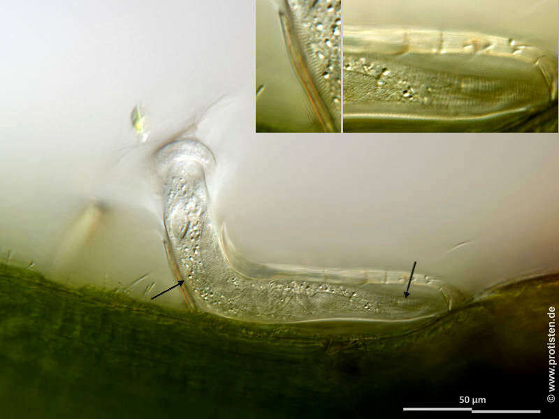
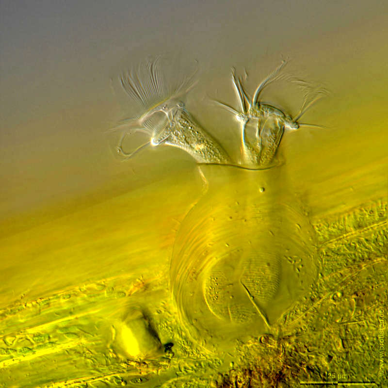
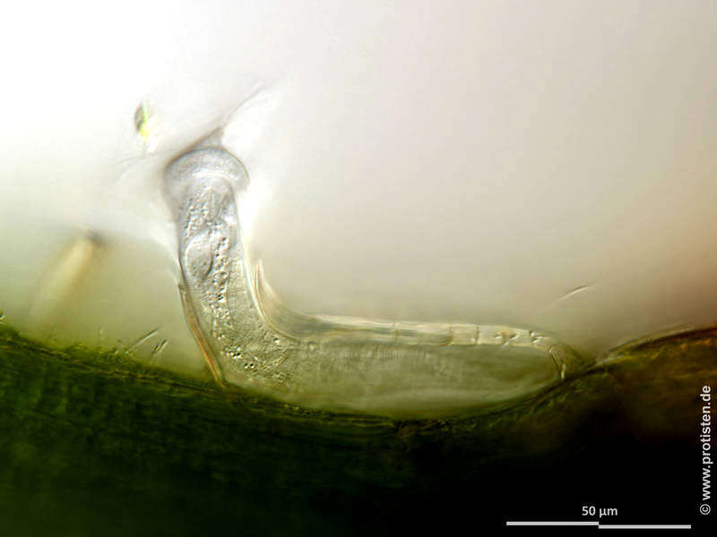
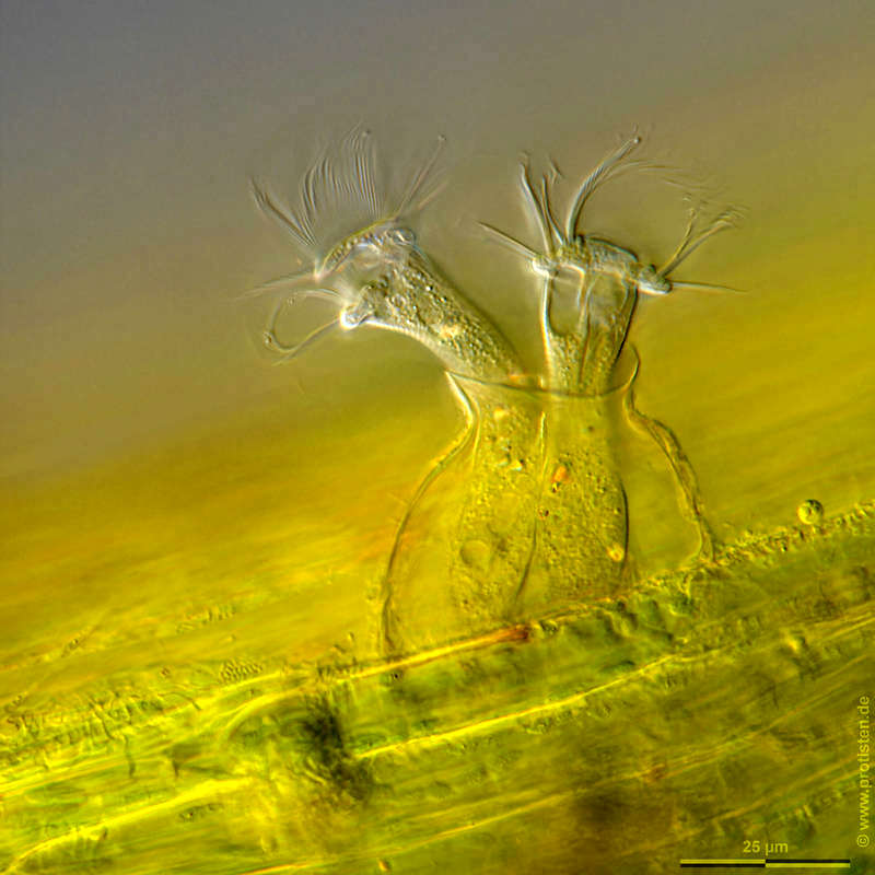
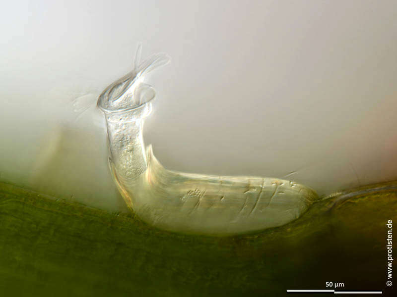
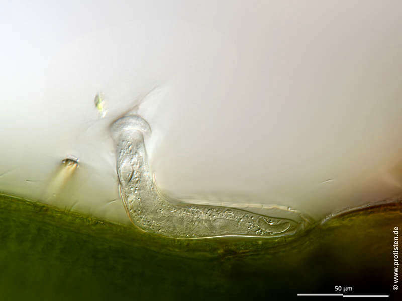
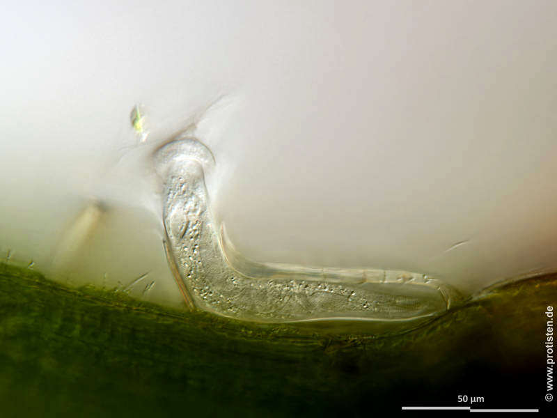
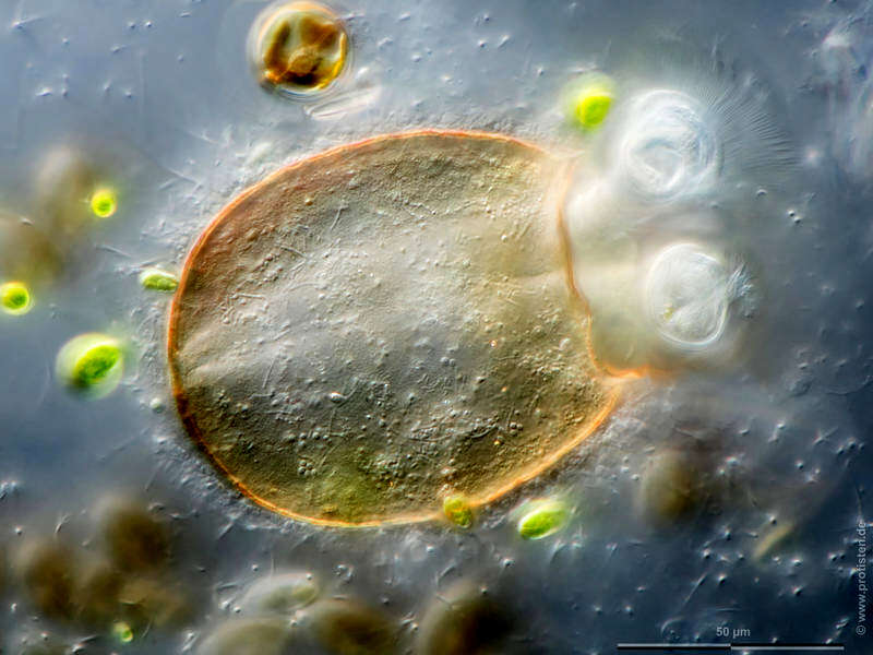
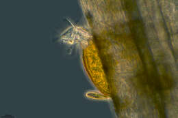
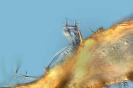
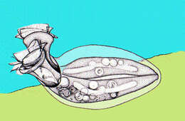
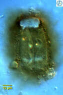
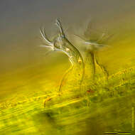
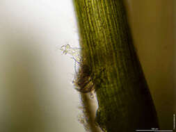
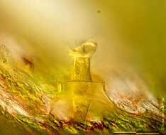
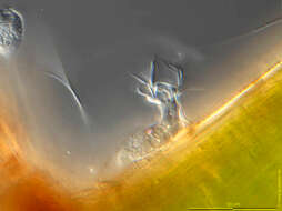
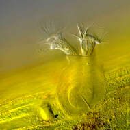
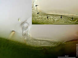
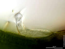
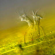
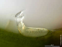
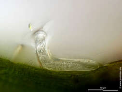
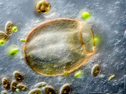
Scale bars indicate 50 µm.Seven images, six of them in a slide changer.First (in the slide changer):Complete representation of the trophont and its lorica.Second:Optical cross-section showing the elliptical shaped vestibulum.Third and fourth:Optical cross-section. The sectional plane is placed in such a way that the striation on the cell body is partially shown (arrows and insets in fourth image). Objective 40x/1.1 water immersion.Fifth and sixth:Optical cross-section through the cell and the lorica showing several parts of the worm-shaped macronucleus traversing almost the entire cell (arrows and insets in sixth image). The brightly glowing dots in the cell body are mitochondria.Please click on < or > on the image edges or on the dots at the bottom edge of the images to browse through the slides!Place name: Tropical freshwater aquariumLatitude: 54.3018013 Longitude: 10.07120132Microscope Zeiss Axioplan, camera Olympus OM-D M5 MKII. DOF images.© Wolfgang Bettighofer,images under Creative Commons License V 3.0 (CC BY-NC-SA).For permission to use of (high resolution) images please contact
postmaster@protisten.de.For further information about the image, please click here:
Link to protisten.de page