-
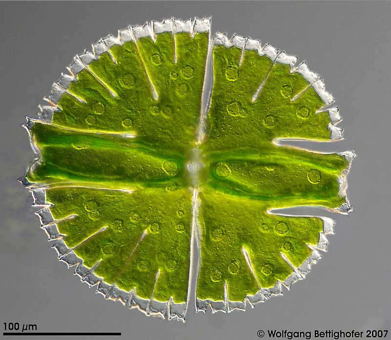
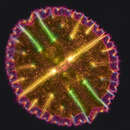
Micrasterias rotata This optical transversal section of the desmid cell shows the outline, the surface texture of the chloroplast with many pyrenoids and the nucleus at the center of the cell. This picture was built up using 9 high resolution DIC frames with manual stacking technique. Sample from sphagnum pond situated in the northern alpine region of Austria near Salzburg. Images were taken using Zeiss Universal with Olympus C7070 CCD camera.Image under Creative Commons License V 3.0 (CC BY-NC-SA). Place name: Bogs near Salzburg (Austria) Latitude: 48.068516 Longitude: 12.954134 Dieser optische Querschnitt der Desmidiaceenzelle zeigt den Umriss und das Oberflächenmuster der Chloroplasten mit vielen Pyrenoiden und den Kern in der Zellmitte. Tiefenschärfe durch Multiebenenabbildung aus 9 Bildebenen, manuell gestapelt. Probe aus einem Moor in den nördlichen Kalkalpen von Österreich in der Nähe von Salzburg. Mikrotechnik: Zeiss Universal, Kamera: Olympus C7070. Creative Commons License V 3.0 (CC BY-NC-SA). For permission to use of (high-resolution) images please contact postmaster@protisten.de.
-
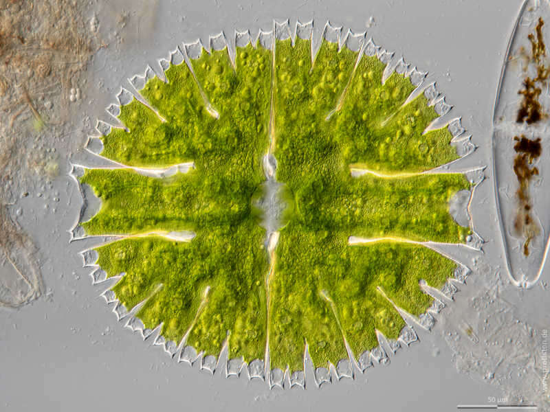
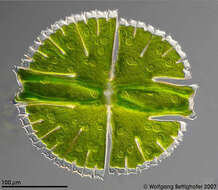
Micrasterias rotata The optical cross section through the cell shows pyrenoids. In the pyrenoids, the cell produces the storage substance starch, which is deposited as a shell around them. The nucleus (light disk) is located in the middle of the cell. Sample from wetland Lauchseemoor, Fieberbrunn, Tyrol, Austria. Sampling date 06/2023. The image was built up using several photomicrographic frames with manual stacking technique. Images were taken using Zeiss Axioplan with Olympus OM-D M5 MKII. Image under Creative Commons License V 3.0 (CC BY-NC-SA). Place name: Wetland Lauchseemoor, Fieberbrunn (Tyrol, Austria) Latitude: 47.46954439 Longitude: 12.53826499 Der optische Querschnitt durch die Zelle zeigt Pyrenoide. In den Pyrenoiden produziert die Zelle den Reservestoff Stärke, dieser wird als Hülle um diese abgeschieden. In der Zellmitte befindet sich der Zellkern (helle Scheibe). Multiebenen-Abbildung, manuell gestapelt. Der Messbalken markiert eine Länge von 50 µm. Probe aus dem Lauchseemoor bei Fieberbrunn, Tirol. Datum der Aufsammlung: 06/2023. Mikrotechnik: Zeiss Axioplan, Kamera: Olympus OM-D M5 MKII. Creative Commons License V 3.0 (CC BY-NC-SA). For permission to use of (high-resolution) images please contact postmaster@protisten.de.
-
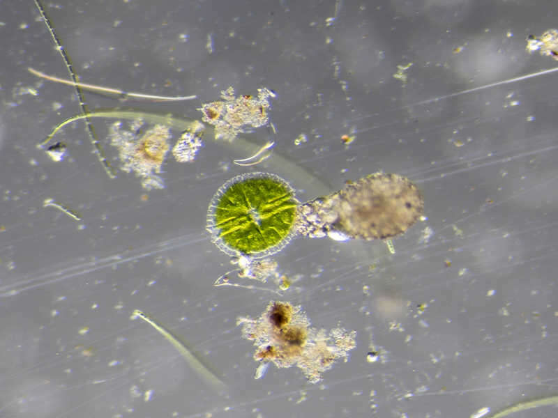
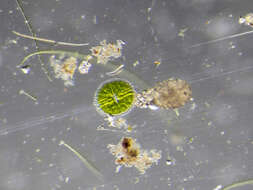
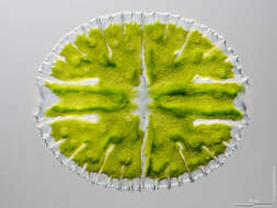
Micrasterias rotata Sample from a pond near Großostheim, Germany. Sampling date 04/2021. Images were taken using stereo microscope Olympus SZX16 with Olympus OM-D M5 MKII. Image under Creative Commons License V 3.0 (CC BY-NC-SA). Place name: Pond near Großostheim (Germany) Latitude: 49.88482168 Longitude: 9.09980822 Probe aus einem Waldteich bei Großostheim. Datum der Aufsammlung: 04/2021. Mikrotechnik: Stereomikroskop Olympus SZX16, Kamera: Olympus OM-D M5 MKII. Creative Commons License V 3.0 (CC BY-NC-SA). For permission to use of (high-resolution) images please contact postmaster@protisten.de.
-
Micrasterias rotata Scale bar indicates 50 µm. Sample from a pond near Großostheim, Germany. Sampling date 04/2021. The image was built up using several photomicrographic frames with manual stacking technique. Images were taken using Zeiss Axioplan with Olympus OM-D M5 MKII. Image under Creative Commons License V 3.0 (CC BY-NC-SA). Place name: Pond near Großostheim (Germany) Latitude: 49.88482168 Longitude: 9.09980822 Multiebenen-Abbildung, manuell gestapelt. Der Messbalken markiert eine Länge von 50 µm. Probe aus einem Waldteich bei Großostheim. Datum der Aufsammlung: 04/2021. Mikrotechnik: Zeiss Axioplan, Kamera: Olympus OM-D M5 MKII. Creative Commons License V 3.0 (CC BY-NC-SA). For permission to use of (high-resolution) images please contact postmaster@protisten.de.