In June, 2014, a 14-year-old immuno-compromised boy from Wisconsin ended up in the hospital with life-threatening encephalitis requiring a medically induced coma, following undiagnosable complaints and on and off hospitalization for headaches and fevers for two years. All tests for specific pathogen suspects returned negative.As a last resort, doctors employed an experimental DNA therapy, which involved “unbiased next generation” sequencing techniques to sequence all the DNA molecules in a series of body fluid samples from the patient.This sequencing technology, coupled with just-developed bioinformatics “sequence-based ultra-rapid pathogen identification,” (SURPI) software, compared eight million DNA sequences in the boy’s samples with all known pathogen DNA sequences from National Center for Biotechnology Information (NCBI) databases, ultimately identifying a Leptospira pathogen within 48 hours. This solves the problem “that your critically ill patient will be dead by the time you make a diagnosis,” says Dr. Charles Chiu, one of the creators of this diagnostic technology.The case is a first in using sequencing to identify and facilitate treatment of microbe-borne illness; although expensive now, use of this technology for broader diagnosis of infections will certainly become more common. This case is “a demonstration that this technology has arrived,” said Dr. DeRisi biochemist and collaborator with Chiu.
(Naccache et al. 2014; Norris 2014; Wilson et al. 2014; Zimmer 2014).
Leptospira is a genus of spiral-shaped, gram-negative bacteria in family Leptospiraceae, order Spirochaetales. Before 1989 the different strains, or serotypes, were organized into two species: L. interrogans, the pathogenic varieties, and L. biflexa, which are harmless.Genetic techniques have since helped group the more than 250 pathogenic known “serovars” (types, based mostly on cell surface proteins, and generally using specific mammalian or marsupial hosts) and more than 50 non-pathogenic known serovars into 21 species for formal classification.Because serovars regularly exchange genetic information, this serotype system is still used, especially in clinical practice.
Leptospira serotypes are found world wide, except in Antarctica.They are common in warm wet climates especially where sanitation is poor, as the bacteria are spread in the urine of infected hosts.Common hosts are rats, and other rodents, cattle and muskrats. In humans and other vertebrates, pathogenetic Leptospira cause leptospirosis.Infection occurs by ingesting the spirochete bacteria in contaminated food, water, or through skin contact, especially when broken skin comes in contact with contaminated water. Outbreaks regularly occur in developing countries especially in wet seasons or after flooding. Most cases are mild or show no symptoms, but leptospirosis is a potentially life-threatening disease which usually manifests as flu-like symptoms in early stage, progressing to symptoms including encephalitis and dysfunction of kidneys, liver, reproductive organs and lungs and ultimately leading to the failure of these organs. Number of human cases is estimated at up to 100 cases per 100,000 persons per year in topical climates, fewer in temperate locales.Though diagnosis can be difficult, once diagnosed leptospirosis can be effectively treated with antibiotics.Leptospirosis also affects a variety of mammals including farm animals, pets (dogs and cats), opposums, rats, deer, South American camelids, and while generally rare among wild animals, wild pinnipeds regularly become infected. Leptospira bacteria cannot multiply outside their hosts but in warm conditions can remain viable in water or wet soil for months (Levett 2001; Norris 2014; Ren et al. 2003; Spickler and Leedom Larson 2013; WHO 2014; Zimmer 2014).
Levett (2001) provides a detailed review of the biology, taxonomy, epidemiology, clinical features, and pathogenesis of leptospires.
Recent high tech sequencing and bioinformatics methods diagnosed a Leptospira infection in a patient whose symptoms could not be identified, a first use of new powerful medical technology (Naccache et al. 2014; Norris 2014; Wilson et al. 2014; Zimmer 2014).
In June, 2014, a 14-year-old immuno-compromised boy from Wisconsin ended up in the hospital with life-threatening encephalitis requiring a medically induced coma, following undiagnosable complaints and on and off hospitalization for headaches and fevers for two years. All tests for specific pathogen suspects returned negative.As a last resort, doctors employed an experimental DNA therapy, which involved “unbiased next generation” sequencing techniques to sequence all the DNA molecules in a series of body fluid samples from the patient.This sequencing technology, coupled with just-developed bioinformatics “sequence-based ultra-rapid pathogen identification,” (SURPI) software, compared eight million DNA sequences in the boy’s samples with all known pathogen DNA sequences from National Center for Biotechnology Information (NCBI) databases, ultimately identifying a Leptospira pathogen within 48 hours. This solves the problem “that your critically ill patient will be dead by the time you make a diagnosis,” says Dr. Charles Chiu, one of the creators of this diagnostic technology.The case is a first in using sequencing to identify and facilitate treatment of microbe-borne illness; although expensive now, use of this technology for broader diagnosis of infections will certainly become more common. This case is “a demonstration that this technology has arrived,” said Dr. DeRisi biochemist and collaborator with Chiu.
(Naccache et al. 2014; Norris 2014; Wilson et al. 2014; Zimmer 2014).
Leptospira (Ancient Greek: leptos, 'fine, thin' and Latin: spira, 'coil')[1] is a genus of spirochaete bacteria, including a small number of pathogenic and saprophytic species.[2] Leptospira was first observed in 1907 in kidney tissue slices of a leptospirosis victim who was described as having died of "yellow fever".[3]
Leptospira, together with the genera Leptonema and Turneria, is a member of the family Leptospiraceae. The genus Leptospira is divided into 20 species based on DNA hybridization studies.[4][5]
Pathogenic Leptospira
Intermediates or opportunistic Leptospira
Non-pathogenic Leptospira
Members of Leptospira are also grouped into serovars according to their antigenic relatedness. There are currently over 200 recognized serovars. A few serovars are found in more than one species of Leptospira.
At its 2002 meeting, the Committee on the Taxonomy of Leptospira of the International Union of Microbiological Societies approved the following nomenclature for serovars of Leptospira. Genus and species names are italicized as usual, with the serovar name not italicized and with an upper case first letter.
For example:
The currently accepted taxonomy is based on the List of Prokaryotic names with Standing in Nomenclature (LPSN)[10] and National Center for Biotechnology Information (NCBI).[11]
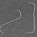
Although over 200 serotypes of Leptospira have been described, all members of the genus have similar morphology. Leptospira are spiral-shaped bacteria that are 6-20 μm long and 0.1 μm in diameter with a wavelength of about 0.5 μm.[18] One or both ends of the spirochete are usually hooked. Because they are so thin, live Leptospira are best observed by darkfield microscopy.
The bacteria have a number of degrees of freedom; when ready to proliferate via binary fission, the bacterium noticeably bends in the place of the future split.
Leptospira have a Gram-negative-like cell envelope consisting of a cytoplasmic and outer membrane. However, the peptidoglycan layer is associated with the cytoplasmic rather than the outer membrane, an arrangement that is unique to spirochetes. The two flagella of Leptospira extend from the cytoplasmic membrane at the ends of the bacterium into the periplasmic space and are necessary for the motility of Leptospira.[19]
The outer membrane contains a variety of lipoproteins and transmembrane outer membrane proteins.[20] As expected, the protein composition of the outer membrane differs when comparing Leptospira growing in artificial medium with Leptospira present in an infected animal.[21][22][23] Several leptospiral outer membrane proteins have been shown to attach to the host extracellular matrix and to factor H. These proteins may be important for adhesion of Leptospira to host tissues and in resisting complement, respectively.[24][25][26]
The outer membrane of Leptospira, like those of most other Gram-negative bacteria, contains lipopolysaccharide (LPS). Differences in the highly immunogenic LPS structure account for the numerous serovars of Leptospira.[18] Consequently, immunity is serovar specific; current leptospiral vaccines, which consist of one or several serovars of Leptospira endemic in the population to be immunized, protect only against the serovars contained in the vaccine preparation. Leptospiral LPS has low endotoxin activity.[18] An unusual feature of leptospiral LPS is that it activates host cells via TLR2 rather than TLR4.[27] The unique structure of the lipid A portion of the LPS molecule may account for this observation.[28] Finally, the LPS O antigen content of L. interrogans differs in an acutely infected versus a chronically infected animal.[29] The role of O antigen changes in the establishment or maintenance of acute or chronic infection, if any, is unknown.
Leptospira, both pathogenic and saprophytic, can occupy diverse environments, habitats, and life cycles; these bacteria are found throughout the world, except in Antarctica. High humidity and neutral (6.9–7.4) pH are necessary for their survival in the environment, with stagnant water reservoirs—bogs, shallow lakes, ponds, puddles, etc.—being the natural habitat for the bacteria.
Leptospira are cultivated at 30 °C in Ellinghausen-McCullough-Johnson-Harris (EMJH) medium, which can be supplemented with 0.21% rabbit serum to enhance growth of fastidious strains.[30] Growth of pathogenic Leptospira in an artificial nutrient environment such as EMJH becomes noticeable in 4–7 days; growth of saprophytic strains occur within 2–3 days. The minimal growth temperature of pathogenic species is 13–15 °C. Because the minimal growth temperature of the saprophytes is 5–10 °C, the ability of Leptospira to grow at 13 °C can be used to distinguish saprophytic from pathogenic Leptospira species.[30] The optimal pH for growth of Leptospira is 7.2–7.6.
Leptospira are aerobes whose major carbon and energy source during in vitro growth is long-chain fatty acids, which are metabolized by beta-oxidation.[31][32] Fatty acids are provided in EMJH in the form of Tween.[30] Fatty acid molecules are bound by albumin in EMJH and are released slowly into the medium to prevent its toxic accumulation.
Like most bacteria, Leptospira require iron for growth.[33] L. interrogans and L. biflexa have the ability to acquire iron in different forms.[34] A TonB-dependent receptor required for utilization of the ferrous form of the iron has been identified in L. biflexa, and an ortholog of the receptor is encoded in the genome of L. interrogans. L. interrogans can also obtain iron from heme, which is bound to most of the iron in the human body. The HbpA hemin-binding protein, which may be involved in the uptake of hemin, has been identified on the surface of L. interrogans[35] Although other pathogenic species of Leptospira and L. biflexa lack HbpA, yet another hemin-binding protein, LipL41, may account for their ability to use hemin as a source of iron.[35] Although they do not secrete siderophores, L. biflexa and L. interrogans may be capable of obtaining iron from siderophores secreted by other microorganisms.[34]
The genome of pathogenic Leptospira consists of two chromosomes. The size of the genomes of L. interrogans serovars Copenhageni and Lai is approximately 4.6 Mb.[36][37] However, the genome of L. borgpetersenii serovar Hardjo is only 3.9 Mb in size with a large number of pseudogenes, gene fragments, and insertion sequences relative to the genomes of L. interrogans.[38] L. interrogans and L. borgpetersenii share 2708 genes from which 656 are pathogenic specific genes. The guanine plus cytosine (GC) content is between 35% and 41%.[39] L. borgpetersenii serovar Hardjo is usually transmitted by direct exposure to infected tissues, whereas L. interrogans is often acquired from water or soil contaminated by the urine of carrier animals harboring Leptospira in their kidneys. The high number of defective genes and insertion sequences in L. borgpetersenii Hardjo together with the poor survival outside of the host and difference in transmission patterns compared to L. interrogans suggest that L. borgpetersenii is undergoing insertion-sequence mediated genomic decay, with ongoing loss of genes necessary for survival outside of the host animal.[38]
Genome sequence determination several strains of Leptospira lead to the development of multilocus VNTR (Variable Number of Tandem Repeats) typing and multilocus sequence typing (MLST) for species level identification of pathogenic Leptospira species.[40] Both methods hold the potential to replace the highly ambiguous serotyping method currently in vogue for leptospiral strain identification.[40]
Leptospira (Ancient Greek: leptos, 'fine, thin' and Latin: spira, 'coil') is a genus of spirochaete bacteria, including a small number of pathogenic and saprophytic species. Leptospira was first observed in 1907 in kidney tissue slices of a leptospirosis victim who was described as having died of "yellow fever".