Microsporaceae

描述:
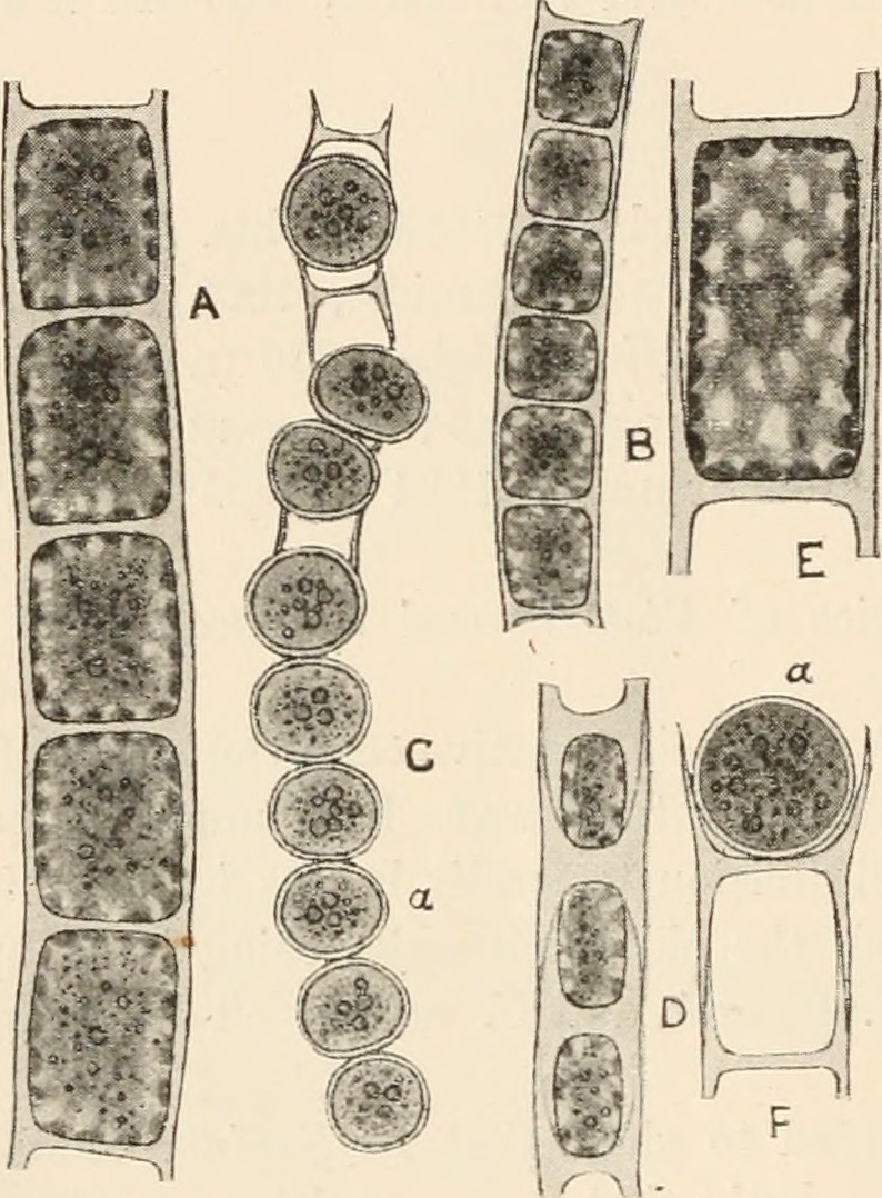
Description: English: Fig. 184. A, Microspora amœna (Kütz.) Lagerh. B and C, ? M. abbreriata (Rabenh.) Lagerh.; B, vegetative filament; C, filament with aplanospores (a). D, M. pachyderma (Wille) Lagerh. E, single vegetative cell of M. amœna var. crassior Hansg., showing the reticulated chloroplast. The indistinct blur in the centre of the cell indicates the position of the nucleus. F, fragment of filament of M. amœna with aplanospore (a). All x 520. Date: 1916. Source: https://www.flickr.com/photos/126377022@N07/14760704711/ page 301 of "Algæ. Vol. I. Myxophyceæ, Peridinieæ, Bacillarieæ, Chlorophyceæ, together with a brief summary of the occurrence and distribution of freshwat4er Algæ" (1916). Author: West, G. S. (George Stephen), 1876-1919.
来源信息
- 许可协议
- cc-publicdomain
- 创作者
- West, G. S. (George Stephen), 1876-1919
- 来源
- Internet Archive Book Images (126377022@N07)
- 原文
- 原始媒体文件
- 访问来源
- 合作网站
- Wikimedia Commons
- ID