Obligate intracellular protozoan parasites of the genus Leishmania are responsible for the vector-borne disease known as Leishmaniasis (transmitted by phlebotomine sandflies). Human infection is caused by about 21 of 30 species that infect mammals. These include the L. donovani complex with 3 species (L. donovani, L. infantum, and L. chagasi); the L. mexicana complex with 3 main species (L. mexicana, L. amazonensis, and L. venezuelensis); L. tropica; L. major; L. aethiopica; and the subgenus Viannia with 4 main species (L. (V.) braziliensis, L. (V.) guyanensis, L. (V.) panamensis, and L. (V.) peruviana). The different species are morphologically indistinguishable, but they can be differentiated by isoenzyme analysis, molecular methods, or monoclonal antibodies.
Leishmaniasis is found in parts of about 88 countries. Approximately 350 million people live in these areas. Most of the affected countries are in the tropics and subtropics. The settings in which leishmaniasis is found range from rain forests in Central and South America to deserts in West Asia. More than 90 percent of the world's cases of visceral leishmaniasis are in India, Bangladesh, Nepal, Sudan, and Brazil. Leishmaniasis is found in Mexico, Central America, and South America—from northern Argentina to Texas (not in Uruguay, Chile, or Canada), southern Europe (leishmaniasis is not common in travelers to southern Europe), Asia (not Southeast Asia), the Middle East, and Africa (particularly East and North Africa, with some cases elsewhere).
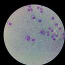
Leishmaniasis is transmitted to humans by the bite of infected female phlebotomine sandflies (Psychodidae:Phlebotominae). The sandflies inject the infective stage (i.e., promastigotes) from their proboscis during blood meals. Promastigotes that reach the puncture wound are phagocytized by macrophages and other types of mononuclear phagocytic cells. Progmastigotes transform in these cells into the tissue stage of the parasite (i.e., amastigotes), which multiply by simple division and proceed to infect other mononuclear phagocytic cells. Parasite, host, and other factors affect whether the infection becomes symptomatic and whether cutaneous or visceral leishmaniasis results. Sandflies become infected by ingesting infected cells during blood meals. In sandflies, amastigotes transform into promastigotes, develop in the gut (in the hindgut for leishmanial organisms in the Viannia subgenus; in the midgut for organisms in the Leishmania subgenus), and migrate to the proboscis.

Leishmania infantum is the causative agent of infantile visceral leishmaniasis in the Mediterranean region[1] and in Latin America, where it has been called Leishmania chagasi.[2][3] It is also an unusual cause of cutaneous leishmaniasis,[4] which is normally caused by specific lineages (or zymodemes). Wild canids and domestic dogs are the natural reservoir of this organism. The sandfly species Lutzomyia longipalpis serves as the primary vector for the transmission of the disease.[5]
Leishmania infantum is closely related to Leishmania donovani, and some authors believe that these two species are so close as to actually be subspecies of each other;[6] however, phylogenetic analyses can easily distinguish between the two groups despite no difference in morphology in the species complex. Some isolates formerly labelled L. donovani may be actually L. infantum.[2][7]
Comparative bioinformatic analyses showed that the size of the L. infantum BRCA2 protein is approximately three times smaller (125 kD) than its human counterpart. Furthermore, analyses revealed that LiBRCA2 possesses key features of the BRCA2 family. The smaller size of the Leishmania BRCA2 DNA repair protein has been exploited to better understand its function in homologous recombination and its interaction with the LiRAD51 recombinase.[8]
Cutaneous leishmaniasis in North Africa; Leishmania infantum infected areas are in green Leishmania infantum is the causative agent of infantile visceral leishmaniasis in the Mediterranean region and in Latin America, where it has been called Leishmania chagasi. It is also an unusual cause of cutaneous leishmaniasis, which is normally caused by specific lineages (or zymodemes). Wild canids and domestic dogs are the natural reservoir of this organism. The sandfly species Lutzomyia longipalpis serves as the primary vector for the transmission of the disease.
Leishmania infantum is closely related to Leishmania donovani, and some authors believe that these two species are so close as to actually be subspecies of each other; however, phylogenetic analyses can easily distinguish between the two groups despite no difference in morphology in the species complex. Some isolates formerly labelled L. donovani may be actually L. infantum.