-
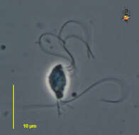
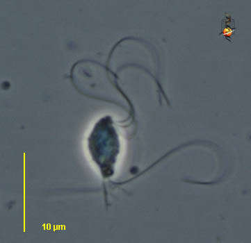
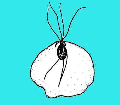
Tritrichomonads are small trichomonads (8-22 µm) with three anterior flagella and a recurrent flagellum forming a conspicuous undulating membrane with a posterior free portion. Costa stout or slender sustaining the undulating membrane; axostyle well developed; sausage-shaped parabasal. At the time of writing, there are about 20 species living in the intestinal tract of rodents, birds, reptiles and amphibians one species T. foetus is a parasite of the uro-genital tract of bovines. This image of Tritrichomonas foetus parasitic from uro-genital tract of the cattle (cows). It has three anterior flagella, a recurrent flagellum forming an undulating membrane with a free posterior part, a thick axostyle (immunofluorescence).
-
-
-
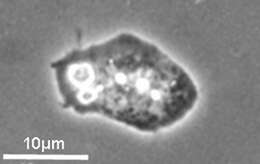
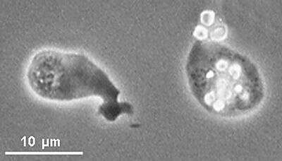
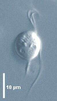
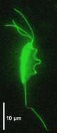
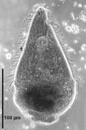
Dientamoeba is an amoeboid and amastigote parabasalid of about 5-20 µm in diameter, containing v-shaped parabasal body close to the nucleus but no flagella and axostyle. In binucleate cells a paradesmose is stretched between the two polar centers at the origin of the parabasal fibers. Anaerobic, contains hydrogenosomal granules, moves by amoeboism and phagocytoses particles such as bacteria and inhabits the intestine of humans. Dientamoeba fragilis, phase contrast.
-
Centers for Disease Control/Division of Parasitic Diseases and Malaria
EOL staff
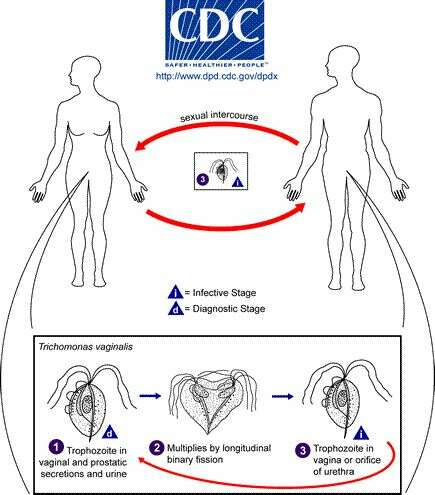
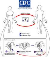
Life cycle of Trichomonas vaginalis, the cause of trichomoniasis in humansTrichomonas vaginalis resides in the female lower genital tract and the male urethra and prostate (1), where it replicates by binary fission (2). The parasite does not appear to have a cyst form, and does not survive well in the external environment. Trichomonas vaginalis is transmitted among humans, its only known host, primarily by sexual intercourse (3).From
Centers for Disease Control Parasites and Health website
-
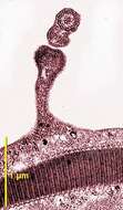
Section of the "rail-type" undulating membrane by transmission EM.
-
Monocercomonas are small trichomonad flagellates (5-15 µm) with three anteriorly directed flagella and a recurrent one slightly adhering on its proximal part to the cell body. Well developed axostyle protruding posterioly. Parabasal body rod-, disc- or V-shaped. About 20 species occurring in the intestinal tract of vertebrates such as M. caviae from the caecum of guinea pigs (Nie 1950) or invertebrates such as termites, roaches, tipulid larvae. Unidentified species from the gut of the cockroach Parasphaeria boleiriana from Brazil (interference contrast)
-
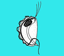
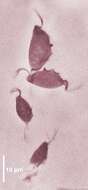
"Tritrichomonads are small trichomonads (8-22 µm) with three anterior flagella and a recurrent flagellum forming a conspicuous undulating membrane with a posterior free portion. Costa stout or slender sustaining the undulating membrane; axostyle well developed; sausage-shaped parabasal. At the time of writing, there are about 20 species living in the intestinal tract of rodents, birds, reptiles and amphibians one species T. foetus is a parasite of the uro-genital tract of bovines. This image of Tritrichomonas muris from mice, three anterior flagella, a recurrent flagellum forming an undulating membrane, axotyle protruding at the posterior end (phase contrast).
-
Dientamoeba is an amoeboid and amastigote parabasalid of about 5-20 µm in diameter, containing v-shaped parabasal body close to the nucleus but no flagella and axostyle. In binucleate cells a paradesmose is stretched between the two polar centers at the origin of the parabasal fibers. Anaerobic, contains hydrogenosomal granules, moves by amoeboism and phagocytoses particles such as bacteria and inhabits the intestine of humans. Dientamoeba fragilis, phase contrast.
-
Centers for Disease Control/Division of Parasitic Diseases and Malaria
EOL staff
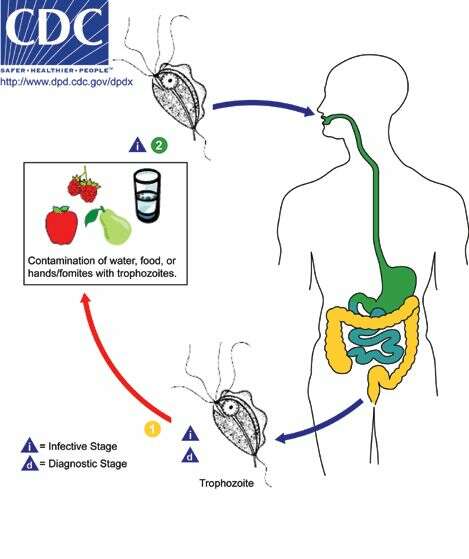
Life cycle of the flagellate Pentatrichomonas hominisPentatrichomonas hominis is a trichomonad flagellate with a worldwide distribution. Only trophozoites are shed in feces (1) as there is no known cyst stage for this species. Infection occurs after the ingestion of trophozoites in fecally-contaminated food or water or on fomites (i.e., other non-living objects or substances that can transmit them) (2). These organisms reside in the large intestine, where they are regarded as commensals (i.e., benefiting from but not harming their host) and are not known to cause disease in humans.From
Centers for Disease Control Parasites and Health website
-
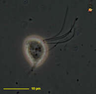
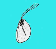
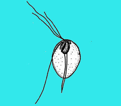
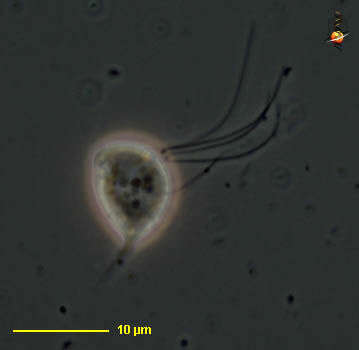
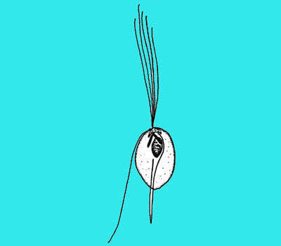
Hexamastix (hex-a-mas-sticks) is a monocercomonad - a member of the trichomonads. It is relatively small. It has five flagella, three can be seen in front of the cell, one passes under the cell and to the left, and one curves round to the right of the cell. The point projecting from the back of the cell is a skeletal element called an axostyle. From the termite Zootermopsis. Phase contrast.
-
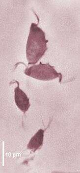
Tritrichomonads are small trichomonads (8-22 µm) with three anterior flagella and a recurrent flagellum forming a conspicuous undulating membrane with a posterior free portion. Costa stout or slender sustaining the undulating membrane; axostyle well developed; sausage-shaped parabasal. At the time of writing, there are about 20 species living in the intestinal tract of rodents, birds, reptiles and amphibians one species T. foetus is a parasite of the uro-genital tract of bovines. This image of two trichomonad species from the coecum of rodents, Tritrichomonas muris is larger than Tritrichomonas minuta, T. minuta has a narrower undulating membrane (protargol).
-
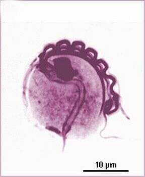
Calonympha are multinucleate polymastigotes parabasalid flagellates of about 70-80 µm long. The anterior part is occupied by several crown of akaryomastigonts and karyomastigonts each of which composed of four anteriorly directed flagella, a posterior axostyle and a nucleus in karyomastigonts. The posterior part contains an axostylar trunk a bundle composed of the axostyles of the karyomastigonts and wood particles. This species, Calonympha grassii, is from Neotermes jouteli. The anterior part is occupied by karyomastigonts bearing flagella and at the posterior end the protruding axostyle is typical (phase contrast).
-
Hexamastix (hex-a-mas-sticks) is a monocercomonad - a member of the trichomonads. It is relatively small. It has five flagella, three can be seen in front of the cell, one passes under the cell and to the left, and one curves round to the right of the cell. The point projecting from the back of the cell is a skeletal element called an axostyle. From the termite Zootermopsis. Phase contrast.
-
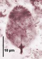
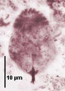
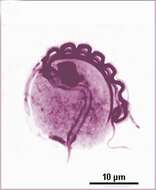
"Tritrichomonads are small trichomonads (8-22 µm) with three anterior flagella and a recurrent flagellum forming a conspicuous undulating membrane with a posterior free portion. Costa stout or slender sustaining the undulating membrane; axostyle well developed; sausage-shaped parabasal. At the time of writing, there are about 20 species living in the intestinal tract of rodents, birds, reptiles and amphibians one species T. foetus is a parasite of the uro-genital tract of bovines. This image of Tritrichomonas muris from guinea pig, axostyle with posterior subterminal rings, recurrent flagellum forming a thick undulating membrane with several layers subtended by the costa fibre, anterior nucleus (Giemsa staining).
-
Calonympha are multinucleate polymastigotes parabasalid flagellates of about 70-80 µm long. The anterior part is occupied by several crown of akaryomastigonts and karyomastigonts each of which composed of four anteriorly directed flagella, a posterior axostyle and a nucleus in karyomastigonts. The posterior part contains an axostylar trunk a bundle composed of the axostyles of the karyomastigonts and wood particles. This species, Calonympha grassii, is from Neotermes jouteli. The anterior part is occupied by karyomastigonts bearing flagella and at the posterior end the protruding axostyle is typical (Giemsa staining).
-
-
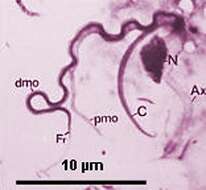
Tritrichomonads are small trichomonads (8-22 µm) with three anterior flagella and a recurrent flagellum forming a conspicuous undulating membrane with a posterior free portion. Costa stout or slender sustaining the undulating membrane; axostyle well developed; sausage-shaped parabasal. At the time of writing, there are about 20 species living in the intestinal tract of rodents, birds, reptiles and amphibians one species T. foetus is a parasite of the uro-genital tract of bovines. This image of Tritrichomonas muris squashed cell showing the recurrent flagellum (Fr) associated with the different layer of the undulating membrane (dmo, pmo) linked to the subjacent costa (C), the axostyle (Ax) and nucleus (N) (Giemsa).
-
-
-
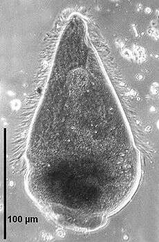
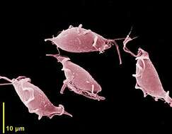
Scanning electron micrograph of cells from a rodent.
-
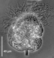
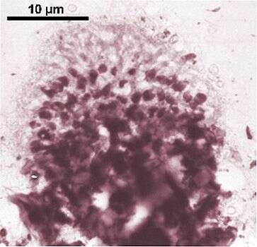
Large hypermastigid (450-500 µm) symbiont in Mastotermes darwiniensis. Body broadly triangular in shape with an anterior dome-shaped rostrum completely flagellated. Cell body covered with longitudinal rows of flagella except the amoeboid posterior part which is separated by a girdle and used for wood ingestion. The nucleus is situated in the anterior region; the axostylar fibres surround the nucleus and some group backwards to form an axial axostylar trunk.
-
-
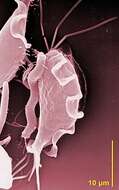
Scanning EM showing the three anterior flagella and the undulating membrane.