Doi.10.1128.JVI.00175-20.F5.large
Опис:
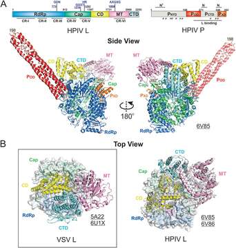
Description: English: The cryo-EM structure of the Paramyxoviridae polymerase. (A) Linear domain representation of the L and P proteins of the human parainfluenza virus (HPIV) polymerase. The side view of the ribbon diagram of the 4.3-Å (PDB: 6V85) cryo-EM structure of the HPIV polymerase complex. (B) The top view of the superimposed VSV L and HPIV L shows the domain switch of the CD-MT-CTD module. The superimposition is based on the RdRp (surface view), and CD, MT, and CTD are shown as the ribbon diagram. The domain colorings are the same as Fig. 2. The VSV L is shown in the left panel (box), and the HPIV L is shown in the right panel. The HPIV L (PDB: 6V85) is colored the same as A, and another stable conformation of the HPIV L (PDB: 6V86) is colored in gray. Note the significant location switch of CTD, facing down (VSV) versus facing up (HPIV L). The PDB accession codes are underlined. Date: 27 October 2020. Source: Liang B. 2020. Structures of the Mononegavirales polymerases. J Virol 94:e00175-20. https://doi.org/10.1128/JVI.00175-20. Author: Bo Liang.
Се јавува на следниве страници:
- Biota
- Virus
- Riboviria
- Orthornavirae
- Negarnaviricota
- Haploviricotina
- Monjiviricetes
- Mononegavirales
- Rhabdoviridae
- Lyssavirus
- Rabies virus
Сликата ја нема во ниедна збирка.
Информации за изворот
- лиценца
- cc-by-3.0
- авторски права
- Bo Liang
- изворно
- изворна податотека
- посети извор
- соработничко мреж. место
- Wikimedia Commons
- ID