-
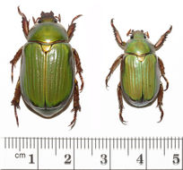
Image showing size differential between two specimens of Chrysina lecontei collected at the same locality on the same night.
-
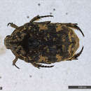
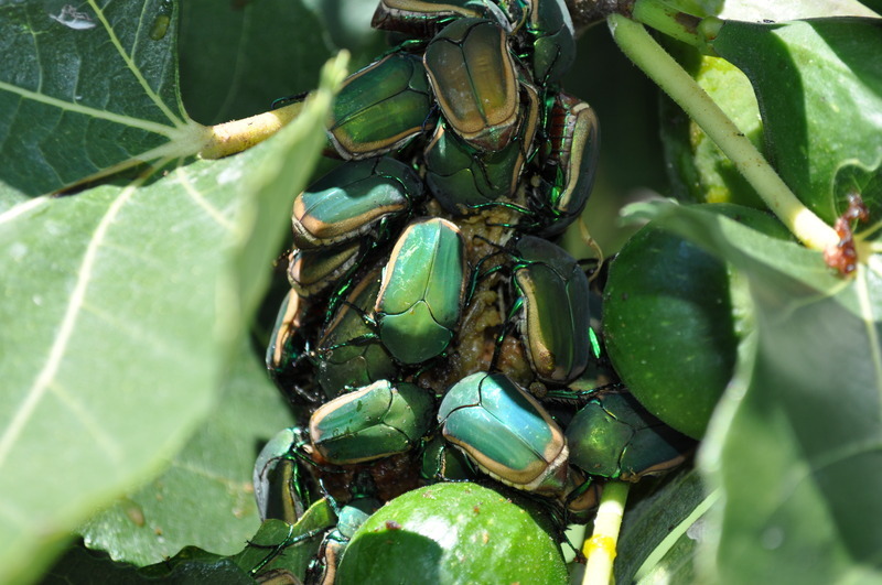
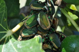
Recorded/photographed #1, 24 beetles upon a single fig.
-
Close up of June beetles on a single fig fruit. Refer to photograph #1 DSC_0259.JPG.
-
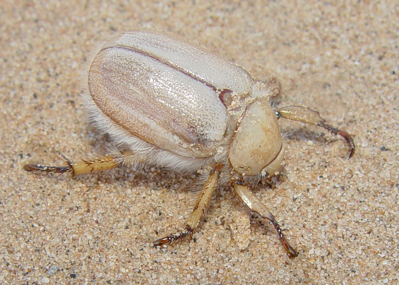
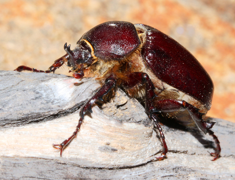
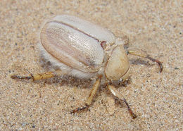
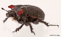
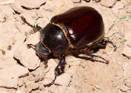
Dug from burrow around mid-morning.
-
Attracted to a light trap at the northern terminus of Farmer Road, Julian, California in the hour before dawn in light rain. Later that day it was photographed in Cuyamaca Woods, Julian.
-
Specimen collected February 12, 1972 by P. Tuskes.
-
-
-
-
-
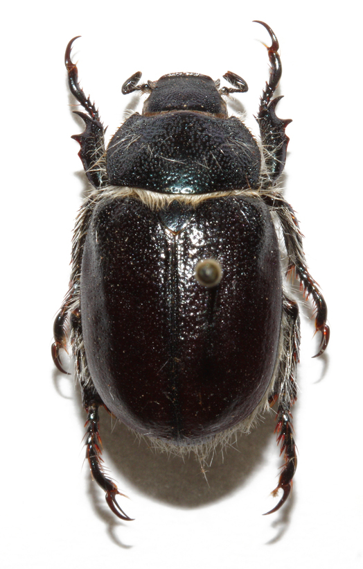
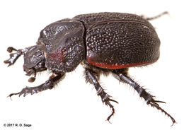
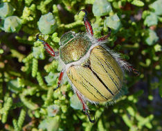
Found with a small number of other beetles of the same species in mid-morning on a sunny day. The beetles were clinging passively to the juniper branches.
-
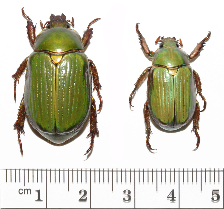
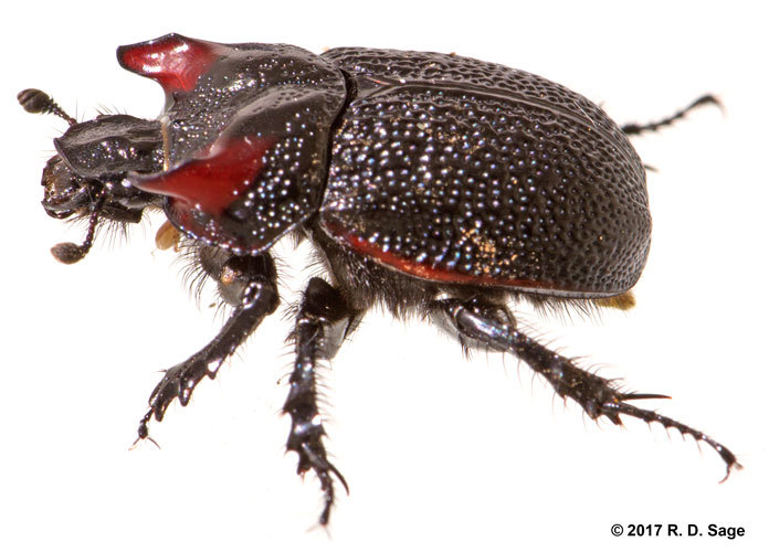
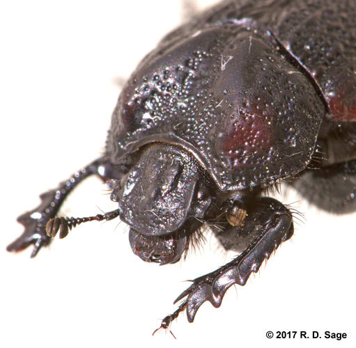
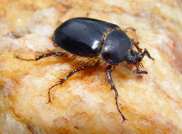
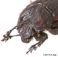
Male, as shown by the presence of a horn. Attracted to light.
-
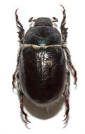
Female, as shown by lack of horn. Attracted to light.
-
At a magnification of 130X, this scanning electron micrograph (SEM) focused on the head region of an adult figeater beetle, Cotinis mutabilis. In this particular view, the transitional area between one of the insects two compound eyes, i.e., right eye, and the vertex of its head is visualized. For even greater magnifications of the surface of the eye, see PHIL 9950, 9951, and 9952.Created: 2007
-
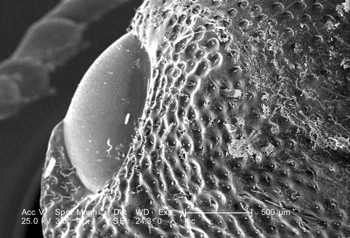
At a low magnification of 47X, this scanning electron micrograph (SEM) focused on the head region of an adult figeater beetle, Cotinis mutabilis. In this particular view, one of the insects two compound eyes, i.e., right eye, is visualized. For even greater magnifications of the surface of the eye, see PHIL 9950, 9951, and 9952.Created: 2007
-
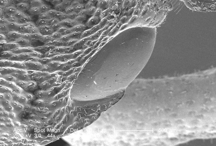
At a low magnification of 44X, this scanning electron micrograph (SEM) focused on the head region of an adult figeater beetle, Cotinis mutabilis. In this particular view, one of the insects two compound eyes, i.e., left eye, is visualized. For even greater magnifications of the surface of the eye, see PHIL 9950, 9951, and 9952.Created: 2007
-
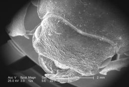
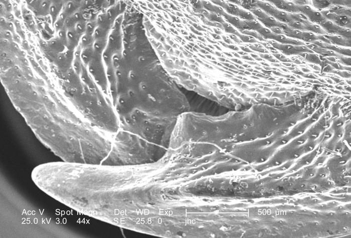
At a low magnification of 44X, this scanning electron micrograph (SEM) focused on the head region of an adult figeater beetle, Cotinis mutabilis. Note the insects two overlapping mandibles. For successively greater magnifications of this region with its exoskeletal features PHIL 9953, 9954, 9956, 9957 and 9958.Created: 2007
-
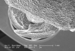
At a low magnification of 21X, this scanning electron micrograph (SEM) focused on the head region of an adult figeater beetle, Cotinis mutabilis. Note the insects two overlapping mandibles. For successively greater magnifications of this region with its exoskeletal features PHIL 9953, 9955, 9956, 9957 and 9958.Created: 2007
-
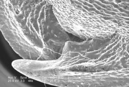
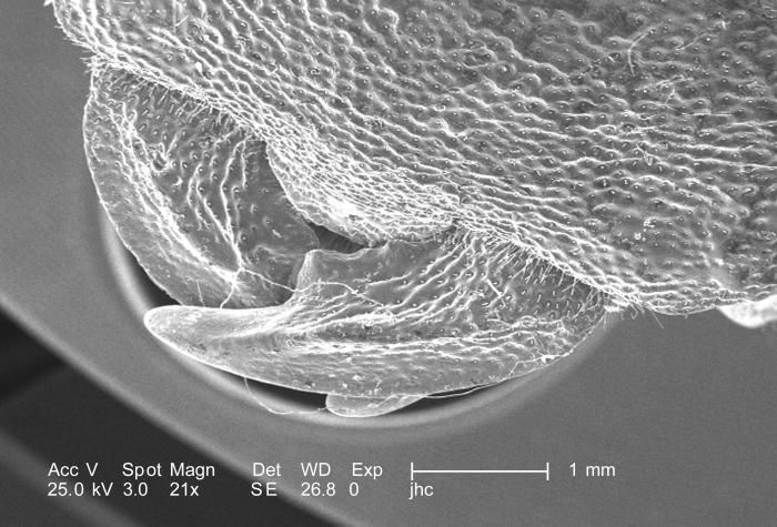
At a very low magnification of 12X, this scanning electron micrograph (SEM) focused on the head region of an adult figeater beetle, Cotinis mutabilis. Note the two laterally-positioned eyes, as well as the insects overlapping mandibles. For successively greater magnifications of this region with its exoskeletal features PHIL 9954, 9955, 9956, 9957 and 9958.Created: 2007
-
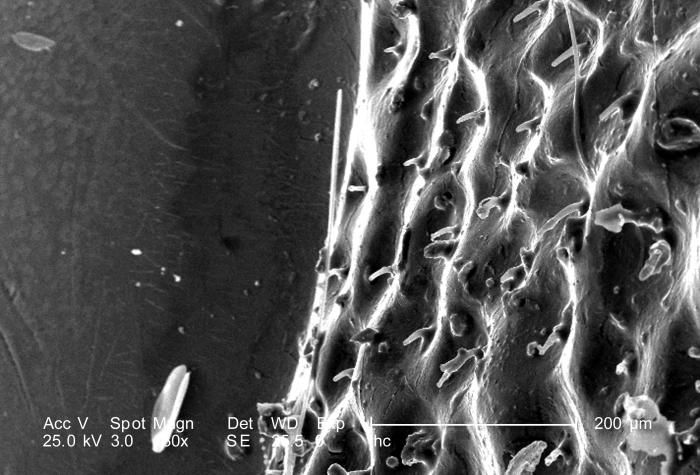
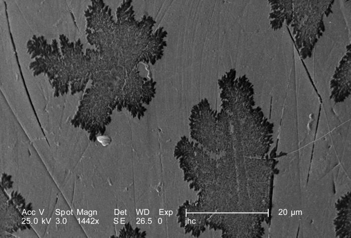
Magnified 1442X, twice that of PHIL 9950 and 9951, this scanning electron micrograph (SEM) revealed some of the ultrastructural details found on the surface of one of the two eyes of this adult figeater beetle, Cotinis mutabilis. The meaning behind the leaf-like pattern seen on the eyes chitinous surface is unknown, however, when carefully scrutinized, it does not appear to be randomized.Created: 2007
-
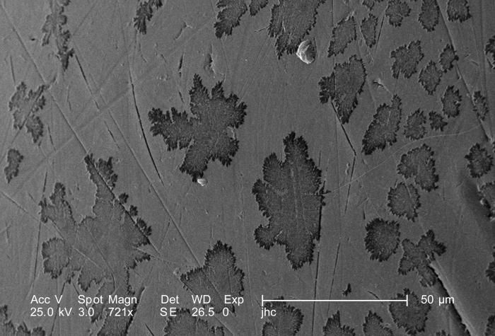
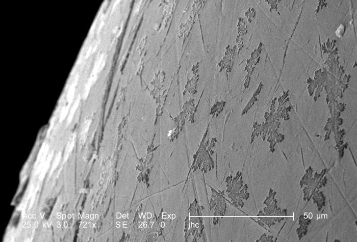
Magnified 721X, this scanning electron micrograph (SEM) revealed some of the ultrastructural details found on the surface of one of the two eyes of this adult figeater beetle, Cotinis mutabilis. The meaning behind the leaf-like pattern seen on the eyes chitinous surface is unknown, however, when carefully scrutinized, it does not appear to be randomized. Note PHIL 9950 and 9952 for other views of this configuration.Created: 2007
-
Magnified 721X, this scanning electron micrograph (SEM) revealed some of the ultrastructural details found on the surface of one of the two eyes of this adult figeater beetle, Cotinis mutabilis. The meaning behind the leaf-like pattern seen on the eyes chitinous surface is unknown, however, when carefully scrutinized, it does not appear to be randomized. Note PHIL 9951 and 9952 for other views of this configuration.Created: 2007
-
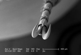
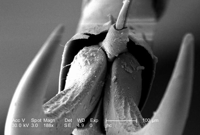
At a magnification of 188X, half that of PHIL 9947, this scanning electron micrograph (SEM) depicted a head-on view of the distal clawed tip of an adult figeater beetles, Cotinis mutabilis leg. Phil 9943, 9944, and 9945 depict this anatomical appendicular relationship from its side. The insect leg is comprised of a variable number of segments, however, there are usually six which predominate, including the most proximal coxa, i.e., attaching the leg to the thorax, followed by the trochanter, femur, tibia, tarsus, and pretarsus, which in the case of this beetle is a claw with its spiked empodium.Created: 2007
-
At a magnification of 188X, twice that of PHIL 9947, this scanning electron micrograph (SEM) depicted a head-on view of the distal clawed tip of an adult figeater beetles, Cotinis mutabilis leg. Phil 9943, 9944, and 9945 depict this anatomical appendicular relationship from its side. The insect leg is comprised of a variable number of segments, however, there are usually six which predominate, including the most proximal coxa, i.e., attaching the leg to the thorax, followed by the trochanter, femur, tibia, tarsus, and pretarsus, which in the case of this beetle is a claw with its spiked empodium.Created: 2007