Comprehensive Description
provided by Smithsonian Contributions to Botany
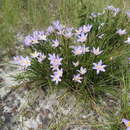
Vellozia angustifolia Goethart & Henrard
Vellozia angustifolia Goethart & Henrard, Blumea, 2:365, 1937.—L. B. Smith, Contr. U.S. Nat. Herb., 35:261, 1962.—Ayensu, Smithsonian Contr. Bot., 15:12, fig. 7d–f, pl. 28a–e, 1974.
TYPE.—Among rocks, Morro Cubatão, near Guariroba, Goiás, Brazil, 11 April 1895, Glaziou 22213 (L, holotype (?); P, isotype; US, photo).
DISTRIBUTION.—Brazil: Minas Gerais: Diamantina, Guinda. Goiás: Guariroba.
- bibliographic citation
- Smith, Lyman B. and Ayensu, Edward S. 1976. "A Revision of American Velloziaceae." Smithsonian Contributions to Botany. 1-172. https://doi.org/10.5479/si.0081024X.30
Comprehensive Description
provided by Smithsonian Contributions to Botany
Vellozia angustifolia Goethart & Henrard
SPECIMENS EXAMINED.—W. A. Archer, 4092; Centro Pesq, (R)01411; Riedel s.n.
SURFACE VIEW.—Hairs: absent. Epidermis: adaxial and abaxial cells mostly rectangular, thin walled. Stomata: tetracylic, 30 × 15 μm; observed on both surfaces.
TRANSVERSE SECTION OF LAMINA.—Dorsiventral; shallowly V-shaped with downturned margins and median adaxial groove. Adaxial surface evenly undulating; abaxial surface furrowed about half of blade thickness. Epidermis: cells more or less rounded or square. One or two rows of thick-walled fibers forming almost a continuous strand subjacent to epidermis. Below strand is a layer of large translucent cells separating it from mesophyll. Cuticle: thin on both surfaces. Stomata: in line with epidermis; mostly confined to abaxial surface, especially in fibrous walls; guard cells thickened with short inner and outer ledges; substomatal chamber present. Mesophyll: adaxial palisade two to four layered, occupying about half of mesophyll and gradually changing into spongy tissue; palisade cells arranged radially above vascular bundles furrows and above midvein distinctly translucent and large; rest of palisade compactly arranged and filled with chloroplast. Vascular bundles: 16–27; commissural bundles present. Large veins each with one to three wide vessels, mostly two. Two phloem units almost coalesing into one lying laterally in abaxial or slightly partial V-shaped girder. Each vascular bundle always accompanied by adaxial cap. Vascular bundle completely surrounded by a distinct bundle sheath with extensions toward adaxial epidermis and short extensions toward abaxial epidermis. Crystals and tannin: not observed.
NOTE: The one row translucent rectangular cells separating the adaxial strands of thick-walled fibers from the palisade cells is very distinctive.
- bibliographic citation
- Ayensu, Edward S. 1974. "Leaf Anatomy and Systematics of New World Velloziaceae." Smithsonian Contributions to Botany. 1-125. https://doi.org/10.5479/si.0081024X.15