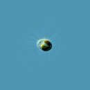
Mallomonas ist eine im Süßwasser vorkommende Algen-Gattung aus der Gruppe der Stramenopilen. Sie besteht aus rund 130 Arten.
Die Einzeller-Gattung aus der Klasse der Chrysomonadea (Engler, 1898) sind etwa zwischen 5 und 20 Mikrometer groß. Am Vorderende des Zellpols befinden sich Geißeln. Das längere Flagellum übernimmt die Aufgaben der Lokomotion und der Anheftung an ein Substrat. An der Geißel befinden sich Mastigonemen. Das zweite Flagellum ist sehr stark reduziert. Es kommen Discobolocysten als Extrusome vor, Extrusome sind schnell ausstülpbare Membranorganellen, die meist der Verteidigung dienen. Diese Protisten haben einen Zellkern und 1–2 pulsierende Vakuolen. Als Reservestoffe sind in den Vakuolen das Polysaccharid Chrysolaminarin und einige Lipide gespeichert. Mallomonas hat zwei große sichelförmige gold-gelbe bis gold-bräunliche Chloroplasten. Die Chloroplasten enthalten Chlorophyll a, c und Fucoxanthin. Es ist bei Mallomonas kein Chlorophyll b vorhanden. Oft ist ein rötlicher Augenfleck auf einem der Chloroplasten zu erkennen, dieses Stigma dient der Wahrnehmung von Licht. Die Art und Weise der Energiegewinnung kann den jeweiligen ökologischen Anforderungen angepasst werden, es ist eine Umstellung auf eine heterotrophe Lebensweise möglich.
Die Zelloberfläche ist von ziegelähnlichen Schuppen bedeckt. Die Schuppen bestehen aus Siliziumdioxid („Kieselsäure“), weisen komplexe, hochgeordnete Strukturen auf und sind wabenartig angeordnet. Die Zelle kann ihr Gehäuse aktiv verlassen.
Arten der Gattung Mallomonas können freischwimmend leben beziehungsweise sich an dem Substrat festheften. Vertreter diese Gattung können auch Gruppen bilden. Sexuelle Vorgänge finden durch die Verschmelzung von Einzelzellen statt, ohne dabei haploide Gameten gebildet zu haben. Nach der Verschmelzung (Isogamie) encystiert sich die Zygote. Dauercysten können auch auf ungeschlechtlichem Wege gebildet werden.
Die Vertreter der Gattung treten besonders im Frühling im Plankton von sauberen, kalten, meist sauren stehenden Gewässern auf.
Mallomonas ist eine im Süßwasser vorkommende Algen-Gattung aus der Gruppe der Stramenopilen. Sie besteht aus rund 130 Arten.
Mallomonas is a genus comprising unicellular algal eukaryotes and characterized by their intricate cell coverings made of silica scales and bristles.[1] The group was first named and classified by Dr. Maximilian Perty in 1852.[2] These organisms live in freshwater and are widely distributed around the world.[3] Some well known species include Mallomonas caudata and Mallomonas splendens.
Mallomonas is a genus of many from the phylum Ochrophyta, which describes organisms as having heterokont flagella in some part of their life history. At first, the family Mallomonadaceae was placed under class Chrysophyceae. However, after finding key biochemical and ultrastructural differences, the family was then placed under the class Synurophyceae. In a broader context, both Chrysophyceae and Synurophyceae are referred to as “chrysophytes”, meaning “golden algae”, because of their close similarities. Despite being quite similar, there are various, noticeable differences.
The genus Mallomonas was first named and classified by Dr. Maximilian Perty in 1852.[2] It was assigned its own genus because Mallomonas consisted of individually living cells, while its sister group Synura is composed of colonial cells that are connected to one another through stalks.[4]
Other distinguishing features that differentiate Mallomonas cells from Synura cells are the presence of bristles and V-shaped ridges on their scales.[1][4] Pore-collar complexes on stomatocysts are species differentiating features.[1]
Mallomonas is composed of planktonic, freshwater organisms.[1]
Cells range in many ecological factors. Many species can tolerate pH levels ranging from 4 to 8, and can live in slightly acidic waters. Some species prefer low phosphorus gradients. Temperature tolerance ranges from 1.5 to 25.5 °C.[1]
According to the temperature-size rule, as the temperature increases, the size of protists decreases. As with all Synurophytes, the inorganic scales produced by organisms in the genus Mallomonas also decrease in size as a response to increasing temperature, the same way as the cells themselves decrease. The shape of the scales became less round and more elongated or oval-shaped. Also, higher temperatures negatively affect scale biogenesis, interfering with the cellular processes that produce the scales.[4]
The organisms in this genus have streamlined morphology in spherical, oval, or elliptical forms, and they have a wide range of sizes that vary from the smallest being 10 µm to the largest being 100 µm. Two flagella emerge from the anterior apical flagellar pocket; one can be easily seen in the light microscope as it is longer and covered in hairs (called mastigonemes) while the other is much shorter, with no hairs, and is not as easily seen.[1]
Pigments such as chlorophyll c1 and fucoxanthin within the chloroplasts cause the cells to have a distinct, golden or yellow-brown colour. The chloroplasts themselves are bi-lobed, but some cells have two single-lobed chloroplasts. Cells have a girdle lamella, grouped thylakoids stacked in three, and additional membranes around the chloroplast called the chloroplast endoplasmic reticulum (CER).[1]
The large nucleus is located between the chloroplast and the Golgi complex. Vacuoles full of the storage product known as chrysolaminarin are found near the posterior end of the cell.[1]
Flagella are microtubule-based structures that allow cells to have controlled movement to a certain degree. The root of the microtubule arrangement that is associated with the flagellar basal bodies, R1, which is also the site of cytoskeletal microtubules, is positioned uniquely in the genus Mallomonas. Instead of pulling away from the basal bodies, R1 actually loops around them in a clockwise fashion. There is also a second root of microtubules extending from the point of origin towards the cell’s centre. Both basal bodies, each assigned one of the two flagella, are enclosed in a thick, fibrous capsule.[1]
The genus Mallomonas is based on the organization of its scales, which are silica plates that are intricately designed, species-specific, and cover the cell. There are many parts to the entire structure; it’s made up of anterior spine scales, posterior spine scales, body scales, and some specialized scales. An example of a specialized scale is one that can fit around a flagellar pocket. The scales are accompanied by bristles, which are long, elongated structures that are tucked under the scales with the use of a small bend in the end, which is called a ‘foot’.[5]
Scale size varies from 1 µm to 10 µm with surface area also varying from 1 µm2 to 50 µm2. Scale shapes are circular, elliptical, or ovate, generally rhombic in nature, but they are wider than the scales of their sister genus Synura. Scales without domes exhibit bilateral symmetry, except for the collar scales, which are different in terms of shape altogether. Scales with domes exhibit slight asymmetry due to the dome shape in regards to the poster rim and the V-rib. Instead of being flat, scales are curved so they can conform to the cell shape. Curvature increases with scale size and thickness.[1] More information about specific structures mentioned follow.
Each scale and bristle is produced intracellularly in a vesicle known as the silica deposition vesicle (SDV), which is connected to the chloroplast endoplasmic reticulum. Their production is also associated with the Golgi body, the nucleus, the chloroplast, and how the complex of these organelles is arranged in relation to the SDV within the cell.[6] Formation of the scales and bristles occurs along the chloroplast’s outer surface because of SDV’s close placement to the CER. Directional placement could be parallel to cell length or at an angle away from either the posterior or anterior ends of the cell.[1]
Patterns of perforation and ridge detail vary among species, but all have a general shape consisting of a wide, rough oblong with a dome and posterior rim. The base plate is perforated with pores, and in some species, they are either present or lacking. Usually, they are spaced evenly over the base plate, but the posterior rim, flanges, and domes are not usually perforated. The upturned posterior rim bends forward and encircles about half the scale’s perimeter. The rim can be narrow or broad, equal in length on both sides of the scale or asymmetric, depending upon the species, but the location is consistent throughout the genus and is known as the proximal end.[1]
Majority of species have a V-shaped ridge (V-rib) positioned just ahead of the posterior rim on their scales. Also made of silica, the V-rib extends further towards the distal end and stops close to the dome (if present) or at the perimeter of the scale. Near the distal ends of the V-rib, some species have two additional ridges called anterior submarginal ribs, but they’re quite small and terminate at the distal end. Species with V-ribs also have the two anterior submarginal ribs, and the junction between the two is of taxonomic significance, defining some of the many species within this genus.[1]
Scales that have a dome are associated with bristle attachment. A dome is a raised portion of the base plate at the distal end where the foot of a bristle is tucked underneath. The bristle locks into place under the dome and emerges through an inverted U-shaped opening that is located slightly away from the center of the dome, which causes an asymmetric balance between the bristle and the scale. However, this allows the bristle to rotate with the longitudinal axis of the cell through a wider angle than if it extended straight through the center of the dome. The lip, or rim of the inverted U-shaped opening, could protrude forward in some cases, however, in most, it is slightly off to the right.[1]
The anterior submarginal ribs and the V-rib divide the scale into regions that are in turn ornamented differently for each species. The section of the base plate that is between the submarginal rib and the V-rib is referred to as the shield while the section that is outside both the V-rib and the anterior submarginal ribs is referred to as the posterior flange and the anterior flange, respectively.[1]
The anterior submarginal ribs, V-rib, and posterior rim are considered to be secondary structures in regards to the perforated base plate; however, some species also have other secondary structures such as additional ribs or papillae. Rib placement is dependent upon pore placement, as ribs protrude from between pores. Irregular pore patterns lead to irregular rib placement.[7] Papillae consist of small protrusions sticking out of the dome, shield, or anterior flanges that are regularly spaced, vary in density, and can be solid or hollow.[1]
Bristles are composed of two parts, the foot and the shaft. The foot is at the proximal end, in regards to the bristle, and it is the part of the structure that is tucked under the distal end of the scale, under the dome. The foot is flat and bends at a 30° to 90° angle relative to the shaft. The shaft could be smooth, curved, ribbed, or serrated, and in some species, instead of being a solid bar, it appears rolled up so that the slit running along the shaft length is the point of convergence where both sides meet.[1]
Many variations arise at the distal tip of the bristle, the end that is exposed to the environment. The tips can be sharp, blunt, forked, bifurcated, pseudobifurcated, swollen, expanded, C-shaped, cleft-like (to form helmet bristles), hooked, folded (to form lance bristles), plumed, and serrated - and possibly more.[1] Even within these variations, there are variations, and this diversity gives rise to many different species within this genus.
Each region of the bristle (whether it is the proximal end, the middle, or the distal end) has varying degrees of differences and flexibility in morphology. This variation in characteristics differentiates species.
The resting stage is called a stomatocyst, and it is also composed of silica. It ranges in diameter from 4 µm to 30 µm,[8] can be spherical or oval in shape, and varies considerably in structure and ornamentation between species. Overall texture can vary; some cysts are smooth, some have reticulate designs, while others can have spines, or ridges, or depressions. The pore-collar complex, which is the presence and shape of a silica collar around the pore (or entrance) of the stomatocyst, is taxonomically significant.[9] The cyst wall is also produced in the silica deposition vesicle (SDV), where parts of the wall are produced intracellularly and then excreted into the extracellular complex, where the wall is then formed and stabilized. The standard size and shape is retained throughout the number of species,[10] but the variations between species of ornamentation may be due to physiological influences from the environment.[8]
Development occurs in two phases within the continuous process. The first phase involves the primary inner wall of the cyst being formed before the collar and surface, which is thin and occurs rapidly from a proximal to distal manner.[11] The second phase is more controlled and occurs more slowly while the wall is being thickened, and the collar and surface ornamentation are being produced. Because of this process of making a stomatocyst, cysts can be found at any stage of development. After development has been completed, the cyst sinks down to the sediment, and remains in its resting stage until certain conditions trigger its germination. After germination, a flagellated cell emerges from the cyst through pore-collar complex and produces new siliceous armour of scales and bristles. It is currently unclear if germination occurs in the water column after redistribution or if it occurs while the cyst is sitting in the sediment.[1]
Very little is known about reproduction in Mallomonas. All that is known is that two vegetative cells fuse to produce a zygote, which then encysts and remains in sediment until germination.[3] Vegetative cell division occurs after excystment. In only minutes cytokinesis occurs, beginning from the anterior end and proceeding down the longitudinal axis of the cell. Sexual reproduction is also noted to occur within this genus, although it is rare.[1]
Stomatocyst microfossils are used in the study of lake paleontology.[12]
When comparing bristle formation of species Mallomonas muskokana with the Eocene fossil taxon Mallomonas dispar, it was found that the asymmetrical pattern observed was identical to the extant species.[13] The Eocene Epoch is a part of the Paleogene Period, occurring about 56 to 33.9 million years ago. Studies with this fossil taxon would suggest that parts of its morphology have been retained up to the present.
To date, the genus Mallomonas has 223 species. A list of well-known species is provided in the table below.[2]
Mallomonas is a genus comprising unicellular algal eukaryotes and characterized by their intricate cell coverings made of silica scales and bristles. The group was first named and classified by Dr. Maximilian Perty in 1852. These organisms live in freshwater and are widely distributed around the world. Some well known species include Mallomonas caudata and Mallomonas splendens.
Mallomonas is a genus of many from the phylum Ochrophyta, which describes organisms as having heterokont flagella in some part of their life history. At first, the family Mallomonadaceae was placed under class Chrysophyceae. However, after finding key biochemical and ultrastructural differences, the family was then placed under the class Synurophyceae. In a broader context, both Chrysophyceae and Synurophyceae are referred to as “chrysophytes”, meaning “golden algae”, because of their close similarities. Despite being quite similar, there are various, noticeable differences.

ミノヒゲムシ Mallomonas は、黄金色藻類の1属。細胞表面を珪酸質の鱗片が覆っており、往々にしてこの鱗片から長い棘が出る。
単細胞生の鞭毛藻類[1]。細胞の先端に1本の鞭毛を持つものが多い。一部に2本の不等鞭毛を持つ例があり、それらはマロモノプシス節 Section Mollomonopsis に纏められている。この類では長い羽型と短い鞭型を有するが、それ以外の種は羽型鞭毛1本のみを持つ。
細胞の形は球形、卵形、楕円形、流線型、円筒形など様々で、その表面を珪酸質の鱗片が瓦状に並んだ被殻が覆っている。鱗片からは細い針のような剛刺が出るものがある。剛刺はその先端が鱗片の先端近くに曲がった基部が刺さったような形で外に向かう。一つの鱗片に複数の剛刺を持つ例もあれば、一部の鱗片にのみ剛刺を持つ例もある。外から見れば、細胞全体から長い棘が多数出ているものが多いが、例えば細胞の前端や後端からのみ出る例、まばらにごく少数のみが出る例などもある。
葉緑体は1つだが、細胞壁に沿って広がり、中央が縦に縊れ、2葉に分かれているので、2つあるようにも見える。
和名としては、古くは内田他(1947)にはマロヒゲムシというのがあるが、科名にはマロモナス科を採用している[2]。水野(1964)は科名、属名もそれぞれミノヒゲムシ科、ミノヒゲムシ属とし、幾つかの種にそれぞれ○○ミノヒゲムシという和名を与えている。また、種としては M. caudata の和名をミノヒゲムシとしている。岡田他(1965)もほぼこれに倣っている。他方で学名仮名読みのマロモナスもよく使われている[3]。やや特殊なのは月井(2010)で、マルロモナスとの表記を採っている。和名としてはぶれがないので、この項ではミノヒゲムシを採った。
淡水産であり、世界の淡水域に広く見られ、時に植物プランクトン相の中で優占的な地位を占める[4]。
特に秋から冬にかけてその数を増す[5]。
黄金色藻類に含まれるものとして扱われてきた。その中で、モトヨセヒゲムシ属 Synura と本属は、共に珪酸質の鱗片に覆われることで共通するほか、近年になってこれらが他の黄金色藻類とは鞭毛装置の構造や光合成色素の種類が異なる点などで区別出来ることが明らかになり、合わせてシヌラ科とし、これを独立させてシヌラ綱とすることも提案されている。
この属の分類では、細胞表面にある珪酸質の鱗片の形態がとても重視される。古くは光学顕微鏡によって研究が行われたので、細胞の上で剛刺の出る位置や、せいぜい鱗片の外形程度しか知ることが出来なかった。電子顕微鏡が利用出来るようになり、この分野は飛躍的に進歩した。1982年から1986年までの間だけで、50の新種と変種が追加されている[6]。2011年の段階では記載された種は180に達し、鱗片を持つ黄金色藻類系の属では最大のものとなっている[7]が、その後にも新種は追加されている模様である。
日本では水野(1964)や岡田他(1965)が3ないし4種の名を挙げている。それが水野・高橋(1991)になると28種が取り上げられている。また水野や岡田他で挙がっている名にはこちらには見えないものもあり、同定の見直しがあったようである。
属内の分類体系として、種を幾つかの系列(series)や節(section)に分けることが試みられている。それらの体系は、主として鱗片の形態や構造に基づいて行われてきた。ただし、分子系統の情報から、この様な体系が人為的なものである可能性も示唆されている[8]。
