-
Ribadelago de Franco, Castille and Leon, Spain
-
Alforja, Catalonia, Spain
-
Puerto Montt, Los Lagos, Chile
-
Casas de Fadoncino, Castille and Leon, Spain
-
Puerto Montt, Los Lagos, Chile
-
Puerto Montt, Los Lagos, Chile
-
Casas de Fadoncino, Castille and Leon, Spain
-
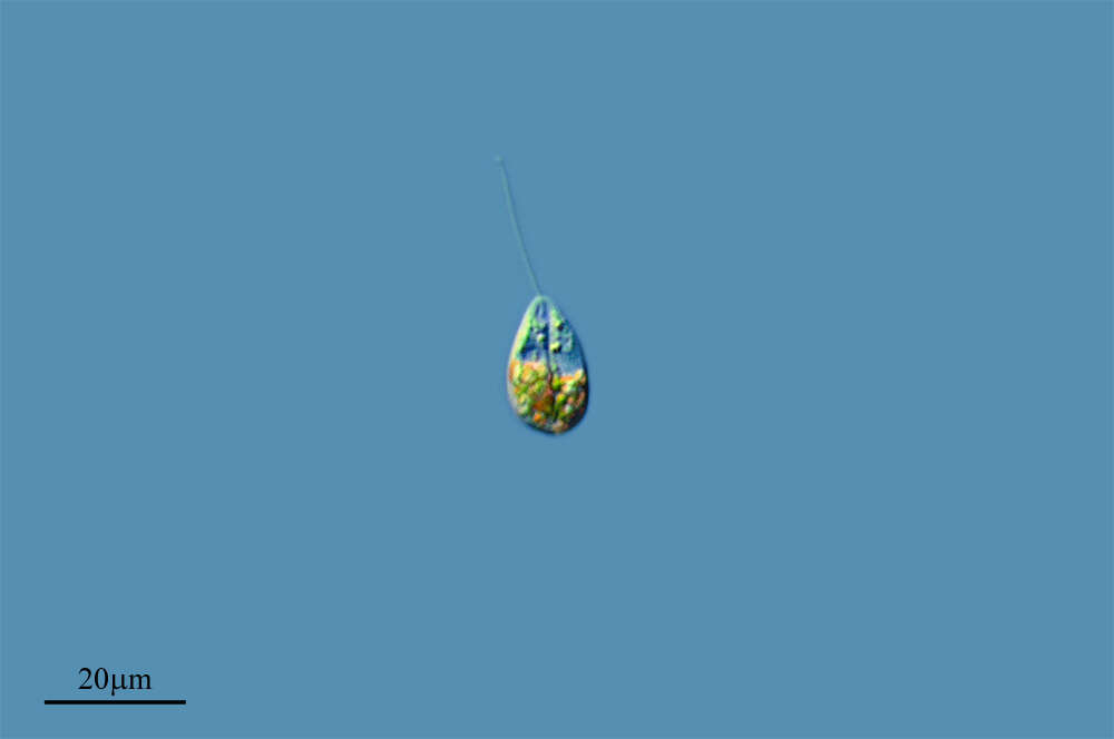
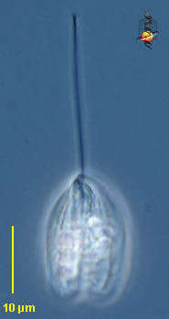
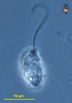
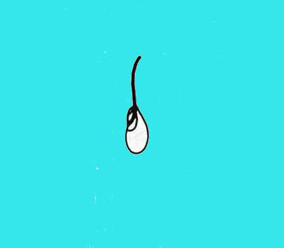
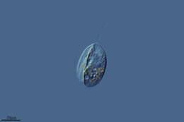
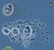
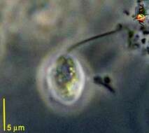
Petalomonas (pet-al-owe-moan-ass), rigid heterotrophic (no plastids) euglenid, one emerging flagellum which points in front of the moving cells. The flagellum starts within the flagellar pocket (also called the reservoir, and seen to the right) extends through a flagellar canal (not visible) and out at or near the apex of the cell. Either absorbs soluble food or consumes small particles although no mouth is visible by light microscopy. This species is probably P. pusillum, one of the smallest species and usually considered to have a smooth surface, although careful microscopy reveals minor ridges and folds. With cysts. Phase contrast.
-
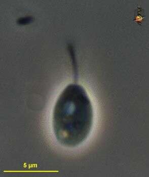
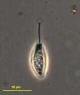
Petalomonas (pet-al-owe-moan-ass), rigid heterotrophic (no plastids) euglenid, one emerging flagellum which points in front of the moving cells. The flagellum starts within the flagellar pocket (also called the reservoir) extends through a flagellar canal and out at or near the apex of the cell. Either absorbs soluble food or consumes small particles although no mouth is visible by light microscopy. Most species have longitudinal or slightly spiral ridges or folds. Mode of feeding is not clear. Common in freshwater and marine ecosystems. Differential interference contrast.
-
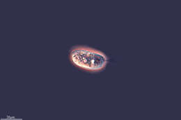
Petalomonas (pet-al-owe-moan-ass), rigid heterotrophic (no plastids) euglenid, one emerging flagellum which points in front of the moving cells. The flagellum starts within the flagellar pocket (also called the reservoir) extends through a flagellar canal and out at or near the apex of the cell. Either absorbs soluble food or consumes small particles although no mouth is visible by light microscopy. Most species have longitudinal or slightly spiral ridges or folds. The structure to the left and near the equator of the cell is its nucleus. Mode of feeding is not clear. Common in freshwater and marine ecosystems. Phase contrast.
-
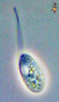
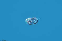
Petalomonas (pet-al-owe-moan-ass), rigid heterotrophic (no plastids) euglenid, one emerging flagellum which points in front of the moving cells. The flagellum starts within the flagellar pocket (also called the reservoir) extends through a flagellar canal (not visible) and out at or near the apex of the cell. Either absorbs soluble food or consumes small particles although no mouth is visible by light microscopy. Most species like this one have longitudinal or slightly spiral ridges or folds. Phase contrast.
-
Petalomonas (pet-al-owe-moan-ass), rigid heterotrophic (no plastids) euglenid, one emerging flagellum which points in front of the moving cells. The flagellum starts within the flagellar pocket (also called the reservoir, and seen to the right) extends through a flagellar canal (not visible) and out at or near the apex of the cell. Either absorbs soluble food or consumes small particles although no mouth is visible by light microscopy. This species is probably P. pusillum, one of the smallest species and usually considered to have a smooth surface, although careful microscopy reveals minor ridges and folds. With cysts. Phase contrast.
-
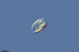
Petalomonas (pet-al-owe-moan-ass), rigid heterotrophic (no plastids) euglenid, one emerging flagellum which points in front of the moving cells. The flagellum starts within the flagellar pocket (also called the reservoir, and seen to the left) extends through a flagellar canal (not visible) and out at or near the apex of the cell. Either absorbs soluble food or consumes small particles although no mouth is visible by light microscopy. This species is P. pusillum, one of the smallest species and usually considered to have a smooth surface, although careful microscopy reveals minor ridges and folds. Phase contrast.
-
Petalomonas (pet-al-owe-moan-ass), rigid heterotrophic (no plastids) euglenid, one emerging flagellum which points in front of the moving cells. and helps hold the cells against the substrate. The flagellum starts within the flagellar pocket (also called the reservoir) extends through a flagellar canal and out at or near the apex of the cell. Either absorbs soluble food or consumes small particles although no mouth is visible by light microscopy. Most species bear longitudinal or slightly spiral ridges or folds. Mode of feeding is not clear. Common in freshwater and marine ecosystems. Phase contrast.
-
Petalomonas (pet-al-owe-moan-ass), rigid heterotrophic (no plastids) euglenid, one emerging flagellum which points in front of the moving cells. The flagellum starts within the flagellar pocket (also called the reservoir, and seen to the left) extends through a flagellar canal (not visible) and out at or near the apex of the cell. Either absorbs soluble food or consumes small particles although no mouth is visible by light microscopy. Phase contrast.
-
-
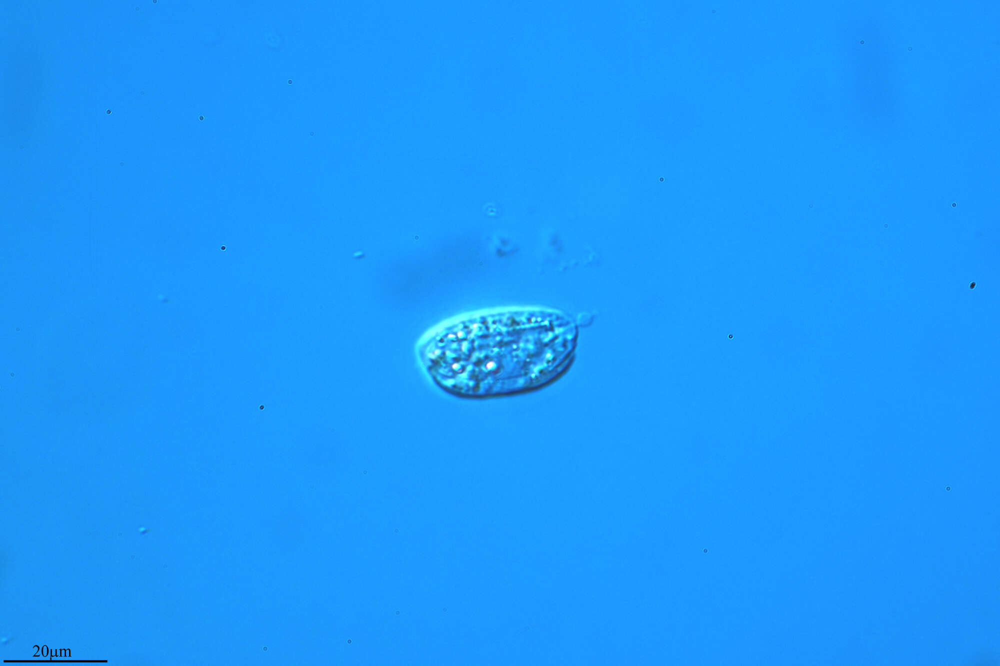
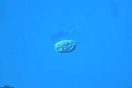
Petalomonas observed in freshwater sediments in the vicinity of Broome, Western Australia in September 2003. This image was taken using phase contrast optics. This work was supported by the Australian Biological Resources Study.
-
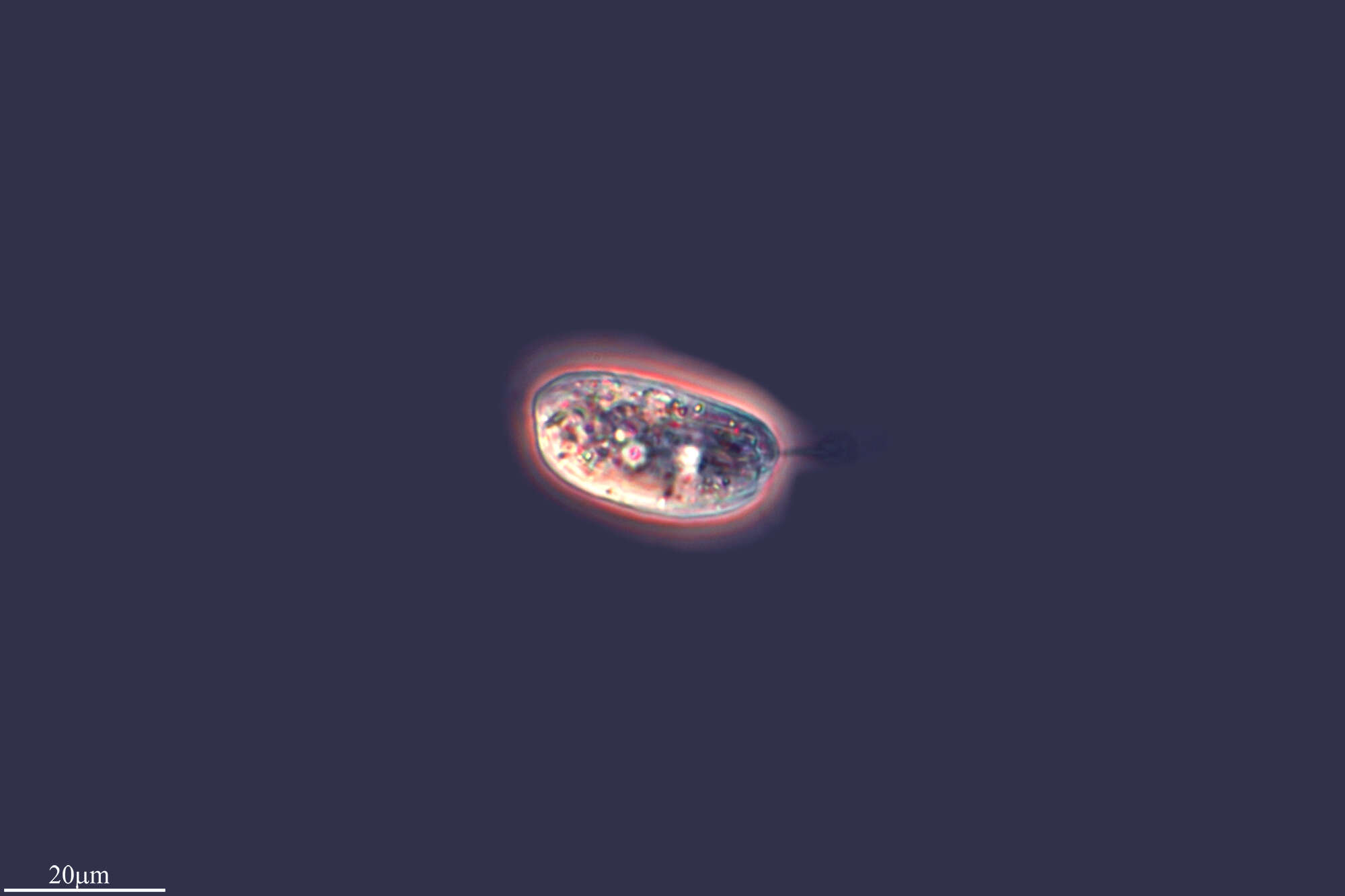
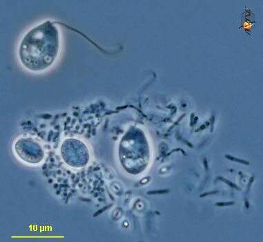
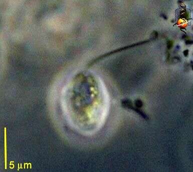
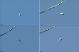
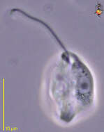
Collected samples from Nobska pond (blackish water), Woods Hole, Massachusetts. Focused on the cell surface (left) and flagellum (right). It is unclear whether this organism has another flagellum (i.e., Notosolenus). Photographed by Hwan Su Yoon.
-
-
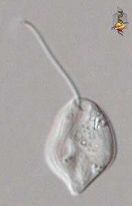
Petalomonas pusilla. Cell observed in sandy and muddy marine sediments in the vicinity of Broome, Western Australia in September 2003. This image was taken using phase contrast optics. This work was supported by the Australian Biological Resources Study.
-
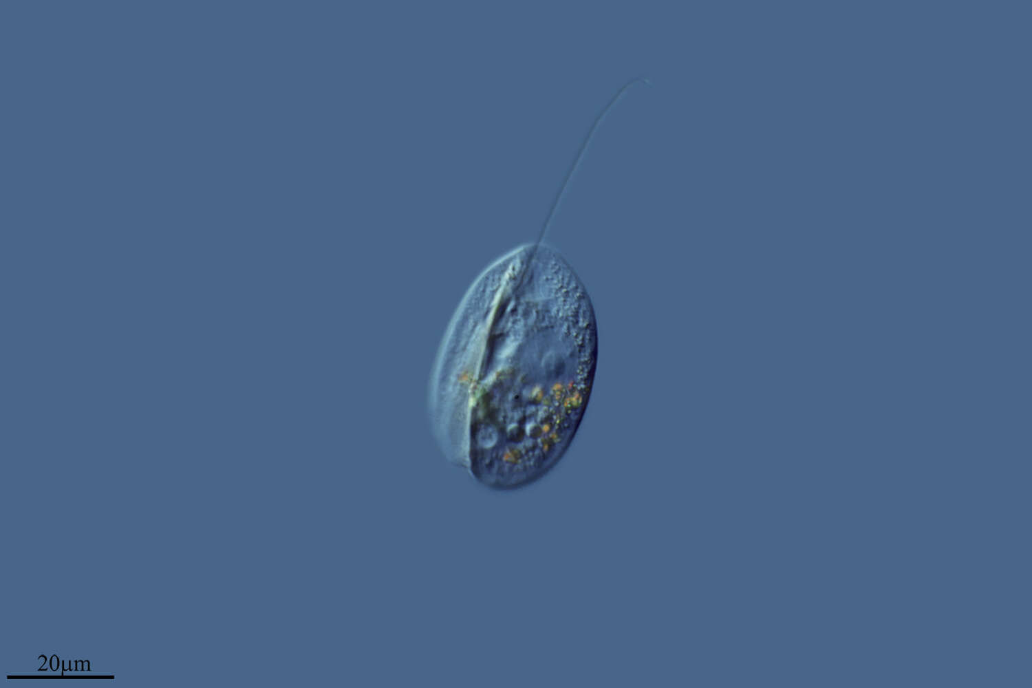
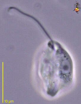
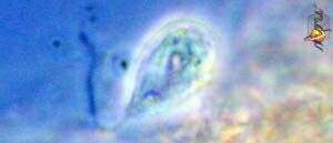
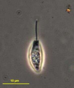
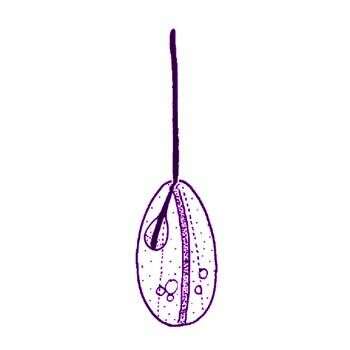
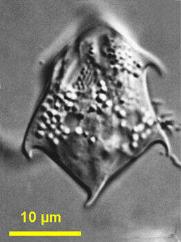
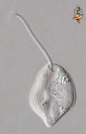
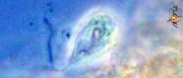
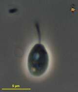
Petalomonas minuta Hollande, 1942. Cell outline is broadly elliptical. Cells are 5.5 to 10 microns long (mostly 6 to 7 microns), 4 to 6 microns wide, flattened, and with a deep longitudinal groove on the dorsal face and two distinct ventral ridges, which were hard to see. The cells have one flagellum inserting into a reservoir in the right hand side of the cell, flagellum about same length as the cell. The nucleus is in the left side of the cell. The cells move by gliding.
-
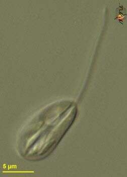
Petalomonas minuta. Cell observed in sandy and muddy marine sediments in the vicinity of Broome, Western Australia in September 2003. This image was taken using differential interference contrast optics. This work was supported by the Australian Biological Resources Study.
-
-
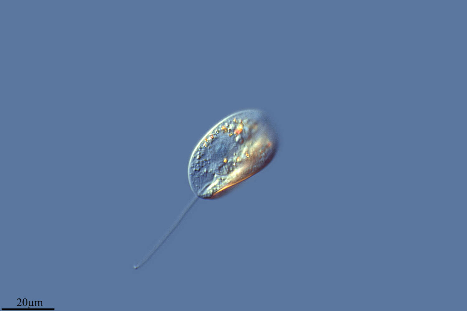
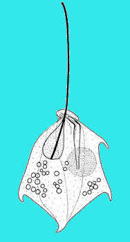
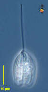
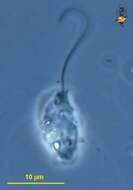
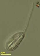
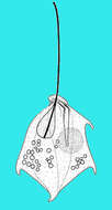
Petalomonas (pet-al-owe-moan-ass) spinifera (Lackey, 1962) Lee and Patterson, 2000. Cell outline is irregular. Cells are about 29 microns long and 21 microns wide, flattened, and with three fine dorsal ridges, two fine ventral ridges and one distinct ridged ventral groove, which extends from the collar around the flagella canal. The cells have four hyaline protrusions: one is located on a wedge-shaped posterior end of the cell, two are in the lateral posterior part, and the last one is in the anterior left hand side or near the nucleus. The one emergent flagellum is slightly shorter than the cell and is anteriorly directed. The large reservoir is situated anteriorly in the right side of the cell and the nucleus is near the midline of the left side. Rarely observed.