-
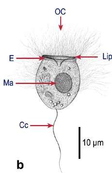
Fig 1a: Balanion comatum Line drawing of protargol stained cell
-
-
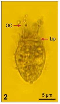
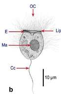
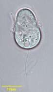
Infraciliature of the planktonic protstomatid ciliate, Apsiktrata gracilis (Penard,1922)Foissner, Berger & Kohmann 1994. Morphologically quite similar to members of the genus Holophrya but lacking a "dorsal brush". The anterior apical cytostome and its circumoral dikinetids is seen here.There is a long caudal cilium in vivo (only its basal body is seen here at the posterior pole). Collected from a freshwater pond near Boise, Idaho. Silver carbonate stain (see Foissner, W. Europ. J. Protistol., 27:313-330;1991). Brightfield
-
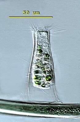
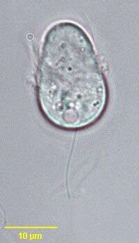
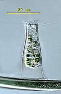
Metacystis (met-ah-sis-tiss) lagenula has a transparent lorica which is formed like an Erlenmeyer flask. The oral aperture is equiped with long pectinelles. There is a conspicuous caudal cilium and the contractile vacuole can be seen in the posterior third of the cell. This specimen was collected in freshwater ponds near Konstanz, Germany. Differencial interference contrast.
-
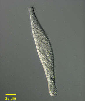
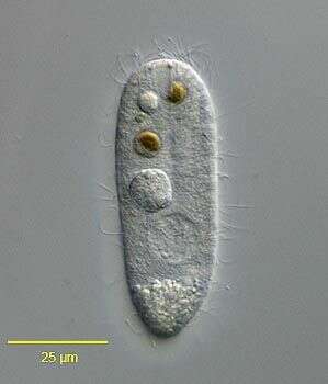
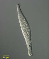
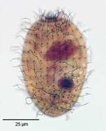
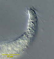
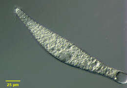
Portrait of extended Lagynus cucumis, a colorless Prostome ciliate found in sapropelic habitats. L. cucumis is synonymous with Lacrymaria cucumis (Penard, 1922) and Lacrymaria putrina (Kahl, 1926). Lagynus cucumis is longer and more slender than L. elegans with about five less pronounced ring-like furrows at the anterior end. The conical head region is much smaller than that of L. elegans. The apical cytostome is supported by fine trichites (seen here). The elongate cell body is flexible, contractile and slightly flattened. Somatic kineties are longitudinal and uniform. Slightly longer cilia surround the anterior end . A bean-shaped macronucleus with a central transverse crease is located in the midportion of the cell (seen well here). There is a single posterior terminal contractile vacuole (not seen in this image). Collected from sapropelic sediments of a freshwater aquaculture tub (pH 7.56) at a Koi farm near Boise, Idaho October 2003. DIC optics.
-
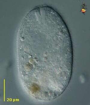
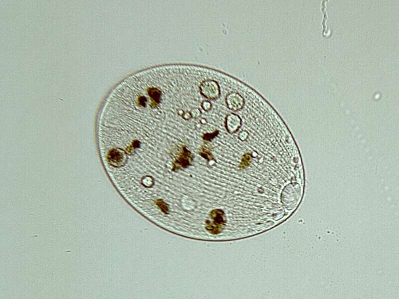
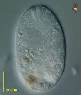
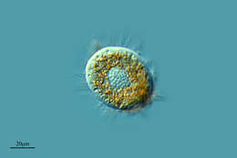
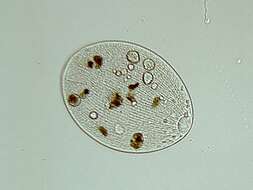
The ciliate Holophrya, with a polar mouth which is surrounded by stiff rods (nemadesmata) that can be used to manipulate food into the body. Normally eats algae and detritus. Differential interference contrast.
-
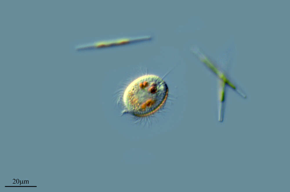
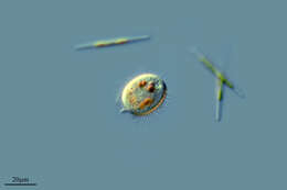
Urozona buetschlii, a small dumb-bell -shaped hymenostome ciliate slightly constricted at the center. Somatic ciliation is restricted to an equatorial girdle. The oral aperture is located in the mid-body. Several very small adoral membranelles are present but not seen in these images. Single long caudal cilium. Posterior contractile vacuole. Single large spherical macronucleus in posterior half of body. Rapid swimmer. Bactiverous. Genus is monospecific. Urozona is somewhat similar in overall shape to Urocentrum turbo but much smaller and without a posterior ciliary tuft. Urozona is similar in size to Mesodinium but without anterior tentacular processes, bristles or furcate ciliary tufts. From freshwater pond near Boise, Idaho. Brightfield.
-
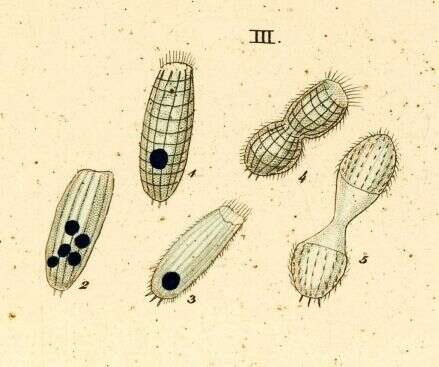
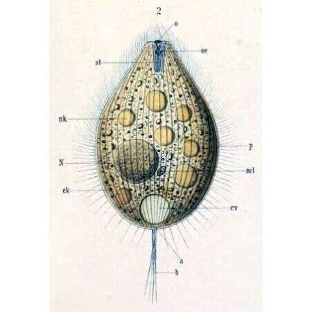
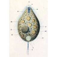
a -- Anus b -- Sensory bristle c.v -- Contractile vacuole ek -- Ectoplasm N -- Macronucleus ncl -- Micronucleus nk -- Food particle o -- Mouth p -- Pellicle st -- Cytopharyngeal basket
-
Lardero, La Rioja, Spain
-
Herrera, Castille and Leon, Spain
-
Villar del Pedroso, Extremadura, Spain
-
Los Cotos, Madrid, Spain
-
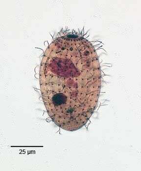
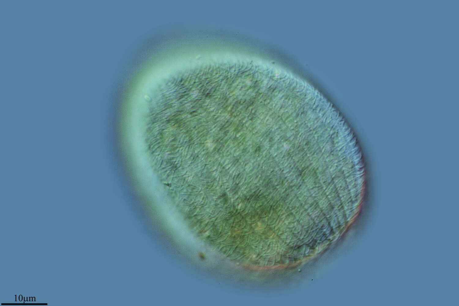
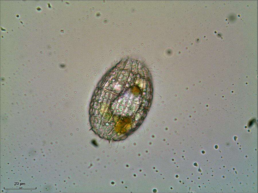
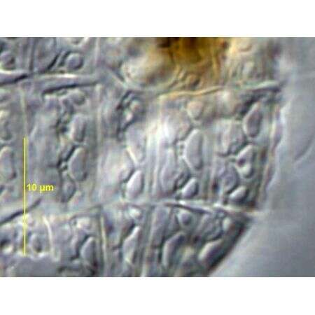
Body with regurarly arranged ectoplasmic plates. Cytostome at anterior end, surrounded by slightly longer cilia . Often spinous projection at or near posterior end.
-
Fig 1b: Balanion comatum Line drawing of live cell (from Wulff, 1919)
-
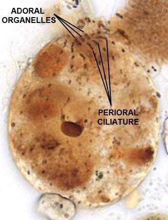
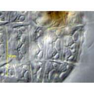
Oral Infraciliature of Nolandia nolandi (KAHL, 1930) SMALL & LYNN, 1985.Collected from a freshwater pond near Boise, Idaho. June 2008.Stained by the Protargol technique (Wilbert modification) (see Foissner, W. Europ. J. Protistol., 27:313-330;1991).Brightfield.
-
Infraciliature of the planktonic protstomatid ciliate, Apsiktrata gracilis (Penard,1922)Foissner, Berger & Kohmann 1994. Morphologically quite similar to members of the genus Holophrya but lacking a "dorsal brush". The anterior apical cytostome and its circumoral dikinetids is seen here.The microfibrillar system associted with the basal bodies of the somatic kineties is visible here. Collected from a freshwater pond near Boise, Idaho. Silver carbonate stain (see Foissner, W. Europ. J. Protistol., 27:313-330;1991). Brightfield
-
Detail view of anterior end of extended Lagynus cucumis, a colorless Prostome ciliate found in sapropelic habitats. L. cucumis is synonymous with Lacrymaria cucumis (Penard, 1922) and Lacrymaria putrina (Kahl, 1926). Lagynus cucumis is longer and more slender than L. elegans with about five less pronounced ring-like furrows at the anterior end. The conical head region is much smaller than that of L. elegans. The apical cytostome is supported by fine trichites (seen here). The elongate cell body is flexible, contractile and slightly flattened. Somatic kineties are longitudinal and uniform. Slightly longer cilia surround the anterior end. A bean-shaped macronucleus with a central transverse crease is located in the midportion of the cell. There is a single posterior terminal contractile vacuole. Collected from sapropelic sediments of a freshwater aquaculture tub (pH 7.56) at a Koi farm near Boise, Idaho October 2003. DIC.
-
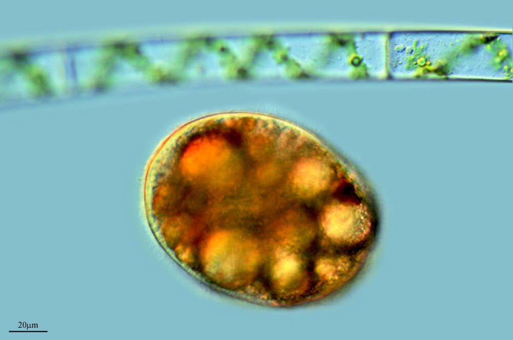
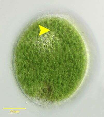
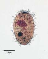
Holophrya ovum (Ehrenberg,1831). The yellow arrowhead indicates the three dorsal brush rows of short cilia. DIC
-
Urozona buetschlii, a small dumb-bell -shaped hymenostome ciliate slightly constricted at the center. Somatic ciliation is restricted to an equatorial girdle. The oral aperture is located in the mid-body. Several very small adoral membranelles are present but not seen in these images. Single long caudal cilium. Posterior contractile vacuole. Single large spherical macronucleus in posterior half of body. Rapid swimmer. Bactiverous. Genus is monospecific. Urozona is somewhat similar in overall shape to Urocentrum turbo but much smaller and without a posterior ciliary tuft. Urozona is similar in size to Mesodinium but without anterior tentacular processes, bristles or furcate ciliary tufts. From freshwater pond near Boise, Idaho. Brightfield.
-
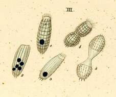
Ovoid to cylindrical; ciliation uniform; oral basket made up of double trichites which end up deep in ectoplasm. Macronucleus ovoid, reniform or ellongate. Cell body 80-200 micron long.
-
Fig. 2: Balanion comatum Lugol's fixed cell, lateral view
-
Calcified armor plates of Nolandia nolandi (KAHL, 1930) SMALL & LYNN, 1985.Collected from a freshwater pond near Boise, Idaho. June 2008.DIC.
-
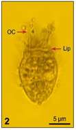
Portrait of the planktonic protstomatid ciliate, Apsiktrata gracilis (Penard,1922)Foissner, Berger & Kohmann 1994. Morphologically quite similar to members of the genus Holophrya but lacking a "dorsal brush". The anterior apical cytostome and its circumoral dikinetids is seen here.The microfibrillar system associted with the basal bodies of the somatic kineties is visible here. Collected from a freshwater pond near Boise, Idaho. DIC.
-
Portrait of extended Lagynus cucumis, a colorless Prostome ciliate found in sapropelic habitats. L. cucumis is synonymous with Lacrymaria cucumis (Penard, 1922) and Lacrymaria putrina (Kahl, 1926). Lagynus cucumis is longer and more slender than L. elegans with about five less pronounced ring-like furrows at the anterior end. The conical head region is much smaller than that of L. elegans. The apical cytostome is supported by fine trichites. The elongate cell body is flexible, contractile and slightly flattened. Somatic kineties are longitudinal and uniform. Slightly longer cilia surround the anterior end. A bean-shaped macronucleus with a central transverse crease is located in the midportion of the cell (seen well here). There is a single posterior terminal contractile vacuole (seen in this image). Collected from sapropelic sediments of a freshwater aquaculture tub (pH 7.56) at a Koi farm near Boise, Idaho October 2003. DIC optics.