-
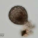
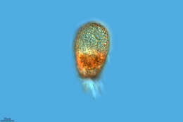
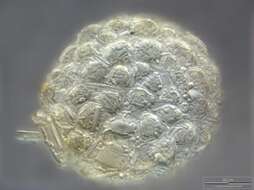
Portrait of Netzelia tuberculata (Wallich,1864) test. The test or shell is composed of quartz particles arranged in small knob-like aggregates on its surface. The approximately circular shell aperture has a narrow collar of quartz particles. Pseudopodia (not seen in this image) are lobose. Collected from freshwater irrigation canal near Boise, Idaho in November 2003. Brightfield illumination.
-
Ribadelago de Franco, Castille and Leon, Spain
-
Logrono, La Rioja, Spain
-
Rumoroso, Cantabria, Spain
-
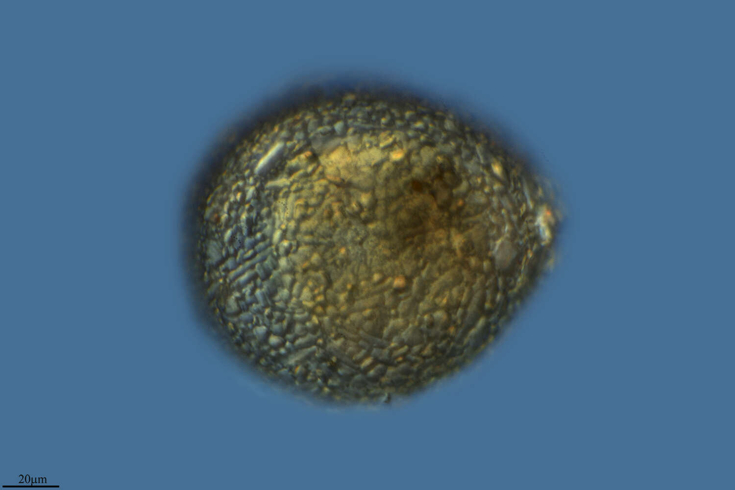
This species has been moved from genus Difflugia to Netzelia (family Lesqueruesiidae). Most of the agglutinated particles are silicious idiosoms, all xenosomes are covered with silica.This multi layer image was built up using 4 brightfield frames with a manual stacking technique using Corel Photopaint. The specimen was gathered in a tiny freshwater pond at the island of Hiddensee (Baltic Sea, Germany) which shows a fascinating biodiversity of naked and testate amoebae. Images were taken using Zeiss Standard with Olympus C7070 CCD camera.
-
Ribadelago de Franco, Castille and Leon, Spain
-
Logrono, La Rioja, Spain
-
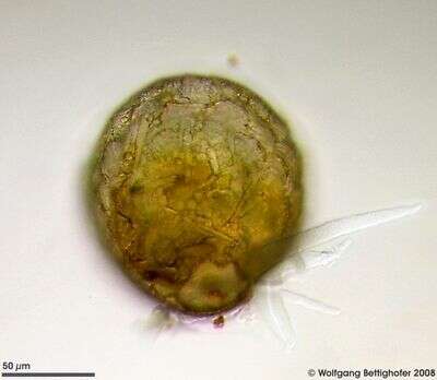
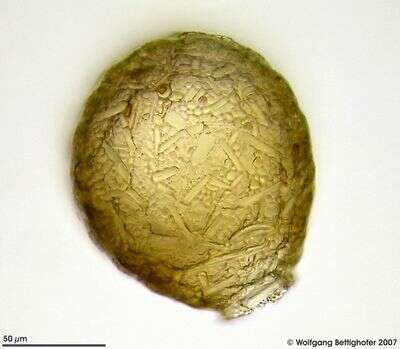
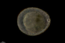
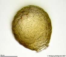
This species has been moved from genus Difflugia to Netzelia (family Lesqueruesiidae). Most of the agglutinated particles are silicious idiosoms, all xenosomes are covered with silica. This multi layer image of Netzelia tuberculata was built up using 12 brightfield frames with a manual stacking technique using Corel Photopaint. The scale bar indicates 50 µm. The specimen was gathered in a tiny freshwater pond at the island of Hiddensee (Baltic Sea, Germany) which shows a fascinating biodiversity of naked and testate amoebae. Images were taken using Zeiss Standard with Olympus C7070 CCD camera.
-
Ribadelago de Franco, Castille and Leon, Spain
-
Logroo, La Rioja, Espaa
-
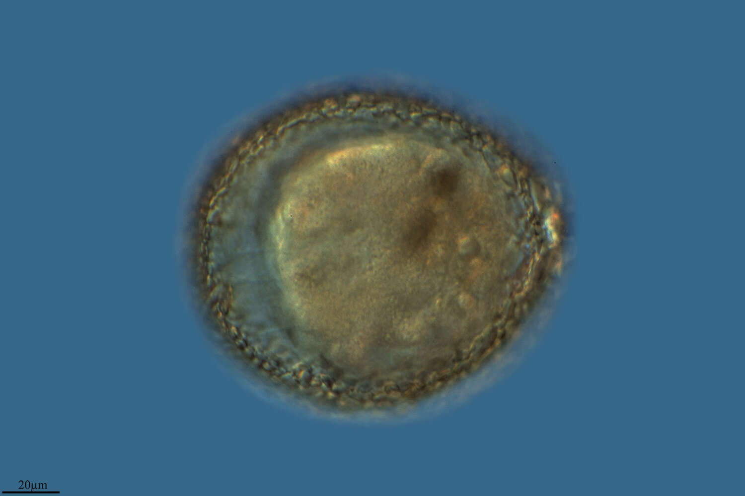
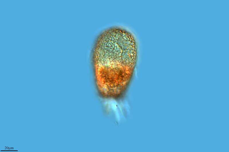
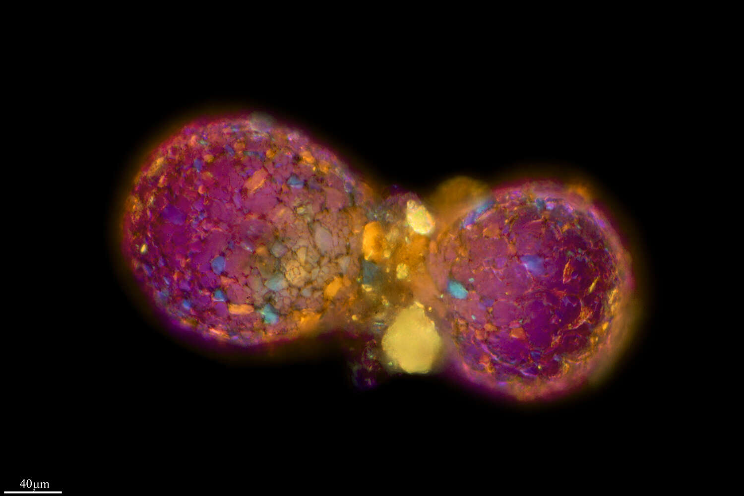
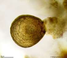
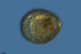
This species has been moved from genus Difflugia to Netzelia (family Lesqueruesiidae). Most of the agglutinated particles are silicious idiosoms, all xenosomes are covered with silica. The texture of the test and the outline of the pseudostome collar of Netzelia tuberculata are (according to R. Meisterfeld) very variable. This multi layer image was built up using 17 brightfield frames with a manual stacking technique using Corel Photopaint. The specimen was gathered in a tiny freshwater pond at the island of Hiddensee (Baltic Sea, Germany) which shows a fascinating biodiversity of naked and testate amoebae. Images were taken using Zeiss Standard with Olympus C7070 CCD camera.
-
Logrono, La Rioja, Spain
-
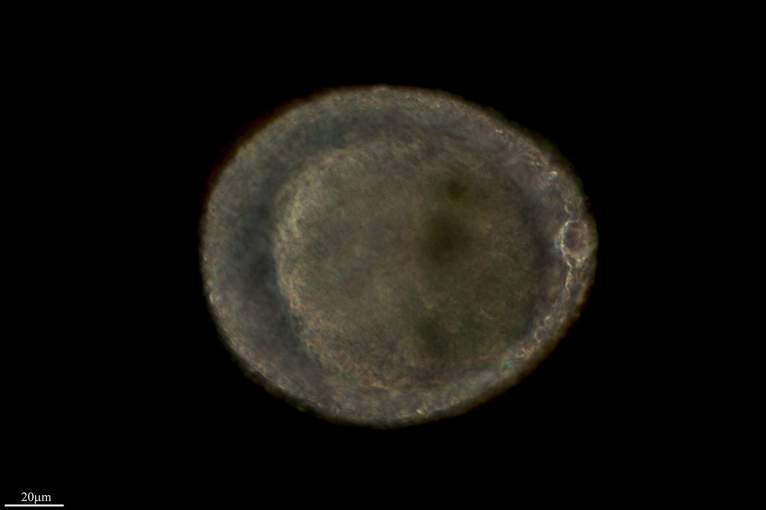
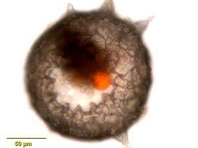
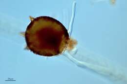
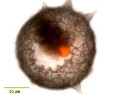
This species has been moved from genus Difflugia to Netzelia (family Lesqueruesiidae). Most of the agglutinated particles are silicious idiosoms, all xenosomes are covered with silica. Multi layer image of Netzelia specimen using 10 frames. Stack assembled with Corel Photopaint. The red dot is garbage waiting for defecation. Sample from sphagnum pond near Bergenhusen (Schleswig-Holstein, Germany). This image was taken using Zeiss Universal with Olympus C7070 CCD camera.
-
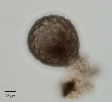
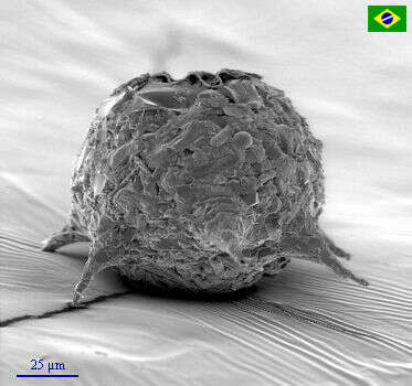
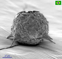
The test of Netzelia tuberculata shows its mulberry surface which gave the name (tuberculata). As all Lesquereusiidae Netzelia builds up the test with self-made siliceous pads (called idiosomes). A few xenosomes (mostly parts of frustule from pennate diatoms) are also visible. All xenosomes are covered with a siliceous coating. Scale bar indicates 25µm Sample from a freshwater pond on the island of Hiddensee (Baltic Sea, Germany). This image was taken using Zeiss Universal with Olympus C7070 CCD camera.
-
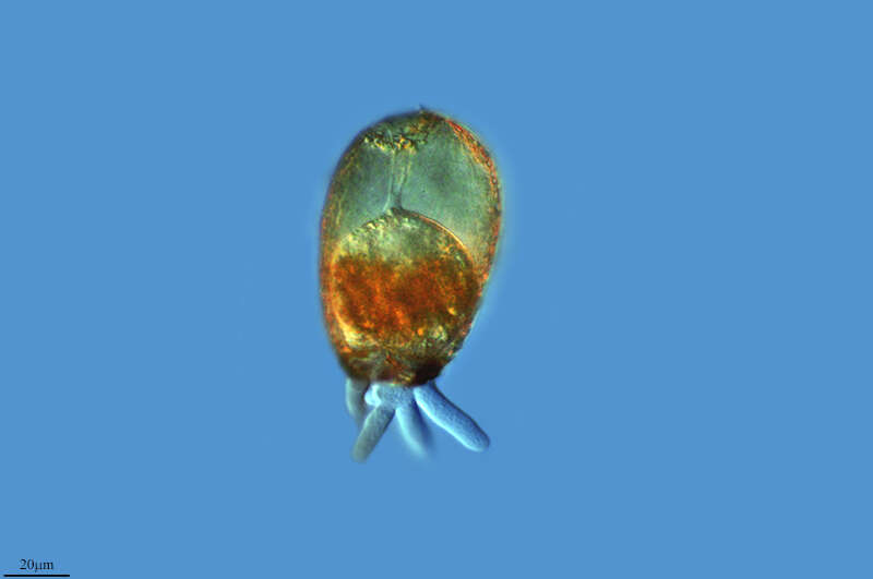
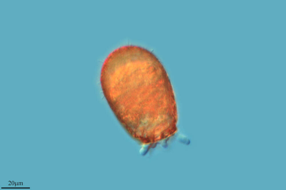
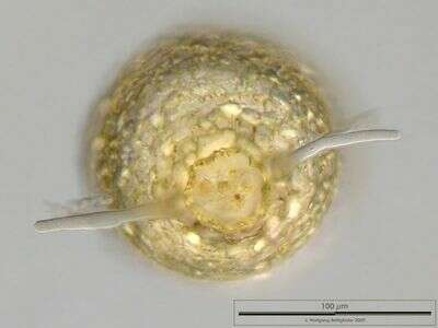
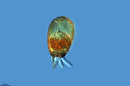
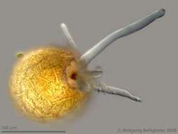
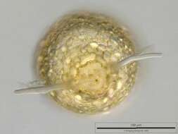
Apertural view on Netzelia tuberculata depicting the pseudostome collar, pseudopods and defecation vacuole. This multi layer image was built up using 42 frames with a manual stacking technique using Corel Photopaint. The specimen was gathered in a tiny freshwater pond at the island of Hiddensee (Baltic Sea, Germany) which shows a fascinating biodiversity of naked and testate amoebae. Images were taken using Zeiss Standard with Olympus C7070 CCD camera.
-
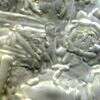
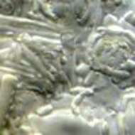
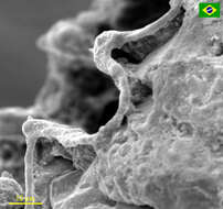
Detail view on the test of Netzelia tuberculata. Idiosomes and (between them, to stabilize the Mulberry texture) a few frustules from tiny pennate diatoms as xenosomes are visible. All xenosomes are covered with a siliceous coating. Sample from a freshwater pond on the island of Hiddensee (Baltic Sea, Germany). This image was taken using Zeiss Universal with Olympus C7070 CCD camera.
-
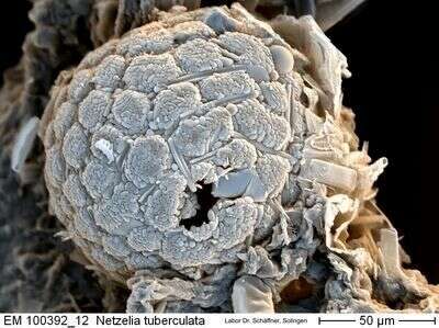
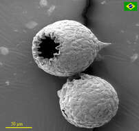
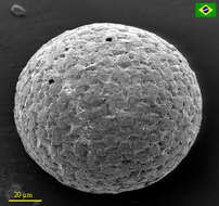
SEM of Netzelia tuberculata test. Courtesy of Lab Dr. Karl-Heinz Schaeffner, Solingen, Germany. Sample from a freshwater pond on the island of Hiddensee (Baltic Sea, Germany).
-
Difflugia corona (Wallich 1864). Portrait of the test, which is composed of quartz particles. The species is identified by the distinctive dentate protrusions around a circular aperture and a variable number of aboral conical spines. From freshwater pond near Boise, Idaho.Brightfield.
-
Scanning electron microscopy side view of D. corona
-
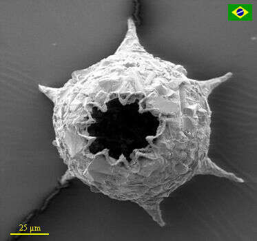
Scanning Electron Microscopy Apertural view of D. corona
-
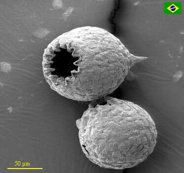
Scanning electron microscopy of two D. corona, one with a spine, and the other spineless.
-
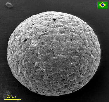
Scanning Electron Microscopy of a Difflugia corona without spines.
-
Scanning Electron Micrograph showing the apertural architecture of D. corona.
-
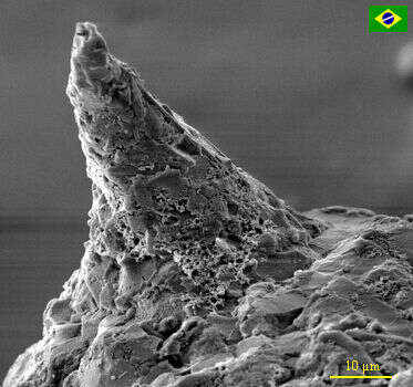
Scanning Electron Micrographs showing a D. corona conical spine.