-
Valea, Galicia, Espaa
-
Grove, O, Galicia, Spain
-
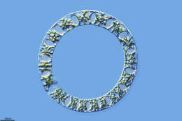
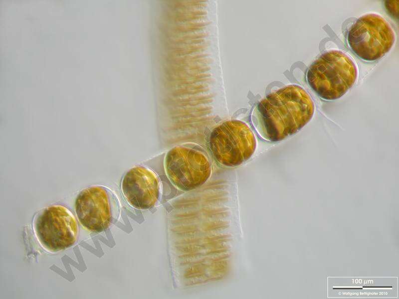
Melosira moniliformis accompanied by Fragilaria islandica. Scale bar indicates 100 m. The image was built up using several photomicrographic frames with manual stacking technique. Sample from North Sea near Heligoland (spring diatom bloom). Images were taken using Zeiss Universal with Olympus C7070 CCD camera.For more look at
www.protisten.de/english/gallery_main/gallery_main.htmlFor high-resolution images please ask postmaster@protisten.de.
-
Galende, Castille and Leon, Spain
-
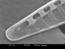
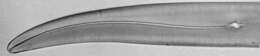
Fig 3: Pseudonitzschia multiseries Scanned electron micrograph image showing detailed stria, fibulae and poroids.
-
All Biocode files are based on field identifications to the best of the researcher’s ability at the time.
-
All Biocode files are based on field identifications to the best of the researcher’s ability at the time.
-
All Biocode files are based on field identifications to the best of the researcher’s ability at the time.
-
All Biocode files are based on field identifications to the best of the researcher’s ability at the time.
-
All Biocode files are based on field identifications to the best of the researcher’s ability at the time.
-
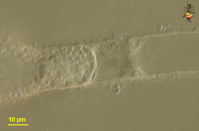
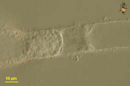
Oomycete fungus. Detail of hypha. Differential interference contrast.
-
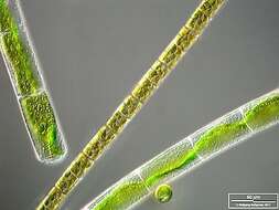
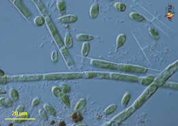
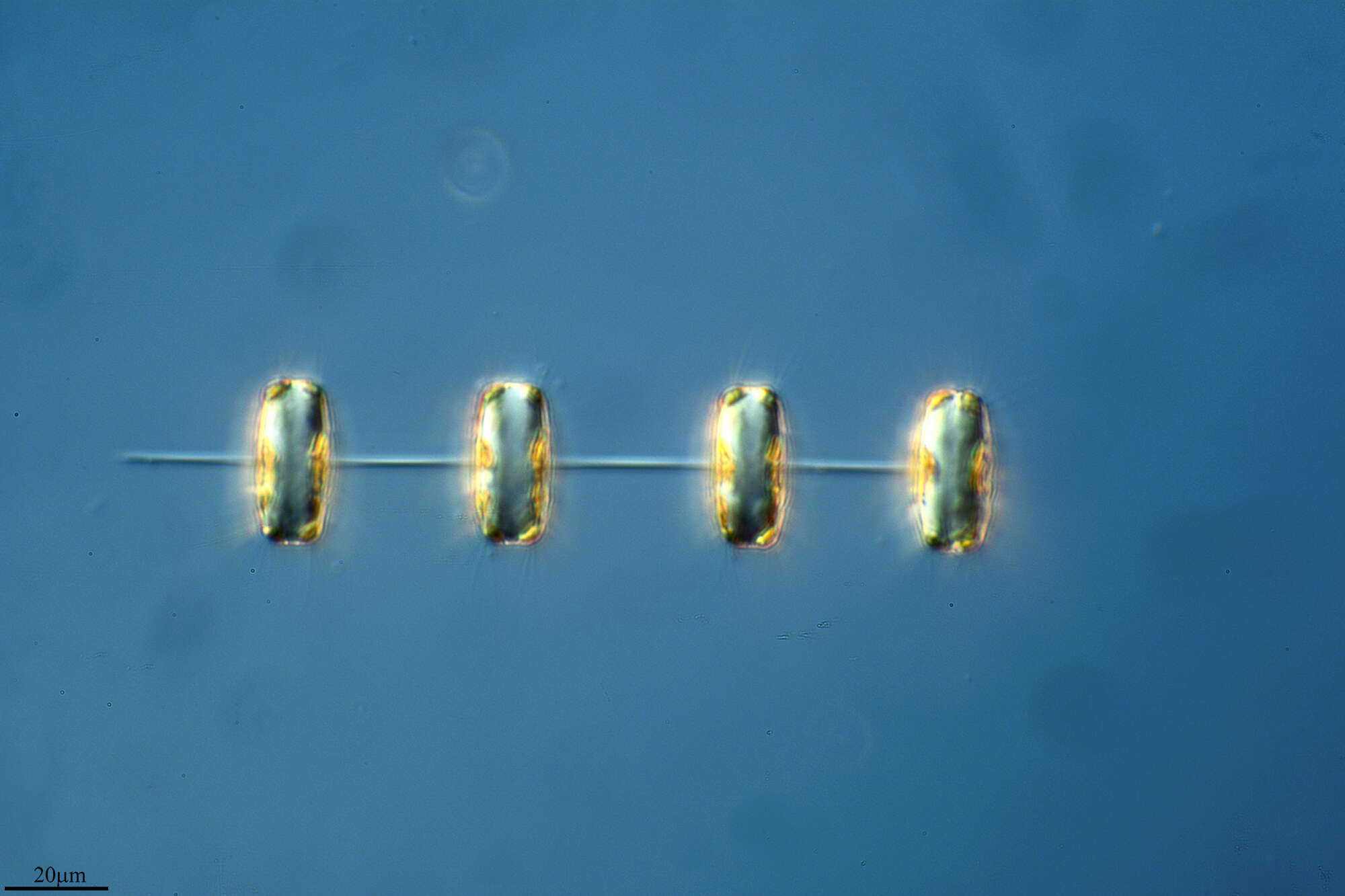
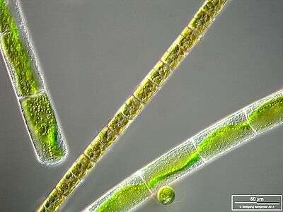
Melosira varians together with Mougeotia and Chlamydomonas. The scale bar indicates 50 µm. The specimen was gathered in the wetlands of Oderbruch (Oder valley 100 km north east of Berlin). The image was built up using several photomicrographic frames with manual stacking technique. Images were taken using Zeiss Universal with Olympus C7070 CCD camera.Image under Creative Commons License V 3.0 (CC BY-NC-SA).
-
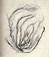
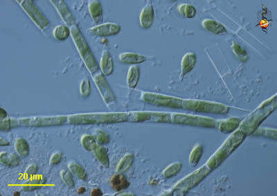
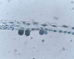
Tribonema (tribe-owe-knee-ma) is a xanthophyte, a group of stramenopiles with chloroplasts. Xanthophytes may adopt a variety of body forms, and the normal form for this genus is the filament. Cell walls often built from H-shaped units, and these overlap in the middle of a cell. Stalked attached cells also occur, as is evident here. Plastids usually discoid, yellow green. Differential interference contrast.
-
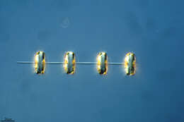
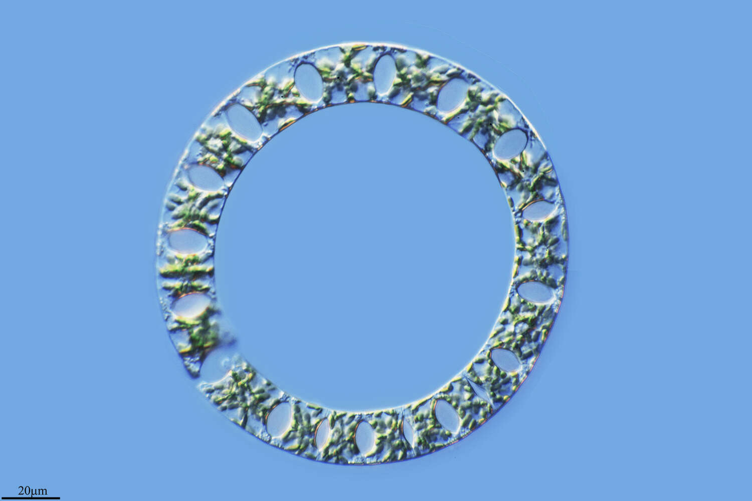
The oblique view exhibits short silicous spines, the so called occluded processes. On the lower left, lower right and central above chitinous spines are visible. Scale bar indicates 50 µm. The image was built up using several photomicrographic frames with manual stacking technique. Sample from North Sea near Heligoland (spring diatom bloom). Images were taken using Zeiss Universal with Olympus C7070 CCD camera.
-
Image from type material.
-
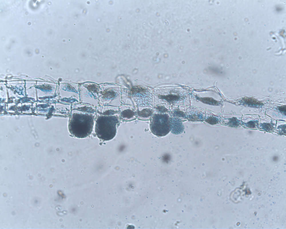
Detail showing plastids inside several cells at the end of a filament. Same filament as included in another picture in this collection.
-
Glatved Strand N
-
Glatved Strand N
-
Dokkedal, Himmerland, Danmark
-
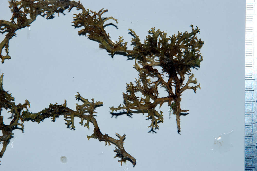
Fejrup, Helgenæs
-
Lendrup S Løgstør, Jylland, Danmark
-
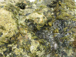
Giant Kelp forest, Santa Barbara County, California (30 September 2008)
-
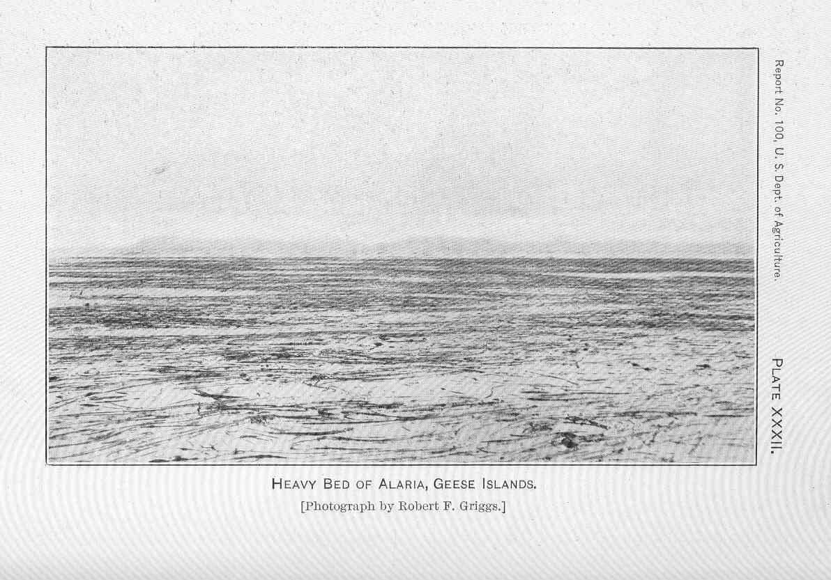
Heavy Bed of Alaria, Geese Islands.
-
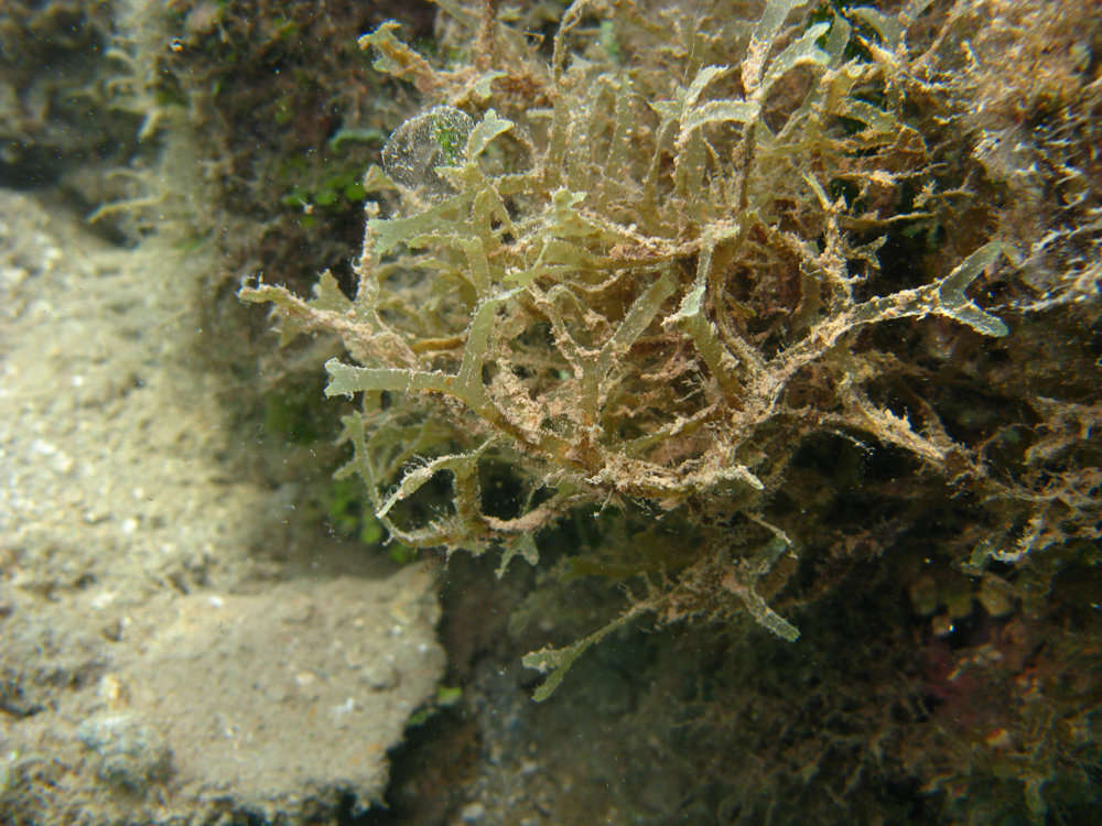
Dictyosiphon foeniculaceus.