-
Galende, Castile and Len, Spain
-
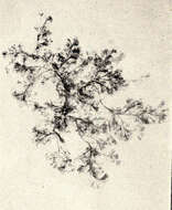
Fig 1: Schematic drawings of Chaetoceros socialis chains; a. vegetative cells, b. cells producing resting spores.
-
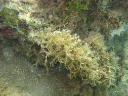
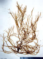
All Biocode files are based on field identifications to the best of the researcher’s ability at the time.
-
All Biocode files are based on field identifications to the best of the researcher’s ability at the time.
-
All Biocode files are based on field identifications to the best of the researcher’s ability at the time.
-
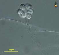
Oomycete fungus. Spore-forming body. Differential interference contrast.
-
-
Tribonema (tribe-owe-knee-ma) is a xanthophyte, a group of stramenopiles with chloroplasts. Xanthophytes may adopt a variety of body forms, and the normal form for this genus is the filament. Cell walls often built from H-shaped units, and these overlap in the middle of a cell. Plastids usually discoid, yellow green. Differential interference contrast.
-
Silicious processes (the labiate and the occluded ones) are visible. Scale bar indicates 25 µm. The image was built up using several photomicrographic frames with manual stacking technique. Sample from North Sea near Heligoland (spring diatom bloom). Images were taken using Zeiss Universal with Olympus C7070 CCD camera.
-
Image from type material.
-
Fejrup, Helgenæs
-
Holmkær, NV-Jylland, Danmark
-
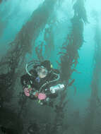
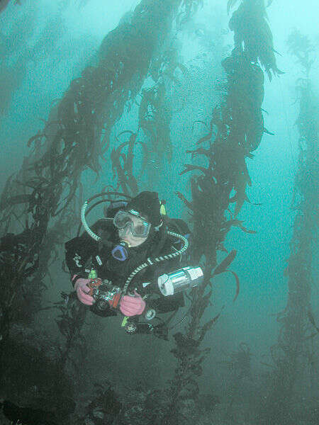
SCUBA diver in kelp forest, Point Lobos, California
-
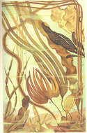
Algues brunes, Pheophycees. 1- Punctaria latifolia, 2- Punctaria plantaginea, 3- Asperococcus bullosus, 4- Scytosiphon tomentarius, 5- Phyllitis fascia, 6- Chorda filum, 7- Chorda tomentosa, 8- Laminaria saccharina, 9- Laminaria digitata, 10- Laminaria hyperborea
-
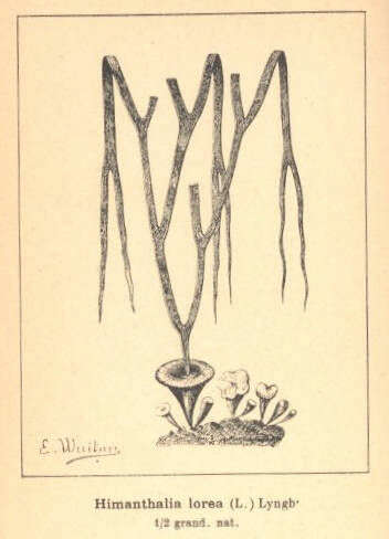
Pheophycees (Algues brunes) Fucacees, HImanthalia lorea (L.) Lyngh..
-
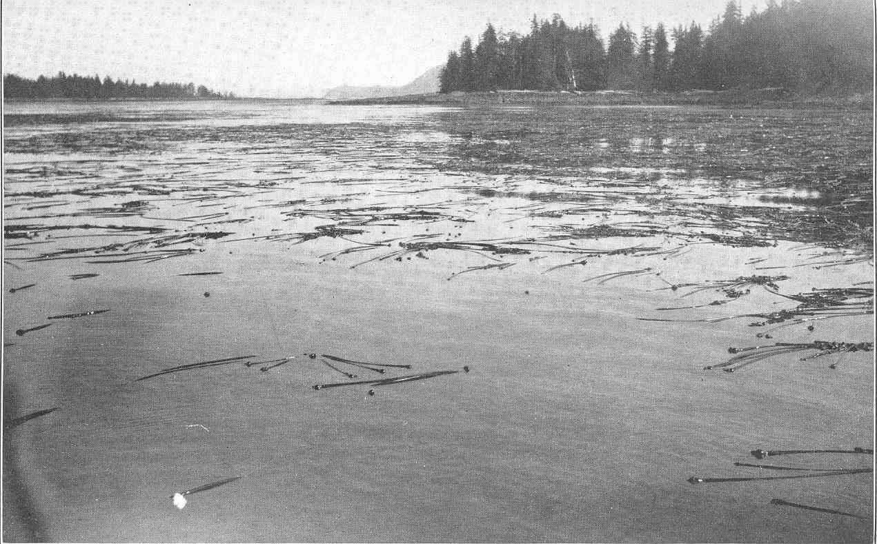
Nereocystis near Barrier Island, in the Neighborhood of Shakan Bay.
-
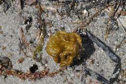
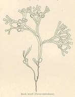
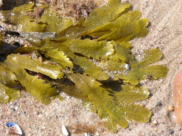
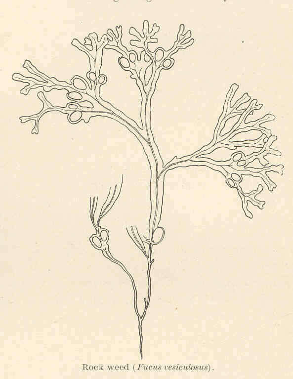
Rock Week (Fucus vesiculosus).
-
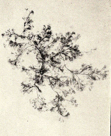
Ectocarpus viridis.
-
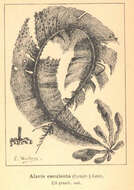
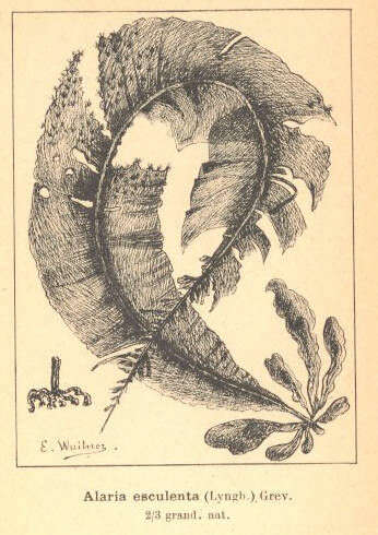
Pheophycees (Algues brunes) Laminariees, Alaria esculenta (Lyngh.) Grev..
-
Ribadelago, Castille and Leon, Spain
-
Covaleda, Castille and Leon, Spain
-
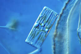
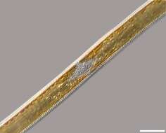
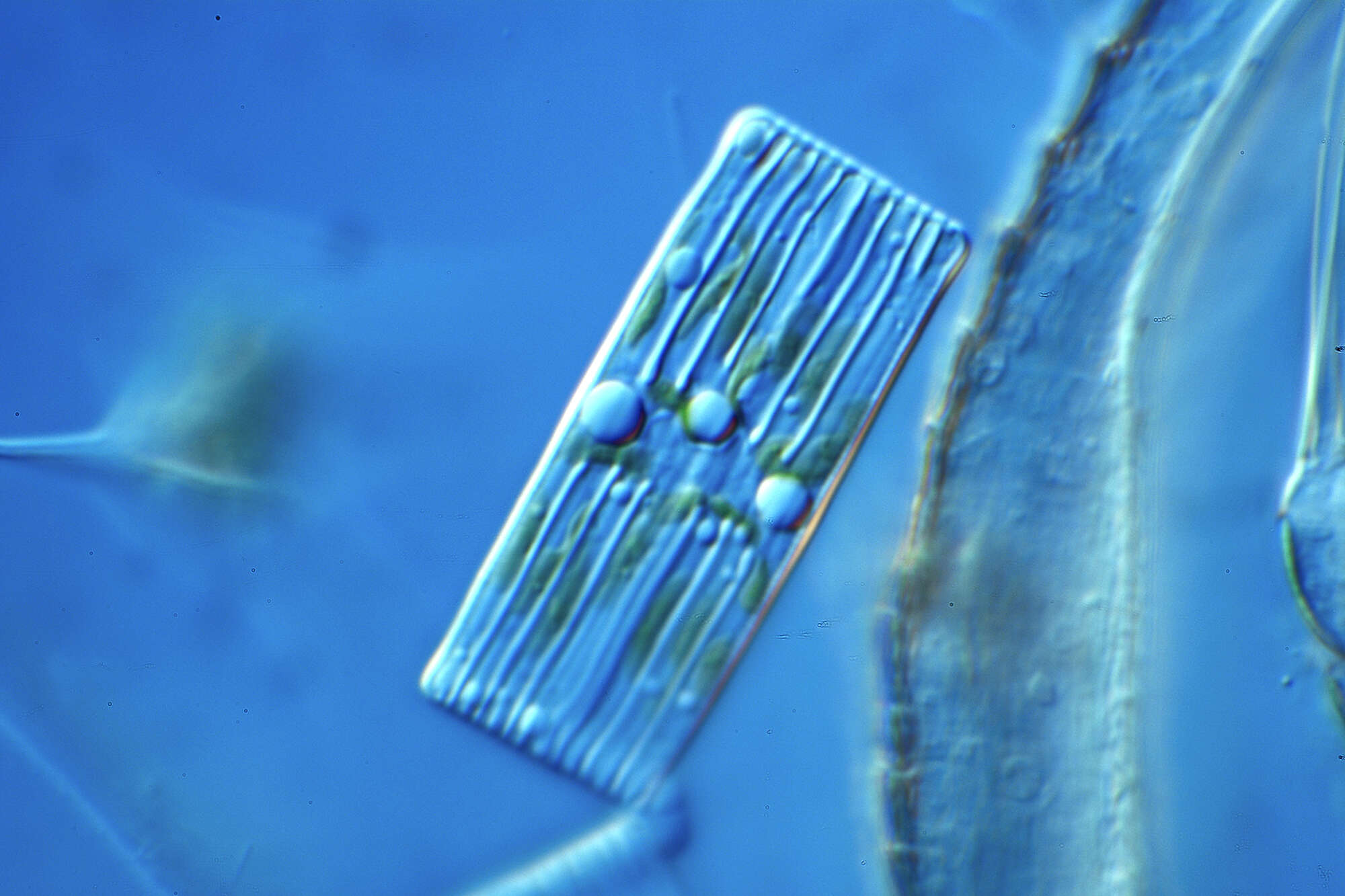
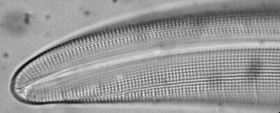
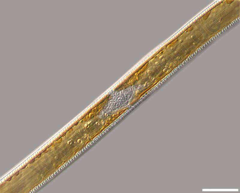
Nitzschia sigmoidea.Detail: Center of the cell in cingular view with nucleus, cloroplasts with oil droplets (energy store) and the canal raphes. Scale bar indicates 25 m.Sample from a wetland at the Pillersee (Tyrol, Austria). The image was built up using several photomicrographic frames with manual stacking technique. Images were taken using Zeiss Universal with Olympus C7070 CCD camera.For more look at
www.protisten.de/english/gallery_main/gallery_main.htmlFor high-resolution images please ask postmaster@protisten.de.
-
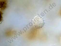
Actinophrys sol.Scale bar indicates 50 m.Collected from Bodden, the brackish water between the isles of Hiddensee an Ruegen (German Baltic Sea). The image was built up using several photomicrographic frames with manual stacking technique. Images were taken using Zeiss Universal with Olympus OM-D M5 MKII.For high-resolution images please ask postmaster@protisten.de.
-
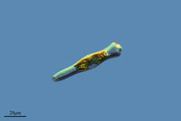
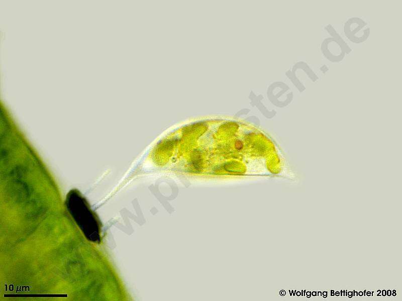
Specimen of Characiopsis spec, probably Ch. acuta. The microscope is focussed on the chloroplast. Digital drawing using 6 frames generating depth of focus, stacked manually using Corel Photopaint . Aufwuchs on Oedogonium which itself was Aufwuchs on roots dangling in a creek. This image was taken using Zeiss Universal with Olympus C7070 CCD camera. For high-resolution images please ask postmaster@protisten.de.