-
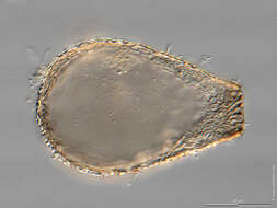
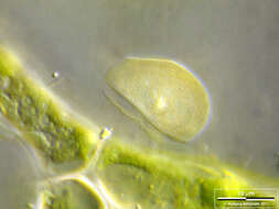
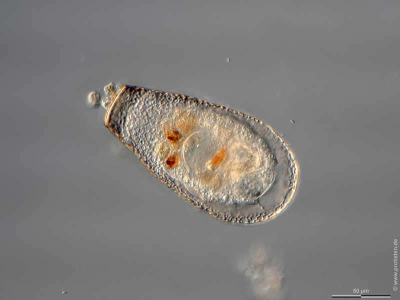
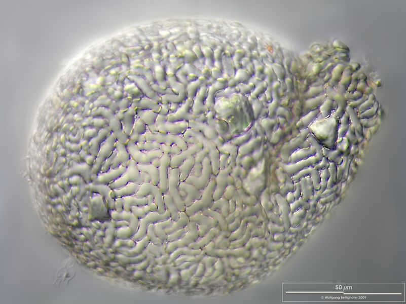
Planocarina marginata Scale bar indicates 50 µm. Sample from wetland Paradieswiese near Kitzbühel, Tyrol, Austria. Sampling date 06/2023. The image was built up using several photomicrographic frames with manual stacking technique. Images were taken using Zeiss Axioplan with Olympus OM-D M5 MKII. Image under Creative Commons License V 3.0 (CC BY-NC-SA). Place name: Wetland Paradieswiese near Kitzbühel (Tyrol, Austria) Latitude: 47.47375067 Longitude: 12.37624884 Multiebenen-Abbildung, manuell gestapelt. Der Messbalken markiert eine Länge von 50 µm. Probe von der Paradieswiese bei Kitzbühel-Steuerberg, Tirol. Datum der Aufsammlung: 06/2023. Mikrotechnik: Zeiss Axioplan, Kamera: Olympus OM-D M5 MKII. Creative Commons License V 3.0 (CC BY-NC-SA). For permission to use of (high-resolution) images please contact postmaster@protisten.de.
-
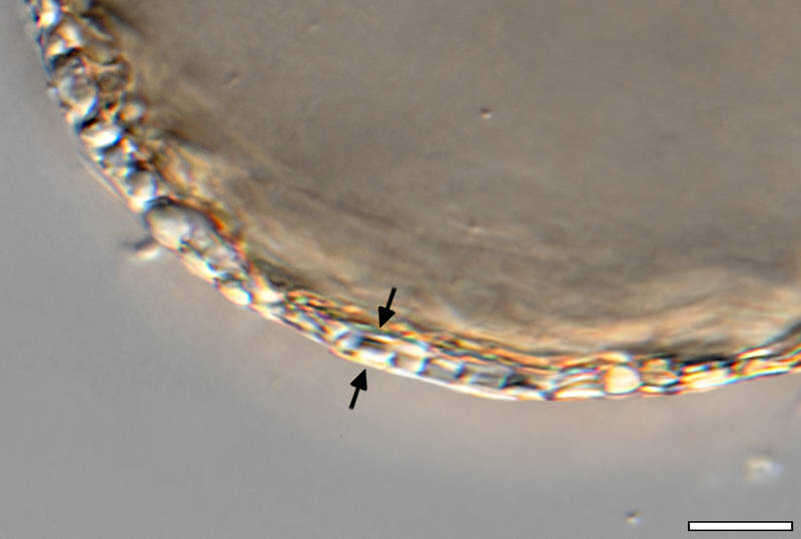
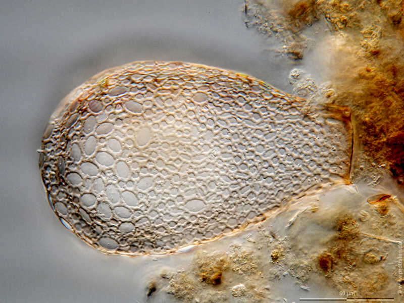
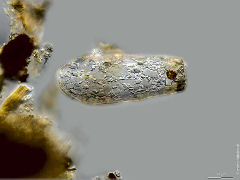
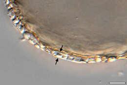
Planocarina marginata The arrows mark the flat zone of the shell at the edge, which is typical for genus Planocarina.Scale bar indicates 10 µm. Sample from wetland Paradieswiese near Kitzbühel, Tyrol, Austria. Sampling date 06/2023. The image was built up using several photomicrographic frames with manual stacking technique. Images were taken using Zeiss Axioplan with Olympus OM-D M5 MKII. Image under Creative Commons License V 3.0 (CC BY-NC-SA). Place name: Wetland Paradieswiese near Kitzbühel (Tyrol, Austria) Latitude: 47.47375067 Longitude: 12.37624884 Die Pfeile markieren die plane Zone der Schale am Rand, welche für die Gattung Planocarina typisch ist. Multiebenen-Abbildung, manuell gestapelt. Der Messbalken markiert eine Länge von 10 µm. Probe von der Paradieswiese bei Kitzbühel-Steuerberg, Tirol. Datum der Aufsammlung: 06/2023. Mikrotechnik: Zeiss Axioplan, Kamera: Olympus OM-D M5 MKII. Creative Commons License V 3.0 (CC BY-NC-SA). For permission to use of (high-resolution) images please contact postmaster@protisten.de.
-

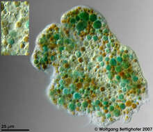
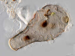
Enteromyxa paludosa Enteromyxa paludosa cell was showing very colorful metabolites and numerous nuclei and contractile vacuoles. In order to deliver depth of focus 50 high resolution frames (Planapo 63/1.4) were processed. The picture inserted shows 4 nuclei and 3 contractile vacuoles in higher magnification. Sample from sphagnum pond situated in the northern alpine region of Austria near Salzburg. Images were taken using Zeiss Universal with Olympus C7070 CCD camera.Image under Creative Commons License V 3.0 (CC BY-NC-SA). Place name: Bogs near Salzburg (Austria) Latitude: 48.068516 Longitude: 12.954134 Die Enteromyxa paludosa -Zelle zeigt sehr farbintensive Metabolite und zahlreiche Kerne und kontraktile Vakuolen. Die ins Bild eingefügten Ausschnittsvergrößerungen zeigen 4 Kerne und 3 kontraktile Vakuolen. Tiefenschärfe durch Multiebenenabbildung aus 50 Bildebenen, manuell gestapelt. Probe aus einem Moor in den nördlichen Kalkalpen von Österreich in der Nähe von Salzburg. Mikrotechnik: Zeiss Universal, Kamera: Olympus C7070. Creative Commons License V 3.0 (CC BY-NC-SA). For permission to use of (high-resolution) images please contact postmaster@protisten.de.
-
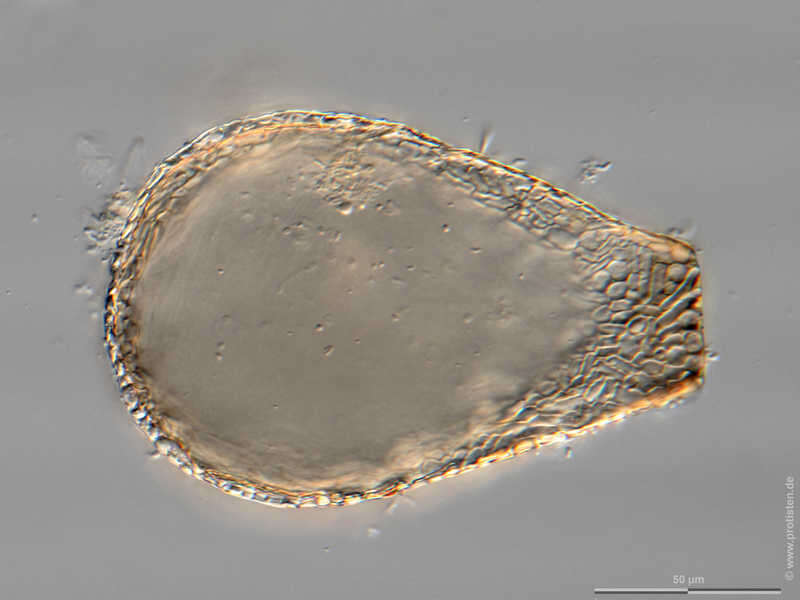
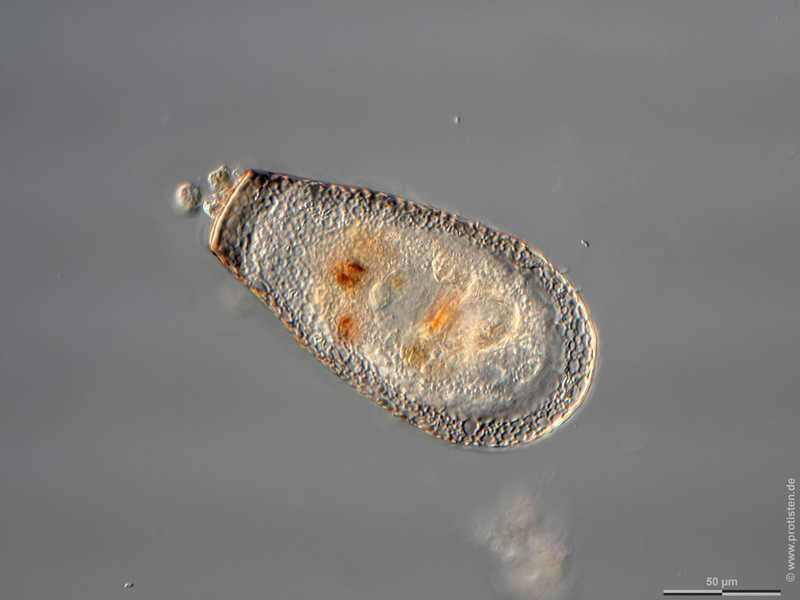
Longinebela penardiana Synopsis of shell structure. Scale bar indicates 50 µm. Sample from wetland Lauchseemoor, Fieberbrunn, Tyrol, Austria. Sampling date 06/2023. The image was built up using several photomicrographic frames with manual stacking technique. Images were taken using Zeiss Axioplan with Olympus OM-D M5 MKII. Image under Creative Commons License V 3.0 (CC BY-NC-SA). Place name: Wetland Lauchseemoor, Fieberbrunn (Tyrol, Austria) Latitude: 47.46954439 Longitude: 12.53826499 Synopse der Schalenstruktur. Multiebenen-Abbildung, manuell gestapelt. Der Messbalken markiert eine Länge von 50 µm. Probe aus dem Lauchseemoor bei Fieberbrunn, Tirol. Datum der Aufsammlung: 06/2023. Mikrotechnik: Zeiss Axioplan, Kamera: Olympus OM-D M5 MKII. Creative Commons License V 3.0 (CC BY-NC-SA). For permission to use of (high-resolution) images please contact postmaster@protisten.de.
-

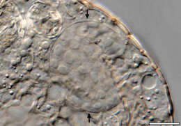
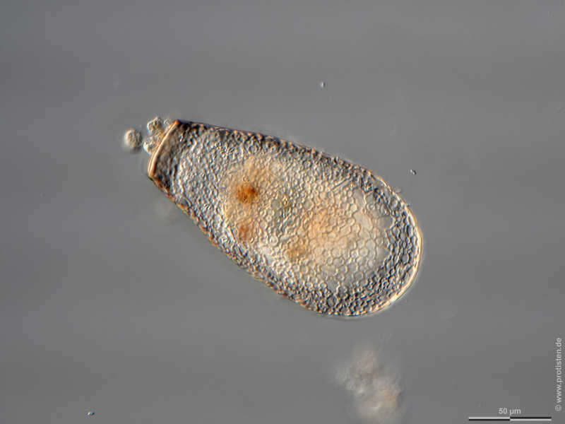
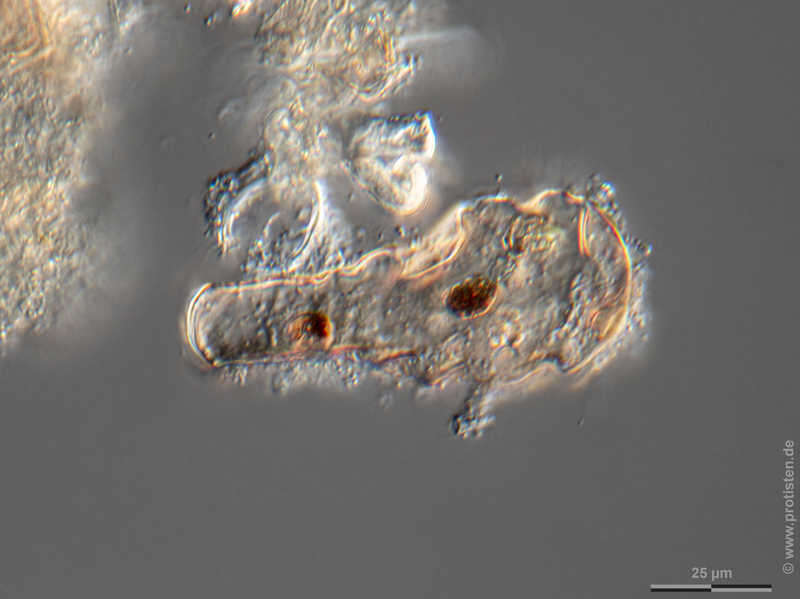
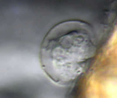
Longinebela penardiana The large nucleus, which is circular in cross section, can be seen in the bulb of the shell. It contains many nucleoli, which are also sphaerical. Scale bar indicates 25/10 µm. Sample from wetland Lauchseemoor, Fieberbrunn, Tyrol, Austria. Sampling date 06/2023. The image was built up using several photomicrographic frames with manual stacking technique. Images were taken using Zeiss Axioplan with Olympus OM-D M5 MKII. Image under Creative Commons License V 3.0 (CC BY-NC-SA). Place name: Wetland Lauchseemoor, Fieberbrunn (Tyrol, Austria) Latitude: 47.46954439 Longitude: 12.53826499 Im Bulbus der Schale ist der große, im Querschnitt kreisrunde Zellkern zu sehen. Er birgt viele, ebenfalls runde Nucleolen. Multiebenen-Abbildung, manuell gestapelt. Der Messbalken markiert eine Länge von 25/10 µm. Probe aus dem Lauchseemoor bei Fieberbrunn, Tirol. Datum der Aufsammlung: 06/2023. Mikrotechnik: Zeiss Axioplan, Kamera: Olympus OM-D M5 MKII. Creative Commons License V 3.0 (CC BY-NC-SA). For permission to use of (high-resolution) images please contact postmaster@protisten.de.
-
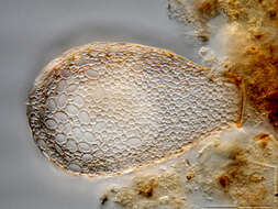
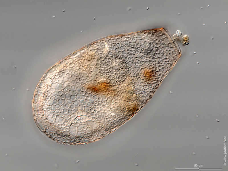
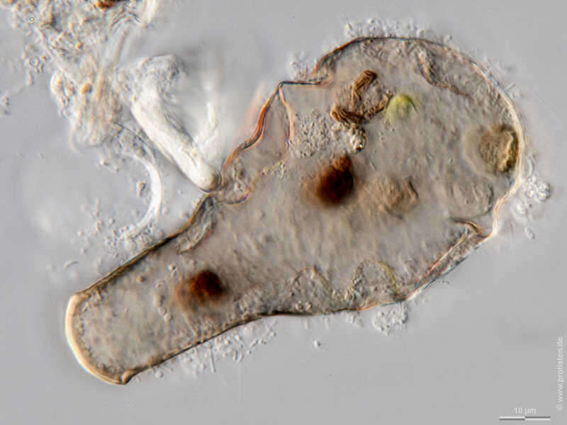
Longinebela penardiana The optical cross section through the cell shows the cytoplasm. Its not filling the shell but is attached to its inner surface by cytoplasmic strands called epipodia. Scale bar indicates 50 µm. Sample from wetland Lauchseemoor, Fieberbrunn, Tyrol, Austria. Sampling date 06/2023. The image was built up using several photomicrographic frames with manual stacking technique. Images were taken using Zeiss Axioplan with Olympus OM-D M5 MKII. Image under Creative Commons License V 3.0 (CC BY-NC-SA). Place name: Wetland Lauchseemoor, Fieberbrunn (Tyrol, Austria) Latitude: 47.46954439 Longitude: 12.53826499 Der optische Querschnitt durch die Zelle zeigt das Cytoplasma. Es füllt die Schale nicht aus, sondern ist durch cytoplasmatische Fäden, sogenannte Epipodien, an deren Innenfläche befestigt. Multiebenen-Abbildung, manuell gestapelt. Der Messbalken markiert eine Länge von 50 µm. Probe aus dem Lauchseemoor bei Fieberbrunn, Tirol. Datum der Aufsammlung: 06/2023. Mikrotechnik: Zeiss Axioplan, Kamera: Olympus OM-D M5 MKII. Creative Commons License V 3.0 (CC BY-NC-SA). For permission to use of (high-resolution) images please contact postmaster@protisten.de.
-
Longinebela penardiana Synopsis of shell structure. Scale bar indicates 50 µm. Sample from wetland Lauchseemoor, Fieberbrunn, Tyrol, Austria. Sampling date 06/2023. The image was built up using several photomicrographic frames with manual stacking technique. Images were taken using Zeiss Axioplan with Olympus OM-D M5 MKII. Image under Creative Commons License V 3.0 (CC BY-NC-SA). Place name: Wetland Lauchseemoor, Fieberbrunn (Tyrol, Austria) Latitude: 47.46954439 Longitude: 12.53826499 Synopse der Schalenstruktur. Multiebenen-Abbildung, manuell gestapelt. Der Messbalken markiert eine Länge von 50 µm. Probe aus dem Lauchseemoor bei Fieberbrunn, Tirol. Datum der Aufsammlung: 06/2023. Mikrotechnik: Zeiss Axioplan, Kamera: Olympus OM-D M5 MKII. Creative Commons License V 3.0 (CC BY-NC-SA). For permission to use of (high-resolution) images please contact postmaster@protisten.de.
-
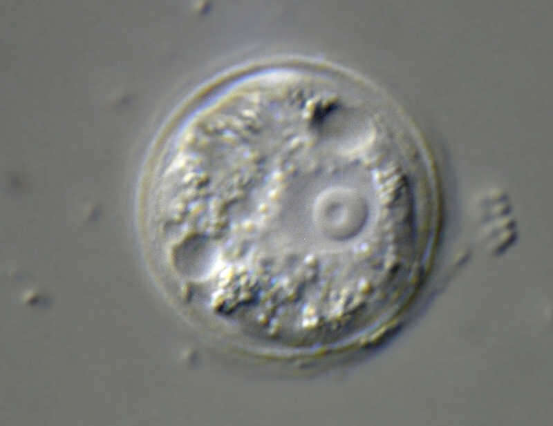
Longinebela penardiana The large nucleus, which is circular in cross section, can be seen in the bulb of the shell. Scale bar indicates 50 µm. Sample from wetland Lauchseemoor, Fieberbrunn, Tyrol, Austria. Sampling date 06/2023. The image was built up using several photomicrographic frames with manual stacking technique. Images were taken using Zeiss Axioplan with Olympus OM-D M5 MKII. Image under Creative Commons License V 3.0 (CC BY-NC-SA). Place name: Wetland Lauchseemoor, Fieberbrunn (Tyrol, Austria) Latitude: 47.46954439 Longitude: 12.53826499 Im Bulbus der Schale ist der große, im Querschnitt kreisrunde Zellkern zu sehen. Multiebenen-Abbildung, manuell gestapelt. Der Messbalken markiert eine Länge von 50 µm. Probe aus dem Lauchseemoor bei Fieberbrunn, Tirol. Datum der Aufsammlung: 06/2023. Mikrotechnik: Zeiss Axioplan, Kamera: Olympus OM-D M5 MKII. Creative Commons License V 3.0 (CC BY-NC-SA). For permission to use of (high-resolution) images please contact postmaster@protisten.de.
-
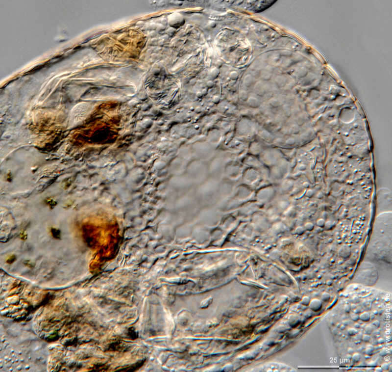
Longinebela penardiana The large nucleus, which is circular in cross section, can be seen in the bulb of the shell. It contains many nucleoli, which are also sphaerical. Scale bar indicates 25/10 µm. Sample from wetland Lauchseemoor, Fieberbrunn, Tyrol, Austria. Sampling date 06/2023. The image was built up using several photomicrographic frames with manual stacking technique. Images were taken using Zeiss Axioplan with Olympus OM-D M5 MKII. Image under Creative Commons License V 3.0 (CC BY-NC-SA). Place name: Wetland Lauchseemoor, Fieberbrunn (Tyrol, Austria) Latitude: 47.46954439 Longitude: 12.53826499 Im Bulbus der Schale ist der große, im Querschnitt kreisrunde Zellkern zu sehen. Er birgt viele, ebenfalls runde Nucleolen. Multiebenen-Abbildung, manuell gestapelt. Der Messbalken markiert eine Länge von 25/10 µm. Probe aus dem Lauchseemoor bei Fieberbrunn, Tirol. Datum der Aufsammlung: 06/2023. Mikrotechnik: Zeiss Axioplan, Kamera: Olympus OM-D M5 MKII. Creative Commons License V 3.0 (CC BY-NC-SA). For permission to use of (high-resolution) images please contact postmaster@protisten.de.
-
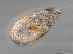
Thecamoeba similis Scale bar indicates 25 µm. Collected from Bodden, the brackish waters lying between the isles of Hiddensee and Ruegen (German Baltic Sea). The image was built up using several photomicrographic frames with manual stacking technique. The images were taken using Zeiss Universal with Olympus C7070 CCD camera.Image under Creative Commons License V 3.0 (CC BY-NC-SA). Place name: Hiddensee Bodden (Germany) Latitude: 54.582633 Longitude: 13.115051 Multiebenen-Abbildung, manuell gestapelt. Der Messbalken markiert eine Länge von 25 µm. Probe aus dem Hiddenseer Bodden, der Brackwasserfläche zwischen den Inseln Hiddensee und Rügen. Mikrotechnik: Olympus BH, Kamera: Olympus C7070. Creative Commons License V 3.0 (CC BY-NC-SA). For permission to use of (high-resolution) images please contact postmaster@protisten.de.
-
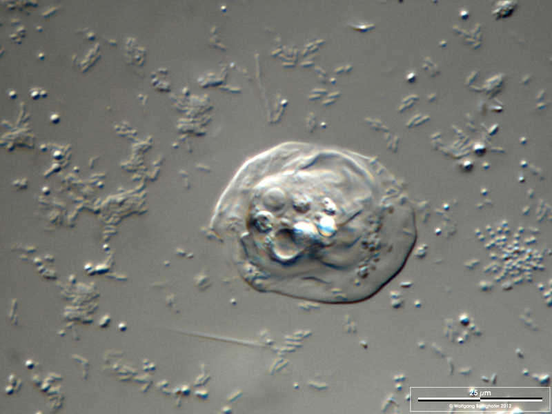
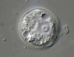
Pyxidicula operculata Cell lived on the neuston, the biofilm on the water surface. The test, the two contractile vacuoles and the nucleus with nucleolus are visible. Dimensions of specimen: 20 µm. Sample from a freshwater pond on the island of Hiddensee (Baltic Sea, Germany). This image was taken using Zeiss Universal with Olympus C7070 CCD camera.Image under Creative Commons License V 3.0 (CC BY-NC-SA). Place name: Pond Suploch, Hiddensee (Germany) Latitude: 54.538638 Longitude: 13.097802 Die Zelle lebte im Neuston, dem Biofilm an der Wasseroberfläche. Die Schale, die beiden kontraktilen Vakuolen und der Kern mit Nucleolus sind sichtbar. Durchmesser des Exemplars: 20 µm. Probe aus einem kleinen Süßwasserteich auf der Insel Hiddensee, welcher eine faszinierende Vielfalt von nackten und beschalten Amöben beherbergt. Mikrotechnik: Zeiss Universal, Kamera: Olympus C7070. Creative Commons License V 3.0 (CC BY-NC-SA). For permission to use of (high-resolution) images please contact postmaster@protisten.de.
-
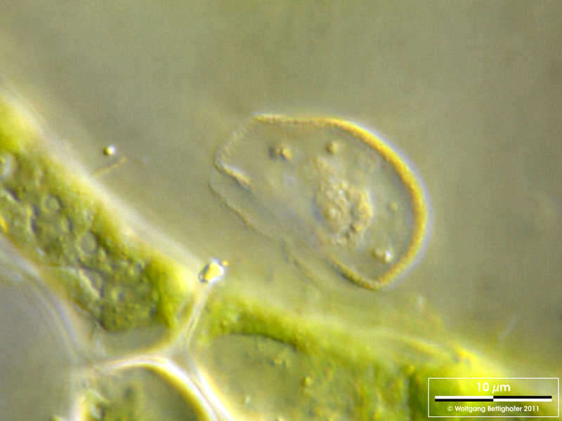
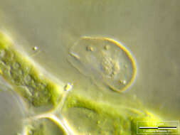
Pyxidicula operculata The lateral view shows an elliptical profile whereas the shape is circular in topview. Pseudopods appear as tapered protuberances of protoplasma. The aperture is almost as wide as the shell diameter, the rim is visible. Dimensions of specimen: 15µm x 20 µm. Sample from a freshwater pond on the island of Hiddensee (Baltic Sea, Germany). This image was taken using Zeiss Universal with Olympus C7070 CCD camera.Image under Creative Commons License V 3.0 (CC BY-NC-SA). Place name: Pond Suploch, Hiddensee (Germany) Latitude: 54.538638 Longitude: 13.097802 In der Seitenansicht zeigt die Schale ein elliptisches Profil, in der Draufsicht zeigt sie sich kreisförmig. Die Pseudopodien erscheinen als vom Protoplasma ausgehende konische Höcker. Der Apertur-Durchmesser ist fast so groß wie der Schalendurchmesser, der umgeschlagene Rand ist sichtbar. Abmessungen des Exemplars: 15 µm x 20 µm. Probe aus einem kleinen Süßwasserteich auf der Insel Hiddensee, welcher eine faszinierende Vielfalt von nackten und beschalten Amöben beherbergt. Mikrotechnik: Zeiss Universal, Kamera: Olympus C7070. Creative Commons License V 3.0 (CC BY-NC-SA). For permission to use of (high-resolution) images please contact postmaster@protisten.de.
-
Pyxidicula operculata Scale bar indicates 10 µm. Sample from a pond situated in the vicinity of Bodman, Lake Constance. The image was built up using several photomicrographic frames with manual stacking technique. Images were taken using Zeiss Universal with Olympus C7070 CCD camera.Image under Creative Commons License V 3.0 (CC BY-NC-SA). Place name: Pond situated in the vicinity of Lake Constance (Germany) Latitude: 47.734945 Longitude: 9.091097 Multiebenen-Abbildung, manuell gestapelt. Der Messbalken markiert eine Länge von 10 µm. Probe aus einem Teich nahe Bodman am Bodensee. Mikrotechnik: Zeiss Universal, Kamera: Olympus C7070. Creative Commons License V 3.0 (CC BY-NC-SA). For permission to use of (high-resolution) images please contact postmaster@protisten.de.
-
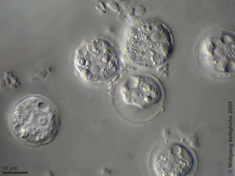
Pyxidicula operculata Synopsis of Pyxidicula´s test and a living cell where focal plane is adjusted to depict the vesicular nucleus with prominent nucleolus. Sample from a freshwater pond on the island of Hiddensee (Baltic Sea, Germany). This image was taken using Zeiss Universal with Olympus C7070 CCD camera.Image under Creative Commons License V 3.0 (CC BY-NC-SA). Place name: Pond Suploch, Hiddensee (Germany) Latitude: 54.538638 Longitude: 13.097802 Synopse einer Pyxidicula-Schale und einer intakten Zelle. Die Fokusebene sind derart angepasst, dass der bläschenförmigen Kern mit auffälligen Nukleolus zu sehen ist. Probe aus einem kleinen Süßwasserteich auf der Insel Hiddensee, welcher eine faszinierende Vielfalt von nackten und beschalten Amöben beherbergt. Mikrotechnik: Zeiss Universal, Kamera: Olympus C7070. Creative Commons License V 3.0 (CC BY-NC-SA). For permission to use of (high-resolution) images please contact postmaster@protisten.de.
-
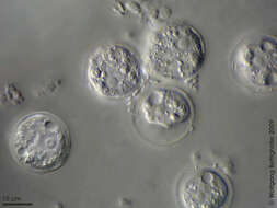
Pyxidicula operculata A group of Pyxidicula living on Hyponeuston. Focal plane on nuclei and contractile vacuoles. Scale bar indicates 10 µm. Sample from a freshwater pond on the island of Hiddensee (Baltic Sea, Germany). This image was taken using Zeiss Universal with Olympus C7070 CCD camera.Image under Creative Commons License V 3.0 (CC BY-NC-SA). Place name: Pond Suploch, Hiddensee (Germany) Latitude: 54.538638 Longitude: 13.097802 Eine Gruppe von Pyxidicula im Hyponeuston. Die Fokusebene ist auf die Kerne und die kontraktilen Vakuolen gelegt. Der Messbalken markiert eine Länge von 10 µm. Probe aus einem kleinen Süßwasserteich auf der Insel Hiddensee, welcher eine faszinierende Vielfalt von nackten und beschalten Amöben beherbergt. Mikrotechnik: Zeiss Universal, Kamera: Olympus C7070. Creative Commons License V 3.0 (CC BY-NC-SA). For permission to use of (high-resolution) images please contact postmaster@protisten.de.
-

Pyxidicula operculata A group of Pyxidicula living on Hyponeuston, the aquatic area closely attached to the water surface, together with bacteria, which are their prey. On the left the nucleus and the two contractile vacuoles are visible, right to this specimen lies an empty test. The water surface has enough tension so that this testate amoebae can walk on it. Sample from a freshwater pond on the island of Hiddensee (Baltic Sea, Germany). This image was taken using Zeiss Universal with Olympus C7070 CCD camera.Image under Creative Commons License V 3.0 (CC BY-NC-SA). Place name: Pond Suploch, Hiddensee (Germany) Latitude: 54.538638 Longitude: 13.097802 Eine Gruppe von Pyxidicula, welche im Hyponeuston wohnt, dem Lebensraum knapp unter der Wasseroberfläche, zusammen mit Bakterien, welche auch ihre Beute darstellen. Auf der linken Seite sind der Kern und die beiden kontraktilen Vakuolen sichtbar, direkt neben der intakten Zelle liegt eine leere Schale. Die Wasseroberfläche hat genug Spannung, so dass diese Schalenamöben auf ihr gehen können . Probe aus einem kleinen Süßwasserteich auf der Insel Hiddensee, welcher eine faszinierende Vielfalt von nackten und beschalten Amöben beherbergt. Mikrotechnik: Zeiss Universal, Kamera: Olympus C7070. Creative Commons License V 3.0 (CC BY-NC-SA). For permission to use of (high-resolution) images please contact postmaster@protisten.de.
-
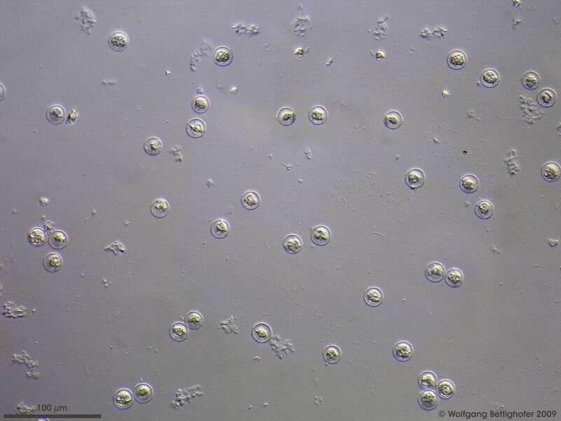
Pyxidicula operculata Pyxidicula-blooming under the water surface of a little tank in the laboratory. Scale bar indicates 100 µm. Sample from a freshwater pond on the island of Hiddensee (Baltic Sea, Germany). This image was taken using Zeiss Universal with Olympus C7070 CCD camera.Image under Creative Commons License V 3.0 (CC BY-NC-SA). Place name: Pond Suploch, Hiddensee (Germany) Latitude: 54.538638 Longitude: 13.097802 Pyxidicula-Blüte unter der Wasseroberfläche eines kleinen tropisches Süßwasseraquariums im Labor. Der Messbalken markiert eine Länge von 100 µm. Probe aus einem kleinen Süßwasserteich auf der Insel Hiddensee, welcher eine faszinierende Vielfalt von nackten und beschalten Amöben beherbergt. Mikrotechnik: Zeiss Universal, Kamera: Olympus C7070. Creative Commons License V 3.0 (CC BY-NC-SA). For permission to use of (high-resolution) images please contact postmaster@protisten.de.
-
Pyxidicula operculata Scale bar indicates 10 µm. Sample from a pond situated in the vicinity of Bodman, Lake Constance. Image was taken using Zeiss Universal with Olympus C7070 CCD camera.Image under Creative Commons License V 3.0 (CC BY-NC-SA). Place name: Pond situated in the vicinity of Lake Constance (Germany) Latitude: 47.734945 Longitude: 9.091097 Der Messbalken markiert eine Länge von 10 µm. Probe aus einem Teich nahe Bodman am Bodensee. Mikrotechnik: Zeiss Universal, Kamera: Olympus C7070. Creative Commons License V 3.0 (CC BY-NC-SA). For permission to use of (high-resolution) images please contact postmaster@protisten.de.
-
Hyalosphenia elegans Scale bar indicates 25 µm. Sample from wetland Paradieswiese near Kitzbühel, Tyrol, Austria. Sampling date 06/2023. The image was built up using several photomicrographic frames with manual stacking technique. Images were taken using Zeiss Axioplan with Olympus OM-D M5 MKII. Image under Creative Commons License V 3.0 (CC BY-NC-SA). Place name: Wetland Paradieswiese near Kitzbühel (Tyrol, Austria) Latitude: 47.47375067 Longitude: 12.37624884 Multiebenen-Abbildung, manuell gestapelt. Der Messbalken markiert eine Länge von 25 µm. Probe von der Paradieswiese bei Kitzbühel-Steuerberg, Tirol. Datum der Aufsammlung: 06/2023. Mikrotechnik: Zeiss Axioplan, Kamera: Olympus OM-D M5 MKII. Creative Commons License V 3.0 (CC BY-NC-SA). For permission to use of (high-resolution) images please contact postmaster@protisten.de.
-
Hyalosphenia elegans Scale bar indicates 10 µm. Sample from wetland Paradieswiese near Kitzbühel, Tyrol, Austria. Sampling date 06/2023. The image was built up using several photomicrographic frames with manual stacking technique. Images were taken using Zeiss Axioplan with Olympus OM-D M5 MKII. Image under Creative Commons License V 3.0 (CC BY-NC-SA). Place name: Wetland Paradieswiese near Kitzbühel (Tyrol, Austria) Latitude: 47.47375067 Longitude: 12.37624884 Multiebenen-Abbildung, manuell gestapelt. Der Messbalken markiert eine Länge von 10 µm. Probe von der Paradieswiese bei Kitzbühel-Steuerberg, Tirol. Datum der Aufsammlung: 06/2023. Mikrotechnik: Zeiss Axioplan, Kamera: Olympus OM-D M5 MKII. Creative Commons License V 3.0 (CC BY-NC-SA). For permission to use of (high-resolution) images please contact postmaster@protisten.de.
-
Difflugia paulii Scale bar indicates 25 µm. The specimen was gathered in the wetlands of Nationalpark Unteres Odertal (100 km north east of Berlin). Sampling date 5/2019. The image was built up using several photomicrographic frames with manual stacking technique. Images were taken using Zeiss Axioplan with Olympus OM-D M5 MKII. Image under Creative Commons License V 3.0 (CC BY-NC-SA). Place name: Creek in Oder valley 100 km north east of Berlin (Germany) Latitude: 53.135032 Longitude: 14.348738 Multiebenen-Abbildung, manuell gestapelt. Der Messbalken markiert eine Länge von 25 µm. Probe aus einem Feuchtbiotop im Nationalpark Unteres Odertal auf halber Strecke zwischen Schwedt und Gartz. Datum der Aufsammlung: 7/2019. Mikrotechnik: Zeiss Axioplan, Kamera: Olympus OM-D M5 MKII. Creative Commons License V 3.0 (CC BY-NC-SA). For permission to use of (high-resolution) images please contact postmaster@protisten.de.
-
Lesquereusia spiralis The test primarily consists of idiosomes (self made siliceous pieces looking like sausages). A few xenosomes (incorporated grains of sand) are visible. All xenosomes are covered with a siliceous coating. Scale bar indicates 50 µm. Sample from a freshwater pond on the island of Hiddensee (Baltic Sea, Germany). This image was taken using Zeiss Universal with Olympus C7070 CCD camera.Image under Creative Commons License V 3.0 (CC BY-NC-SA). Place name: Pond Suploch, Hiddensee (Germany) Latitude: 54.538638 Longitude: 13.097802 Die Schale besteht hauptsächlich aus Idiosomen (selbst erzeugte kieselsäurehaltige, wurstförmige Teilchen). Einige Xenosomen (Fremdkörper, meist aus Quarz) sind auch zu sehen. Alle Xenosomen werden von den Lesquereusiiden mit einer Kieselschicht bedeckt. Der Messbalken markiert eine Länge von 50 µm. Probe aus einem kleinen Süßwasserteich auf der Insel Hiddensee, welcher eine faszinierende Vielfalt von nackten und beschalten Amöben beherbergt. Mikrotechnik: Zeiss Universal, Kamera: Olympus C7070. Creative Commons License V 3.0 (CC BY-NC-SA). For permission to use of (high-resolution) images please contact postmaster@protisten.de.