“Prosorhochmus nelsoni (Sanchez, 1973),
new combination
Figs. 1–8, Tables 1, 2
Amphiporus nelsoni Sanchez, 1973:208– 213, Figs. 10–13.
External appearance. —Prosorhochmus nelsoni is relatively small, with maximum
Table 1.—Measurements of stylet apparatus of immature specimens of Prosorhochmus nelsoni (Sanchez, 1973) from Coquimbo, Chile.
Specimen
Body length (anesthetized, mm)
Stylet length
(S, µm)
Basis length
(B, µm)
Basis diameter (µm)
S/B ratio
1
15
88.2
132.3
49
0.67
2
9.5
73.5
98
34.3
0.75
3
10
78.4
107.8
39.2
0.73
4
12
98
122.5
53.9
0.8
5
8
73.5
112.7
49
0.65
6
10
73.5
73.5
24.5
1
7
13
88.2
107.8
34.3
0.82
8
15
102.9
127.4
29.4
0.81
9
15
93.1
83.3
58.8
1.1
10
12
78.4
122.5
34.3
0.64
11
10
49
73.5
29.4
0.67
12
13
107.8
122.5
49
0.88
13
12
88.2
117.6
34.3
0.75
Average
11.88
84.05
107.80
39.95
0.79
SD
2.29
15.48
20.00
10.74
0.14
Min
8
49
73.5
24.5
0.64
Max
15
107.8
132.3
58.8
1.1
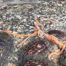
recorded length of reproductive specimens 45–50 mm and width 1.2–1.5 mm. The color in life is yellowish-orange dorsally, paler toward the ventral side. The body is slender and compact, dorso-ventrally flattened, wide at the anterior end, gradually tapering toward the posterior to end in a bluntly rounded tip (Fig. 1). The head is somewhat spatulate in shape and is wider than the remaining body, with a characteristic vertical anterior notch giving it a distinct bilobed appearance. An antero-dorsal horizontal epidermal fold anterior to the eyes separates two ventral apical lobes from a median dorsal lobe, creating the appearance of a ‘‘smile’’ (Fig. 2A), characteristic of the genera Prosorhochmus and Pantinonemertes. The four reddish-brown eyes are situated in front of the brain; the anterior pair is slightly larger than the posterior. The distance between the eyes of the anterior pair and the posterior pair is larger than between the two pairs. The rudimentary cerebral organ furrows, also referred to as the anterior cephalic grooves, appear as a pair of inconspicuous latero-ventral, whitish, semi-circular grooves approximately at the level of the anterior pair of eyes (Fig. 2B); these are not visible from the dorsal side. The shallow posterior cephalic furrow is indistinct and forms a dorsal, posteriorly directed ‘‘V’’ immediately behind the brain and a ventral, incomplete anteriorly directed ‘‘V’’ immediately anterior to the brain (Fig. 2A, B). The rhynchopore is subterminal.
Body wall, musculature and parenchyma.—Epidermis is of typical hoplonemertean structure (Figs. 3A, 6K). Dermis is represented by a thin layer of extracellular matrix. Body-wall musculature consists of an outer circular layer and an inner longitudinal layer. Diagonal (oblique) muscle fibers situated between the circular and longitudinal musculature of the body wall form a thin but distinct layer. This layer is best visualized on longitudinal sections (Fig. 3A) but also is visible on cross-sections (Fig. 3B). The precerebral septum is of split (Kirsteuer 1974) or mixed type (Chernyshev 2002) and corresponds to the description given by Gibson & Moore (1985:150, fig. 2). It is formed by individual muscle fibers emerging from the body wall longitudinal musculature at several levels. Behind the brain, separate bundles of oblique fibers diverge from the inner margins of the longitudinal muscle layer and lead forward toward the proboscis insertion. Here the oblique fibers are joined by additional (radial) fibers, which turn inward from the main layer (Fig. 3C, D). There is no clear demarcation between the radial and oblique fibers forming the septum. A few individual fibers from the inner portion of the longitudinal musculature continue into the head as cephalic retractors. Dorso-ventral muscles are strongly developed between the gonads and intestinal diverticula (Figs. 3E, 8F). Anteriorly, thick bundles of dorso-ventral musculature are found between the lateral pouches of the caecum, between dorsal
lobes of the mucus glands, and between the lateral nerve cords and lateral lobes of the mucus glands (Fig. 3F). Muscle fibers oriented dorso-ventrally, obliquely, and horizontally are abundant in the precerebral region (Fig. 3G). The musculature associated with foregut, in the literature often referred to as ‘‘splanchnic musculature,’’ is very well developed and is continuous with the fibers surrounding the rhynchodeum. A layer of longitudinal muscles surrounds the esophagus from the point of its separation from the rhynchodeum to the beginning of the brain (Fig. 3H). The esophageal musculature becomes surrounded at the brain level by longitudinal fibers originating at the proboscis insertion and running parallel to the esophagus (Fig. 3I). These longitudinal muscles continue as a thin layer surrounding the stomach and are particularly obvious between the folds of the stomach wall (Fig. 3J). The amorphous extracellular matrix, the so-called ‘‘parenchyma,’’ is developed somewhat better than in other species of Prosorhochmus (particularly in the foregut region) but is otherwise unremarkable.
Proboscis apparatus.—The proboscis pore opens terminally. It leads into a short, thin-walled rhynchodeum. Rhynchodeal epithelium comprises squamous cells with small elongated nuclei (Fig. 3K). Just anterior to the proboscis insertion, it is comprised of cells with acidophilic cytoplasm and large nuclei (Fig. 3L). It was not possible with light microscopy to determine whether rhynchodeal epithelial cells bear cilia or not. The rhynchodeal musculature is rather well developed and comprises both longitudinal and circular muscle fibers (Fig. 3K). There is no localized concentration of circular muscle fibers representing a distinct rhynchodeal sphincter. The rhynchocoel reaches almost to the posterior end of the body. Its wall is of typical distromatonemertean (Thollesson & Norenburg 2003) structure and contains separate outer circular and inner longitudinal muscle layers (Fig. 4A). The thickness of the layers changes dramatically with the state of contraction of the animal. The proboscis is thin, longer than the body, somewhat translucent, and whitish or, occasionally, dull cream. The wall of the anterior chamber is composed of a tall glandular epithelium, arranged into conical papillae, a thin layer of extracellular matrix, an outer circular muscle layer, a longitudinal muscle layer, that is divided into concentric layers by the neural sheath, a delicate layer of inner circular muscles, and a thin endothelium (Fig. 4B). The neural sheath bears 10–12 proboscis nerves (n 5 3; Fig. 4C). The proboscis armature consists of a central stylet, mounted on a characteristically truncated basis, and two pouches each containing 1–3 accessory stylets (Fig. 4E). Measurements taken on 13 immature specimens (average length about 12 mm), collected during November 2003 at La Pampilla Beach near Coquimbo (Chile) are presented in Table 1. The average length of the central stylet (S) was 84 mm, average basis length was (B) 2107.8 mm and average S/B ratio was 0.79. Sanchez (1973) reported the central stylet to be 80 mm long, but it is not known whether she measured the stylet in the mature or immature specimen. The wall of the posterior chamber of the proboscis consists of a glandular epithelium organized into papillae, outer longitudinal muscle layer, thin inner circular muscle layer, and a delicate endothelium (Fig. 4D). Similar to other described species of Prosorhochmus, but unlike P. claparedii from Anglesey [Gibson & Moore 1985:151–152, plate II(a)], there is no distinct nerve supply in the longitudinal muscle layer of posterior proboscis of P. nelsoni.
Alimentary canal.—The esophagus opens into the rhynchodeum in front of the precerebral septum. It is enclosed by longitudinal muscle fibers, which are confluent
with the rhynchodeal musculature and continue posteriorly as the musculature of the stomach. The posterior part of the esophagus is ciliated, but lacks acidophilic or basophilic glands present in the wall of the stomach (Fig. 4F). The stomach is of typical hoplonemertean structure with densely ciliated, deeply folded epithelium, containing numerous basophilic and acidophilic glands (Fig. 4G). Presence or absence of the posteriorly directed pouches in the stomach had been used to differentiate between the species of the genus (Frutos et al. 1998). Although it appears quite plausible that these pouches are merely an artifact of fixation, a more thorough study of the intraand interspecific variation of this character would be necessary to reject this character altogether. In P. nelsoni there appears to be intraspecific variation in this character: four specimens studied from series of cross-sections lack a ventral posterior pouch in the stomach, while one additional specimen (USNM 1019785, anterior c.s. slide 5) possesses two consecutive postero-lateral diverticula on the left side of the stomach, each 80–100 mm long (Fig. 4H). The intestinal caecum is well developed, anteriorly divided, up to 5.3 mm long (measured in the largest sectioned specimen), and bears numerous lateral diverticula throughout its length. The anterior lobes or pouches reach the posterior portion of the dorsal cerebral ganglia. Intestinal diverticula are lobed.
Blood system.—The blood system is of a common hoplonemertean type. A cephalic suprarhynchodeal loop crosses just behind the posterior chamber of the frontal organ and continues posteriorly as the body’s paired lateral vessels. The middorsal blood vessel originates near the ventral cerebral commissure from the left cephalic vessel (observed in one specimen) (Fig. 4L) and immediately penetrates the rhynchocoel floor to form a single vascular plug. The wall of the vascular plug consists of endothelium of the blood vessel, a thin layer of extracellular matrix, and a modified rhynchocoel endothelium (Fig. 4M). Similar to other described Prosorhochmus species, but unlike P. claparedii from Anglesey (Gibson & Moore 1985), P. nelsoni lacks transverse connectives linking mid-dorsal and lateral blood vessels in the intestinal region. The blood vessels are thin-walled with a well defined extensive lumen and irregular thickenings of the wall (Fig. 4I). During contraction of the blood vessel wall, depending on whether the muscle fibers of the wall lie toward the inner or outer side of the thickening, the latter appear as ‘‘pouches’’ or ‘‘valves’’ (Fig. 4J, K). This is confirmed by the fact that neither distinct valves nor pouches are visible in relaxed regions of blood vessels, while both appear in the contracted regions (Fig. 4I–K). In addition, many kinds of intermediate states can be observed between the valves and the pouches. Perhaps these regions of thickened extracellular matrix serve to add rigidity to the otherwise elastic walls of the blood vessels.
Nervous system.—The brain is of typical nemertean construction and consists of two ventral and two dorsal ganglia, joined by ventral (subrhynchodeal) and dorsal (suprarhynchodeal) commissures, respectively. The dorsal ganglia are more widely separated than the ventral. A thin but distinct outer neurilemma encloses the brain as a whole, but there is no inner neurilemma dividing the fibrous and ganglionic tissues. There are no neurochord cells in the brain ganglia (as defined by Bürger 1895:355) and no neurochords in the lateral nerve cords. The redescription by Gibson & Moore (1985) of Prosorhochmus claparedii, which served as a basis for the generic diagnosis, appears to misidentify type III neurons (as defined by Bürger 1895:320) as neurochord cells. As in other species of Prosorhochmus, the lateral nerve cords contain a single fibrous core throughout their length. However, in one specimen an unpaired accessory nerve, derived from the dorsal cerebral ganglion, was found in the right lateral nerve cord in the region adjacent to the brain (Fig. 5A). This unpaired accessory nerve cord later came to lie close to the dorsal part of the main fibrous core, forming the so-called ‘‘upper nerve,’’ a bundle of nerve fibers in the dorsal part of the fibrous core of the lateral nerve cord, distinguished by their lighter color, which we observed in all species of Prosorhochmus (Fig. 5B). The difference between this
Table 2.—Comparison between stylet measurement of Prosorhochmus nelsoni and other described species of Prosorhochmus.
Species
Stylet length (µm)
Central stylet length to basis length (S/B) ratio
P. claparedii Keferstein, 1862 (after Gibson & Moore 1985)
30–45
0.25–0.33
P. americanus Gibson et al., 1986
90
0.45–0.5
P. adriaticus Senz, 1993
?
0.25
P. chafarinensis Frutos et al., 1998
99
0.5
P. nelsoni (Sanchez, 1973)
84
0.8
upper nerve and a real accessory nerve is that the upper nerve is never separated from the main fibrous core by cell bodies, as is the accessory nerve, and it is derived from the ventral cerebral ganglion. As observed in most monostiliferans studied in the last three decades, each lateral nerve cord contains a single delicate muscle bundle, consisting of 3–7 fibers and running within or adjacent to the fibrous core, near its dorsal border. In addition, there are several less conspicuous muscle fibers running along the inner lateral side of the fibrous core (Fig. 5C). Muscle fibers associated with the lateral nerve cords can be traced to their extracerebral origin near the proboscis insertion. Numerous cephalic nerves lead anteriorly from the brain lobes to supply various structures of the head. A stout nerve joins each ventral lobe with the appropriate cerebral sensory organ. A single proboscis nerve trunk originates from the ventral ganglion on each side near the ventral cerebral commissure and branches before entering the proboscis (Fig. 5D).
Eyes.—The four eyes are of typical pigment cup structure and well developed (Fig. 5E). The eyes of the anterior pair are slightly larger in diameter than the posterior (90 mm and 75 mm, respectively). The pigment cups of the anterior pair are directed anterolaterally, while those of the posterior pair are directed posterolaterally (Fig. 5F).
Frontal organ.—The frontal organ, opening at the tip of the head, is of typical structure for the genus Prosorhochmus. Its structure, although described slightly differently for different species of Prosorhochmus (Gibson & Moore 1985, Gibson et al.1986, Frutos et al. 1998), appears to be remarkably uniform within the genus. The frontal organ of this species consists of a ciliated canal about 80–100 mm long, lined by a regionally differentiated epithelium. Initially narrow and somewhat dorsoventrally flattened (Figs. 6H, 7A), the canal quickly expands to form a rounded chamber (Figs. 6G, 6I–J, 7B). The ventral wall of the canal comprises a strongly acidophilic epithelium, clad in densely-arranged short cilia, whereas the dorsal wall of the canal and ventral, dorsal,
and posterior walls of the chamber, through which the basophilic mucus cephalic glands discharge, has a vacuolated appearance and bears much longer, sparsely distributed cilia. The acidophilic epithelium divides just in front of the chamber to run on its lateral borders. The lateral part of the lumen of the chamber is dorsoventrally compressed (Fig. 5I). There are no subepidermal acidophilic glands associated with the acidophilic epidermis of the frontal organ. It seems that the acidophilic appearance comes from the densely arranged elongated nuclei of the ciliated cells. The apparent higher degree of development of the frontal organ (comprising the supposed 90° bend) in Prosorhochmus claparedii seems to be a misinterpretation by Gibson & Moore (1985), carried over to the description of Prosorhochmus chafarinensis (Frutos et al., 1998). They misinterpreted a
slightly oblique cross-section as a vertical longitudinal (i.e., sagittal) section of the
frontal end (Gibson & Moore 1985:149, plate I, fig. e; Frutos et al. 1998:296, fig. 3a). As a result, they believed that the canal of the frontal organ first runs horizontally
and then turns ventrally through 90° to end in a flask-shaped chamber. Our re-investigation of the original slides of P. claparedii and P. chafarinensis shows that what they believed to be a vertical portion of the canal is actually a ventral ‘‘groove’’ of the canal.
Cephalic glands.—Cephalic glands are extremely well developed. As in other
members of the genus, they include three distinct types of cells: basophilic lobules with vacuolated appearance (mucus glands), coarsely granular proteinaceous gland cells staining golden-yellow to brown with Mallory trichrome or orange-G with Crandall’s method (orange-G glands), and finely granular proteinaceous acidophilic
cells, staining red with Mallory or Crandall’s technique (acidophilic or red glands). However, the degree of development and distribution of the different types of cephalic glands is somewhat different from that described for other species of the genus. We refer to these cells as ‘‘glands’’ for the sake of briefness and also because they are traditionally referred to as ‘‘glands’’ in nemertean literature. These cells are clearly of secretory nature, but as opposed to real glands they do not form discrete collectives, functioning as units. Moreover, gland cells of different types may intermix with each other. Basophilic (mucus) glands are well developed and, as in other species of the genus, they open through the dorsal and posterior epithelium of the frontal organ. The mucus glands are restricted to the dorsal hemisphere precerebrally in some other prosorhochmids. In this species, the mucus glands are situated centrally, filling most of the precerebral region, whereas the acidophilic glands are situated on the periphery, both in dorsal and ventral hemispheres (Fig. 6A, B). As acidophilic glands become increasingly more abundant in the ventral hemisphere toward the cerebral region, mucus glands become interspersed with acidophilic glands ventrally and with orange-G glands dorsally and laterally (Fig. 6D–F). Mucus glands in the cerebral region are distributed on the perimeter—dorso-lateral of the rhynchocoel, lateral of and under the brain lobes (Figs. 6F, 7A). Mucus glands are also well developed postcerebrally and retain essentially the same arrangement as in the cerebral region (Fig. 7B, C). Lateral lobes of the mucus glands together with the orange-G glands partially surround excretory ducts of the nephridia (Figs. 6L, 7B, C). Dorsal and ventral lobes gradually disappear in the stomach-pylorus region (Figs. 7D, E, 8), while lateral lobes gradually diminish in numbers but reach into the intestinal region (Fig. 7D–F). Orange-G glands are well developed. Overall, they demonstrate typical prosorhochmid distribution in two dorso-lateral tracts starting around the frontal organ canal and reaching as far back as the end of the rhynchocoel. Precerebrally, orange-G glands are restricted to the dorso-lateral regions above the cephalic blood vessels and below the mucus gland lobes (Fig. 6C–E). In the cerebral region they mostly lay dorso-laterally above the brain lobes, either forming dense clusters or interspersing with the mucus gland lobes. They rarely surround mucus gland lobes and mostly lie below the mucus glands (Figs. 7F, 8A). Postcerebrally, orange-G glands become less abundant than in the pre-cerebral or cerebral region. They are interspersed with the lobes of mucus glands and occupy dorso-lateral regions lying above the lateral nerve cords and nephridia. Acidophilic glands are well developed and most abundant just anterior to the brain (at the level of the cerebral organs). Anteriorly (at the level of frontal organ), they are located marginally, inside the perimeter of the body wall, surrounding the mucus gland lobes (Fig. 6A, B). Behind the frontal organ they gradually become more abundant in the ventral hemisphere (Fig. 6C, D). At the level of the cerebral organs acidophilic glands are richly distributed and form clusters among the lobes of mucus glands in the ventral hemisphere (Fig. 6D, E). They also are present as a thin layer dorsally and laterally just under the body-wall musculature. Acidophilic glands become far less numerous behind the proboscis insertion, remaining as a thin layer underneath the body wall musculature around the perimeter of the body, more prominent ventrally and ventro-laterally (Fig. 6F). The distribution evens out in the postcerebral region (Figs. 7A–C, 8). Dorsal and ventral components of the acidophilic glands gradually disappear in the stomach and anterior pyloric region, while the lateral components persist (although also diminished) beyond the end of the pylorus (Fig. 7D–F). There are no purple cephalic glands, associated with the acidophilic glands in the precerebral region described for P. americanus (Gibson et al., 1986). Although the purple glands cells had been described in P. chafarinensis (Frutos et al., 1998), our re-investigation of the type material showed that the authors apparently mistook acidophilic gland cells for the purple cephalic glands.
Cerebral organs.—Relatively small and compact paired cerebral organs reach a maximum diameter of about 145 mm in the adults and are situated in front of the brain, between the anterior and posterior pairs of eyes. Each organ opens anteriorly near the anterior pair of eyes into reduced cephalic furrows. The latter are shallow semi-circular grooves lined by a modified ciliated epithelium, which appears strongly acidophilic in sections stained with Crandall’s method (Figs. 6K, 7C–E). The cerebral organ canals are unbranched. Two types of glandular cells [described by Gibson & Moore (1985) in the Anglesey specimens] can be distinguished in the cerebral organs. The coarsely granular cells, staining dark reddish-brown with Crandall’s method form two clusters, on the dorso- and ventro-anterior margins of the ciliated canal (Fig. 5K). The second type of cell is finely granular, stains less intensively red to brownish purple and forms a single cluster on the inner-lateral and posterior side of each organ. Both types of cells open into the cerebral organ canal (Figs. 5K, 6E).
Excretory system.—The excretory system extends from a short distance behind the dorsal brain lobes back to the anterior pyloric region, most of it lying dorsal and close to the lateral nerve cords and the lateral blood vessels, which at this point lie dorsal to the lateral nerve cords. It consists of thick-walled ciliated collecting tubules, up to 60–65 mm in diameter in the distal part. Collecting tubes are not regionally specialized, although they are smaller in the proximal part (10–15 mm in diameter). Nephridial ducts are surrounded by the mucus gland lobes and orange-G glands (Figs. 6L, 8A–C, 9). They open dorso-laterally, about halfway along the length of the system, via a single nephridiopore on each side (Figs. 5L, 7B). Mononucleate flame cells can be observed embedded in the extracellular matrix in the vicinity of the lateral blood vessels. They possess indistinct and irregular transverse support bars, which can be easily missed.
Reproductive system and life history.—Unlike all other described species of Prosorhochmus, P. nelsoni is gonochoric and oviparous. Reproductive males and females were observed in July–August near Coquimbo. Gonads are distributed irregularly between the lobes of the intestinal diverticula. Up to 20–30 mature oocytes can be observed within the same ovary (Fig. 7F). Females maintained in the laboratory laid eggs in cocoons. Eggs are approximately 100–150 mm in diameter. Females feed again following spawning, possibly indicating iteroparity. The embryonic development from fertilization until the formation of the planuliform swimming larvae takes about 20 days at room temperature.
Habitat and ecology.—Prosorhochmus nelsoni is fully marine and occurs in the mid-intertidal zone on the rocky shore, among the mytilid Perumytilus purpuratus and barnacles Jehlius cirratus, according to Sanchez (1973), and on rocks and boulders covered by algal turf, inhabited by a wide variety of marine invertebrates (polychaets, bivalves, and crustaceans representing the most abundant groups; Thiel et al. 2001). One of us (MT personal observations) considers P. nelsoni to be the most common nemertean species in the rocky intertidal of the Chilean coast. It is commonly found during morning low tides, particularly under overcast conditions.
Geographic distribution.—The Pacific coast of South America. Only known from Chile. Type locality: Quintero (32°46’S, 71°31’W) (Sanchez 1973); other known localities: La Pampilla Beach near Coquimbo (29°57’S, 71°21’W) (Thiel et al. 2001), Arica (Arica: 18°29’S, 70°20’W) and Concepción (36°50’S, 73°03’W) (MT, personal observations).
Conclusions
Characters such as bilobed head with a dorsal epidermal fold (the ‘‘smile’’), truncated stylet basis, well-developed tubular frontal organ with laterally differentiated epithelium, well-developed cephalic glands, combining mucus and proteinaceous components and nephridial system with thick excretory tubules, and a single pair of nephridioducts place this species into the genus Prosorhochmus. This species differs from all other described species of Prosorhochmus in being gonochoric and oviparous (vs. hermaphroditic and ovoviviparous). Additionally, P. nelsoni differs from other described species of Prosorhochmus in having a greater S/B ratio (Table 2). However, the possibility exists that this is because we measured stylets in sexually immature individuals and that the S/B ratio is smaller in mature specimens. Our unpublished sequence data from the two mitochondrial genes 16S rDNA and Cytochrome Oxidase Subunit I show that the average sequence divergence between P. nelsoni and other species of Prosorhochmus (P. americanus, P. claparedii, P. cf. adriaticus and an undescribed species from Belize) is comparable to that between the other species: 6.82% for 16S and 8.82% for COI. Lastly, P. nelsoni is found only on the Pacific side of South America (coast of Chile), whereas the other described species are known only from the Atlantic and Mediterranean. A critical observation for prosorhochmids is that presence vs. absence of a 90° bend in the frontal organ is a misinterpretation and cannot be used to distinguish members of the genus. Reinvestigation of the material of all described species of Prosorhochmus also revealed that no member of this genus possesses neurochord cells, in contrast to the generic diagnosis (Gibson & Moore 1985:146). Finally, we suggest that the so-called valves and pouches in the blood vessels represent artifacts of fixation and their presence or absence has no diagnostic significance. Morphological characters previously used in Prosorhochmus systematics, such as presence/absence of dorso- ventral musculature in the foregut region, esophageal musculature, ciliation in the posterior esophageal region, neural supply in the posterior proboscis chamber, posterior stomach pouch, and number of muscle fibers in the lateral nerve cords must be used with caution and need to be thoroughly re-evaluated, as they might be inconsistent with species boundaries. That is outside the scope of this article but is work in progress.”
(Maslakova et al., 2005; 483-498)