-
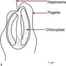
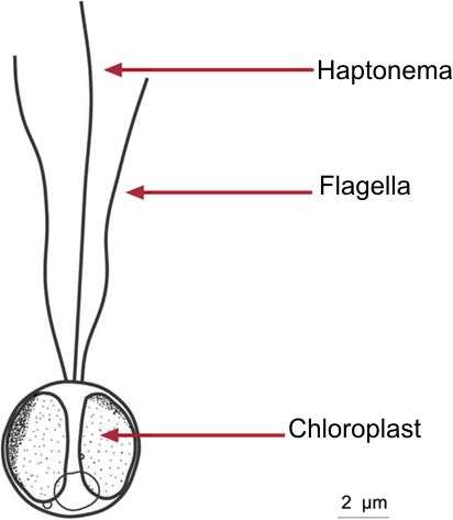
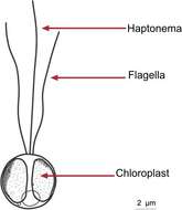
Fig 1: Schematic drawing of Prymnesium parvum
-
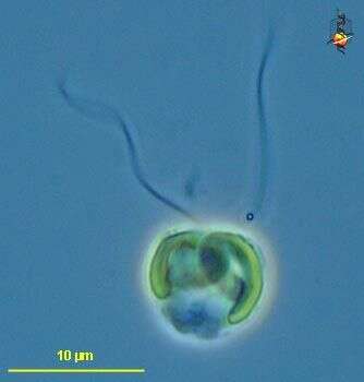
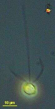
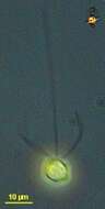
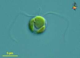
Chrysochromulina (cry-so-crumb-you-line-a) a single-celled haptophyte, with two similar flagella, a short haptonema lying between the flagella, and golden plastids. This may be C. herdlensis. Differential interference microscopy.
data on this strain.
-
Chrysochromulina (cry-so-crumb-you-line-a) ericina a single-celled haptophyte, with two similar flagella, long anterior haptonema and a golden colour from two yellow-brown chloroplasts. Small scales lie on the surface of the cell but these are not evident in this image. Phase contrast microscopy.
data on this strain.
-
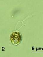
Fig 2: Prymnesium parvum Lugol's preserved cell showing the two flagella but not the haptonema.
-
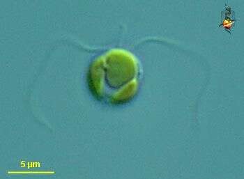
Chrysochromulina (cry-so-crumb-you-line-a) a single-celled haptophyte, with two similar flagella, a short haptonema lying between the flagella, and two golden plastids. This may be C. herdlensis. Differential interference microscopy.
data on this strain.
-
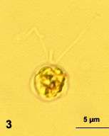
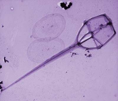
Fig 3: Prymnesium parvum Formalin preserved cell showing the flagella and haptonema
-
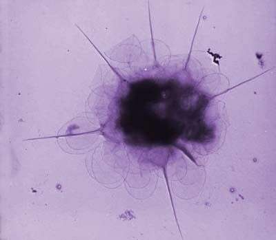
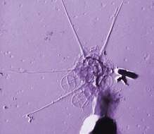
This image was made from samples taken during a scientific cruise in the Pacific. Water was filtered to concentrate the organisms that were present, then dried onto a thin sheet of plastic and then shadowed with a fine layer of metal to provide contrast. The preparation was then observed with an electron-microscope. This technique has been used to document the diversity of marine microbes, especially, protists in the oceans.
-
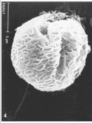
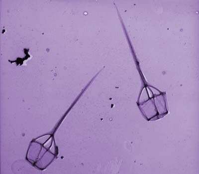
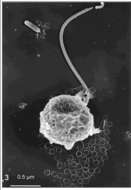
Fig 4: Prymnesium parvum SEM of whole cell showing scale plates and one of the flagella
-
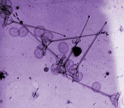
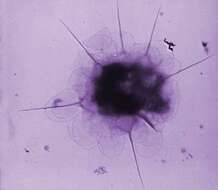
This image was made from samples taken during a scientific cruise in the Pacific. Water was filtered to concentrate the organisms that were present, then dried onto a thin sheet of plastic and then shadowed with a fine layer of metal to provide contrast. The preparation was then observed with an electron-microscope. This technique has been used to document the diversity of marine microbes, especially, protists in the oceans.
-
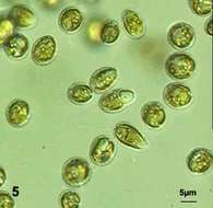
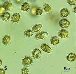
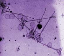
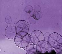
Fig 5: Prymnesium parvum Live cells in culture
-
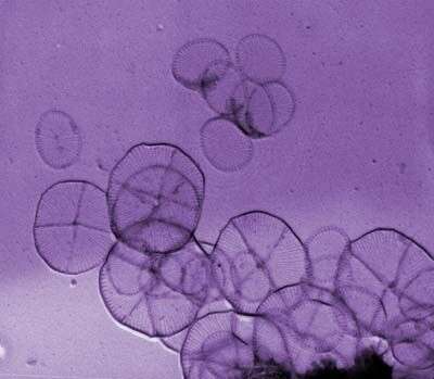
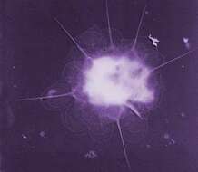
This image was made from samples taken during a scientific cruise in the Pacific. Water was filtered to concentrate the organisms that were present, then dried onto a thin sheet of plastic and then shadowed with a fine layer of metal to provide contrast. The preparation was then observed with an electron-microscope. This technique has been used to document the diversity of marine microbes, especially, protists in the oceans.
-
Fig 1: Chrysochromulina leadbeateri Line drawing of a flagellated cell
-
This image was made from samples taken during a scientific cruise in the Pacific. Water was filtered to concentrate the organisms that were present, then dried onto a thin sheet of plastic and then shadowed with a fine layer of metal to provide contrast. The preparation was then observed with an electron-microscope. This technique has been used to document the diversity of marine microbes, especially, protists in the oceans.
-
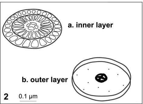
Fig 2: Chrysochromulina leadbeateri Schematic drawing of the two sclae types
-
This image was made from samples taken during a scientific cruise in the Pacific. Water was filtered to concentrate the organisms that were present, then dried onto a thin sheet of plastic and then shadowed with a fine layer of metal to provide contrast. The preparation was then observed with an electron-microscope. This technique has been used to document the diversity of marine microbes, especially, protists in the oceans.
-
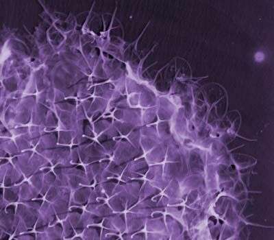
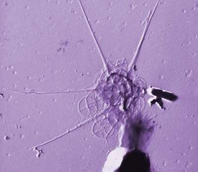
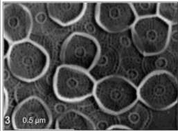
Fig 3: Chrysochromulina leadbeateri SEM of whole cell
-
This image was made from samples taken during a scientific cruise in the Pacific. Water was filtered to concentrate the organisms that were present, then dried onto a thin sheet of plastic and then shadowed with a fine layer of metal to provide contrast. The preparation was then observed with an electron-microscope. This technique has been used to document the diversity of marine microbes, especially, protists in the oceans.
-
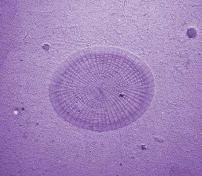
Fig 4: Chrysochromulina leadbeateri SEM inner scales
-
This image was made from samples taken during a scientific cruise in the Pacific. Water was filtered to concentrate the organisms that were present, then dried onto a thin sheet of plastic and then shadowed with a fine layer of metal to provide contrast. The preparation was then observed with an electron-microscope. This technique has been used to document the diversity of marine microbes, especially, protists in the oceans.
-
This image was made from samples taken during a scientific cruise in the Pacific. Water was filtered to concentrate the organisms that were present, then dried onto a thin sheet of plastic and then shadowed with a fine layer of metal to provide contrast. The preparation was then observed with an electron-microscope. This technique has been used to document the diversity of marine microbes, especially, protists in the oceans.