Distribution
provided by Echinoderms of Panama
In Panama this species has been collected from Portobelo (no depth data) and Limon Bay, Colon (USNM E 26639; Centroid Latitude: 9.30, Centroid Longitude: -80.42), Caribbean Sea, by the R. V. Oregon, from a depth of 137 m.
References and links
provided by Echinoderms of Panama
Mortensen T. (1928b): A monograph of the Echinoidea. Vol.1 Cidaroidea. C.A. Reitzel, Copenhagen: 1-551, pages: 336-341.
The Echinoid Directory
World Echinoidea Database
LSID urn:lsid:marinespecies.org:taxname:124268
Synonymised taxa
provided by Echinoderms of Panama
Comprehensive Description
provided by Smithsonian Contributions to Zoology
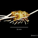
Stylocidaris affinis (Philippi)
Stylocidaris affinis.—For a complete synonymy and additional description see Mortensen, 1928, p. 336.
Test flattened above and below, circumference round, basicoronal plates slightly incurving at the edge of the peristome.
Ambulacra are moderately sinuate; marginal tubercles in uniform series. Commonly there is only one inner tubercle per plate, located on the lower portion. A very small granule is commonly found just above it. Two additional granules are on the lower edge of the plate just below the marginal tubercle (Plate 20: figure 5). All these granules may be indistinguishable without special preparation of the test. The midline suture is white and very naked (Plate 19: figure 3).
The areoles of the interambulacra are quite large, well separated, even the lowermost two in a column are commonly not confluent. Areoles in the ambital region are moderately deep. Large specimens lack crenulation on the tubercles. I have observed crenulation on very small specimens up to 5 mm in diameter, but found it absent on specimens 10 mm and larger. The scrobicular ring of tubercles is rather inconspicuous. Interambulacral and ambulacral midline sutures are naked and white.
The apical system is covered with coarse round granular tubercles (Plate 19: figure 1) except on very small specimens only a few millimeters in horizontal diameter, which have radially elongate tubercles. Specimens large enough to be easily studied with the unaided eye have developed the round tubercles.
The length of the primary spines is from 1 to 1.5 times the horizontal diameter of the test. Some young specimens have spines up to twice the horizontal diameter of the test. The spines are rather slender tapering toward the tip, rows of rather coarse spinules can be observed by the unaided eye. There is a short naked neck between the collar and the rows of spinules. The scrobicular spines are very diagnostic. The most important feature is the dense reddish to reddish brown stripe of pigment running through the midline on the exposed side of the spine. The edges of these spines are white or white with a slight greenish tint. These spines taper in the distal portion toward a cutoff or blunt tip (Plate 18: figure 4). The oral spines have rows of blunted spinules and lack serrate edges, are only slightly flattened and terminate in a blunt tip.
The large globiferous pedicellariae lack an end tooth.
The test is white to slightly olive. Red to reddish-brown pigment is abundant in the apical system and in a midline stripe on the scrobicular and marginal spines. The collar and neck of primary spines commonly have the reddish pigment, especially on specimens with reddish-brown banded spines. Some specimens have white or slightly olive spines without color bands. Color banding is most prevalent on small specimens.
COMPARISON WITH OTHER SPECIES.—The tests of Tretocidaris bartletti and Stylocidaris affinis are quite similar, but the strong crenulation on the upper side of the upper tubercles on Tretocidaris bartletti (Plate 21: figure 4) distinguishes it from Stylocidaris affinis, which lacks crenulation except on very small specimens.
The granules around the periphery of the apical system tend to elongate in a radial direction on T. bartletti (Plate 21: figure 2) while those of S. affinis are round (Plate 19: figure 1).
The ambulacra of the two species are very similar (Plate 20: figure 5; Plate 21: figure 1).
The spinules on the upper side of the primary spines are more fully developed than those on the underside on T. bartletti (Plate 21: figures 5, 6; Plate 22: figures 1, 2). This feature is helpful also in identifying very small specimens only a few millimeters in diameter. The white scrobicular spines are an added identifying feature. Stylocidaris affinis has spinules of uniform size around the primary spines and a reddish midline stripe on the scrobicular and marginal spines.
Stylocidaris affinis and Stylocidaris lineata are superficially similar but are easily distinguished. The more colorful is S. affinis (Plate 20: figure 4), with reddish pigment throughout the plates of the apical system, reddish stripe down the midline of the scrobicular and marginal spines (Plate 19: figure 3), and commonly especially in small specimens reddish to brown banding of the primary spines (Plate 18: figures 5, 6). The rest of the echinoid is white to slightly olive. Stylocidaris lineata is white or almost so. The only noticeable color is the reddish-brown naked midline sutures of the ambulacra (Plate 19: figure 4), the sutures of the interambulacra, and the reddish-brown ring in the apical system (Plate 19: figure 5). These reddish-brown sutures and ring in the apical system are the most distinguishing features. The sutures of S. affinis are white (Plate 19: figure 3).
The apical system of S. affinis is covered with round granules (Plate 19: figure 1), whereas S. lineata lacks granules in the region of the naked reddish ring. The granules on either side of the ring are radially elongate (Plate 19: figure 2).
Many specimens of these species were studied at the U.S. National Museum and at the Museum of Comparative Zoology. Only one specimen of S. affinis was found with an apical system similar to S. lineata. All specimens of S. lineata have the apical system here described. Collections commonly have both of these species identified as S. affinis, and the worker must be careful to identify specimens himself rather than to rely on existing labels.
The primary spines of S. lineata are somewhat longer than those of S. affinis. The length of the naked neck and number of spinule rows are too variable to distinguish the species.
The large globiferous and large tridentate pedicellariae of S. lineata have longer more slender valves (Plate 19: figure 6) than those of S. affinis (Plate 19: figure 7).
Eucidaris tribuloides is usually found in fairly shallow water, but it extends into the upper limits of the distribution of S. affinis. The two species are commonly collected at the same station especially off the west coast of Florida. Specimens of both species only a few millimeters in diameter are strikingly similar, both having light and dark banding of the primary spines with well-developed thornlike spinules (Plate 18: figures 1–3, 5). The wartlike spinules of E. tribuloides first appear in the dark bands nearest the base of the primary spines. Wart development progresses more rapidly in the dark bands as the individual grows. The thorns remain only in the light bands on slightly larger specimens (Plate 18: figures 2, 3). These also change to warts and the color banding is commonly lost. The spinules do not develop into warts on S. affinis. A crownlike tip commonly develops on at least one primary spine even on extremely small individuals of E. tribuloides (Plate 18: figure 3). A distinguishing feature is that one or two very broad tipped large scrobicular spines (Plate 18: figure 2) are commonly present even in the smallest specimens of E. tribuloides. Stylocidaris affinis has a reddish midline stripe on the scrobicular and marginal spines even when very small (Plate 18: figure 5).
DISTRIBUTION.—Bermuda, the West Indies, Caribbean Sea, Gulf of Mexico, eastern Atlantic, and the Mediterranean Sea at depths of 30 to 1,000 meters.
- bibliographic citation
- Phelan, Thomas Francis. 1970. "A field guide to the Cidaroid echinoids of the northwestern Atlantic Ocean, Gulf of Mexico, and the Caribbean Sea." Smithsonian Contributions to Zoology. 1-67. https://doi.org/10.5479/si.00810282.40