Description: Body wide and relatively thick with a sloping forehead and a large round eye and large terminal mouth. Dorsal-fin base long and anal-fin base short. Prominent dorsal, anal, and pelvic-fin spines and a large non-serrated preopercular spine.
Pretransitional mostly-unmarked stage, usually from 10-15 mm SL: Body: Pretransitional larvae can have a few melanophores on the body just below the dorsal fin base where the future bars will develop: at the mid-spinous dorsal fin, the end of the spinous dorsal fin and under the mid-soft dorsal fin. There is a patch of melanophores along the anterior third of the dorsal midline of the caudal peduncle and a full-length band of melanophores along the ventral midline of the caudal peduncle extending forward and ending just before a single large melanophore underlying the pterygiophores of the last anal-fin rays. A bar pattern begins on the lower caudal peduncle as a patch without melanophores between two bars. There are a few deep melanophores at the very end of the lateral midline of the caudal peduncle. Head: Melanophores on the head consist of a patch overlying the brain and on the surface braincase and small melanophores around the tips of both the upper and lower jaw (in similar numbers). The opercular area is covered in iridescence extending down to the pelvic-fin insertion. The inner cleithral surface of the gill cavity is speckled with large melanophores and there are internal melanophores lining the dorsal aspect of the peritoneum extending down to the vent and overlain by a silvery camouflage layer. Fin Spines: The dorsal and anal-fin spines are relatively stout, with prominent internal reticulations. There are fine serrations along the anterior aspect of the anal-fin spines at this stage, but disappearing during transition. Fins: Melanophores on the dorsal-fin membranes are concentrated between the third and eighth dorsal-fin spines, predominantly on the distal half of the fin-ray membranes and typically sparing the membranes adjacent to the base of the fin. Some individuals have melanophores spreading down to meet the dorsal midline, but only behind the fourth and fifth spines and later the ninth and tenth spines (at the site of the future dark bars on the body). On the anal fin, there are melanophores along the base of the spines and membranes, spreading almost half-way up the second and third anal-fin spines. There are melanophores on the lower portion of the membrane between the last anal-fin spine and the first ray and the next membrane or two, followed by melanophores at the base of the membrane for the next few rays. There can be a few melanophores between the bases of the uppermost of the lower segmented caudal-fin rays and also on the lowest two or three segmented rays, often extending out along the rays. Pretransitional analogues: Pretransitional larvae (mostly-unmarked stage, usually from 10-15 mm SL) are separated from some other Lutjanus by having distinct serrations persisting on the anterior profile of the anal and dorsal-fin spines (but shared by L. griseus and L. jocu). L. apodus larvae are distinguished from the L. griseus type at this stage by having the dorsal-fin membrane melanophores concentrated on the distal portion of the membranes, only a small patch on the anterior third of the dorsal midline of the caudal peduncle, usually an incipient bar pattern on the lower caudal peduncle, similar numbers of melanophores on the tip of the upper and lower jaws, and often pigmentation along the longest pelvic-fin membrane. It is likely that pretransitional L. jocu cannot be separated from L. apodus.
Transitional stage: Early transitional L. apodus develop a pattern of bars on the body, beginning at the lower caudal peduncle where two dark bars first separate and then bars progressively develop from the caudal peduncle anteriorly. Each bar starts below the base of the dorsal fin and extends down with development. The mid-body bars begin with three patches of melanophores: the first under the fourth to sixth dorsal-fin spines, the second under the last two dorsal-fin spines and first dorsal soft rays and the third under the middle of the soft dorsal fin. Melanophores are limited to the outer half of the spinous-dorsal-fin membranes at first, but progressively extend down during transition. Before any stripes develop on the head, the tips of the upper and lower jaws are similarly speckled with small melanophores. Even on lightly-marked transitional larvae, there are some small melanophores on the thorax and the pelvic fin membranes. Early transitional larvae have serrations on the anterior aspect of the first two anal-fin spines, but these are usually lost midway through transition. Late transitional larvae have melanophores covering much of the body, but now the bars are made up of alternating areas of smaller and larger melanophores. The lower caudal peduncle at this stage has filled-in with melanophores and no longer has bars separated by non-pigmented skin. By this point, melanophores have advanced down the spinous-dorsal-fin membranes and do reach the base where they merge with the melanophores of the darker bars. On the head, a stripe develops between the eye and the tip of the upper jaw and two stripes diverge behind the eye. Small melanophores fill in and uniformly speckle the cheek, operculum, and thorax as well as the pelvic fins. Transitional analogues: Transitional L. apodus larvae develop bars on the body with no lateral spot, distinguishing them from the spotted species. In the early stages of transition, L. apodus larvae can be separated from L. griseus by having incipient melanophore bars forming on the lower caudal peduncle (vs. uniform) and having melanophores mostly on the distal half of the spinous-dorsal-fin membranes (vs. proximal and base). Late transitional L. apodus larvae differ from L. griseus by having distinct vertical bars of larger melanophores on the body vs. uniform speckling over the upper body (and lighter over the lower half of the head and abdomen) or indistinct bars at most on the upper half of the body, numerous melanophores speckling the cheek, thorax, and pelvic fins (vs. those areas relatively lightly-marked). Transitional recruits of L. jocu can overlap in appearance and can show a similar bar pattern to that of transitional L. apodus, although the lighter bars are narrower and the bar pattern becomes less distinct with development. There is also some overlap in appearance when both have a mostly-uniform speckling pattern, although uniform L. apodus have large melanophores and L. jocu have a fine speckling of melanophores (for example, in the space below the eye, there are about 100 melanophores in an area equal to the pupil in L. jocu vs. about 10 in L. apodus). Transitional recruits of L. apodus and L. griseus can sometimes overlap in appearance. If the bars are distinct from the base of the dorsal fin down to the anal fin, it is L. apodus; when L. griseus have bars, they are apparent on the upper body but fade towards the anal-fin base. As they grow, L. griseus develop distinct striping patterns on the lower body that do not occur on L. apodus.
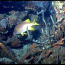
Juveniles: Juvenile L. apodus have prominent vertical bars and no lateral spot. Rare individuals have a uniform pattern with only indistinct bars, but, notably these individuals do not show any striping pattern. Juvenile analogues: The absence of a lateral spot separates L. apodus from most other juvenile snappers ((L. analis, L. mahogoni, L. synagris, and the deep-water snappers). Virtually all juvenile L. apodus have prominent vertical bars which are absent or indistinct in L. jocu, L. griseus, and L. cyanopterus. Rare individuals of L. apodus that have a uniform appearance or indistinct bars can be difficult to separate from juvenile L. jocu, but juvenile L. griseus of the same size would show some evidence of body stripes. Juvenile L. cyanopterus are narrower-bodied, have a wider caudal peduncle, and do not share the blue line under the eye.
Diagnosis: Modal fin-ray counts of D-X,14 A-III,8 are shared among most of the regional Lutjanus species, including L. analis, L. apodus, L. cyanopterus, L. griseus, L. jocu and the deep-water snappers L. buccanella, L. campechanus, and L. vivanus. Transitional and juvenile L. apodus have a prominent pattern of vertical bars without a lateral spot. (DNA)