Comprehensive Description
provided by Smithsonian Contributions to Zoology
Histocidaris sharreri (A. Agassiz)
Histocidaris sharreri.—For a complete synonymy and additional description see Mortensen, 1928, p. 86.
The test is almost globular, moderately elevated at the apical system (Plate 13: figure 1; Plate 14: figure 1), and only very slightly flattened adorally.
The ambulacra are moderately sinuate, and have large marginal tubercles in uniform series. The most significant feature of the ambulacra is the near naked interporiferous zones (Plate 12: figures 9, 11). Many plates lack even a single inner tubercle. I believe that the deep ambulacral midline suture which Mortensen (1929, p. 87) suggests as being diagnostic of this species is only an irregularity or growth deformity of the specimen he studied as it is not continuous on the specimen. The upper portion of Plate 12: figure 11 shows the region of the ambulacrum illustrated by Mortensen (1928, pl. 68: fig. 4). Peristomial ambulacral plates bear an internal prolongation as in other histocidarids (Plate 10: figure 3).
Each large intermediately deep areole of the interambulacral plates is surrounded by a prominent scrobicular ring which crowds the plate sutures. The extrascrobicular area is therefore very limited, with few secondary tubercles and spines (Plate 14: figure 1). The primary tubercles are strongly crenulate even on tubercles below the ambitus (Plate 14: figures 1, 2).
There is a marked difference in the relative size of the genital pores of the two specimens studied. The very large genital pores of the smaller specimen, MCZ 253 (Plate 12: figure 1), indicate that it is a female. The smaller genital pores of MCZ 362 (Plate 12: figure 10) suggest that it is a male. Tubercles of the genital plates are most abundant around the genital pores, and on the female form a distinct ring around the large pores. Small tubercles are also scattered along the periproctal edge of the genital plates.
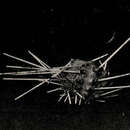
The two specimens were too few to permit determination of the normal variation in primary spine form and only the lower portion of two large primary spines remain attached to MCZ 253. The length of the uppermost primary spines is approximately 1.5 times the horizontal diameter of the test. These spines lack or rarely possess thorns. The tips flare out (Plate 12: figure 4) due to radial expansion of the longitudinal ridges. At the ambitus the tips differ in that the ridges expand to form an elongate swollen tip (Plate 12: figures 2, 3). At or just below the ambitus the primary spines develop two rows of serrations which become progressively larger on transitional spines approaching the peristome (Plate 13: figures 1, 3). Below the ambitus the spines curve adorally. The curvature increases with adoral position. The oral primaries are broadly rounded at the tip, transversely flat adorally, and convex adapically. The spines are strongly serrate and curve longitudinally toward the mouth. Specimen MCZ 362 has suffered considerable loss of spines but a drawing in Agassiz (1883, pl. 3) shows the spine forms and position. The tips of spines from this specimen are shown in Plate 12: figures 2–4. Only the lower portion of two primary spines remain attached to MCZ 253. Well-developed thoms (Plate 12: figure 1) occur on the shafts of these broken spines. There are no thorns on the primary spines of MCZ 362. The presence or absence of thorns is a normal variant in the histocidarids and their absence on MCZ 362 is not significant.
The large tridentate pedicellariae are the most diagnostic feature. They are very large, and easily observed with the unaided eye, and have three rather long broad blades (Plate 12: figure 5). No globiferious pedicellariae occur in any species of the histocidarids.
The specimens are brown with a small amount of yellow pigment. The primary spines are white with a yellow-brown collar.
The three histocidarids are compared and discussed in detail following the description of Poriocidaris purpurata (Wyville Thomson).
COMMENTS ON THE HISTORY AND CONFUSION ASSOCIATED WITH Histocidaris sharreri.—The echinoid that is currently recognized as Histocidaris sharreri was named Porocidaris sharreri in the original description (A. Agassiz, 1880, p. 71). Three specimens, two large males and a small female, were mentioned in this description, but it was not until the more complete description (A. Agassiz, 1883, p. 12, pl. 3; pl. 4: figs. 1, 2), again only mentioning the three specimens, that drawings of this species were published. Mortensen (1903, pp. 23, 28) recognized that more than one species was represented in the descriptions and illustrations of A. Agassiz (1880, 1883). The current name of the species that Mortensen separated from H. sharreri is Calocidaris micans. In A. Agassiz (1883) only the illustration in Plate 3 and a portion of the description on pages 12 and 13 represent the species currently recognized as H. sharreri. The specimen that A. Agassiz (1883) illustrated on plate 3 is MCZ 362.
Following the listing of MCZ 362 in the catalog of recent echinoid type specimens (Downey, 1968, p. 62) is the entry “(= Calocidaris micans).” Downey in personal communication stated that it was not intended to indicate that this specimen (MCZ 362) was a specimen of Calocidaris micans. Specimen MCZ 362 is herein selected as the lectotype of H. sharreri, and specimen MCZ 253 is designated a paralectotype.
The two specimens in the Museum of Comparative Zoology (MCZ 362 and MCZ 253) are the large male and small female mentioned by A. Agassiz (1880, 1883). The third specimen mentioned by Agassiz was later renamed Calocidaris micans.
DISTRIBUTION.—Leeward Islands: Nevis and St. Kitts. The distribution of all histocidarids of the northwestern Atlantic, Caribbean, and Gulf of Mexico is inadequately known.
- bibliographic citation
- Phelan, Thomas Francis. 1970. "A field guide to the Cidaroid echinoids of the northwestern Atlantic Ocean, Gulf of Mexico, and the Caribbean Sea." Smithsonian Contributions to Zoology. 1-67. https://doi.org/10.5479/si.00810282.40