Description
provided by Zookeys
Body length 3.62 mm (range 3.3–4.2; N = 5) for females. Body uniformly reddish brown and shiny; the toruli area, flagellomeres of antenna, area above clypeus, occiput, dorsolateral margin of pronotum, anteroadmedian signa area, parapsidal signa, mesopleuron, metapectal-propodeal complex and anteromedial area of scutellum dark brown to black. Legs with all coxae and femora yellowish; tibia and tarsomeres dark brown to black. Forewing hyaline with some very light infumation; veins dark brown to black.
Asexual female. Head moderately pubescent with piliferous punctures, in dorsal view about 3.5 times wider than long. POL 1.5 as long as OOL, posterior ocellus separated from inner orbit of eye by 1.7 times its longest diameter. Head in anterior view (Fig. 6A) transversely ovate, 1.15 times wider than high, gena not expanded behind eyes. Vertex frons and gena slightly alutaceous. Head moderately pubescent, with relatively long white setae, except vertex, frons with sparse, shorter setae. Clypeus more or less trapezoidal, 1.6 times wider than high, mostly smooth and moderately pubescent; ventral margin sinuate, slightly projecting over mandibles. Anterior tentorial pits visible; epistomal sulcus indicated, clypeo-pleurostomal lines visible. Malar space 0.3 times height of compound eye, without malar sulcus; some irradiating striae from clypeus present, reaching ventral margin of compound eye, absent medially above clypeus. Toruli situated slightly above mid-height of compound eye; distance between antennal rim and compound eye 1.1 times width of antennal socket including rim. Ocellar plate not raised. Head, posterior view (Fig. 6B) without occipital carina. Gula short; distance between occipital and oral foramina 0.5 times height of occipital foramen (Fig. 6B). Hypostomal sulci well separate at oral fossa.
Mouthparts (Figs 6A–B): mandibles exposed, with dense setae in base, right mandible with three teeth, left with two teeth. Cardo of maxilla not visible, maxillary stipes about 2.3 times longer than wide. Maxillary palp five-segmented. Labial palp three-segmented.
Antenna (Fig. 7A-C) of moderate length, as long as 1/2 body length, with 15 flagellomeres, but F15 only partially separated from F14 dorsally (Fig. 7C); flagellum not broadening towards apex; with relatively long, erect setae, and elongate placodeal sensilla well visible (Fig. 7C). Relative lengths of antennal segments: 16:12:43:32:24:20:19:17:13:12:11:11:10:9:9:8:14. Pedicel sub-globose, 0.8 as long as scape; F1-F9, gradually decreasing in length. F1 1.34 times as long as F2. F10-F13 short and wide, F15 1.8 times longer than wide, 1.7 as long as F14 (Fig. 7C). Placodeal sensillae on F4-F15 disposed in one row of 5–6 sensillae in half dorsal area of each flagellomere.
Mesosoma. Smooth, moderately pubescent with piliferous punctures, in lateral view 1.3 times as long as high, slightly convex dorsally. Pronotum, moderately pubescent; lateral surface of pronotum with some longitudinal wrinkles dorsally; with long and dense setae (Fig. 6D). Pronotum short medially, ratio of length of pronotum medially/laterally = 0.16. Pronotal plate indistinct dorsally (Fig. 6C).
Mesonotum (Fig. 6E). Mesoscutum smooth, moderately pubescent with piliferous punctures medially and along notauli; slightly broader than long in dorsal view. Notauli complete, smooth, broad, deep and convergent posteriorly, without median mesoscutal impression, anteroadmedian signa and parapsidal signa indistinct. Transscutal fissure narrow, clearly visible, deeply impressed, and slightly sinuate. Scutellar foveae shallow, confluent, indistinctly margined posteriorly and rugose. Mesoscutellum (Fig. 6E), about 0.7 times length of mesoscutum, 1.2 times as long as wide, strongly reticulate-rugose and moderately pubescent, in lateral view extended posteriorly over dorsellum. Axillula moderately pubescent, their anterior margins marked and posterior margins indistinct. Mesopleuron smooth, moderately pubescent except in speculum (Fig. 6D).
Metanotum (Fig. 6F). Metapectal-propodeal complex. Metapleural sulcus reaching posterior margin of mesopectus at about mid-height of metapectal-propodeal complex (Fig. 6D). Lateral propodeal carinae moderately divergent posteriorly, reaching the nucha; median propodeal area longer than broad, smooth, with some setae anteriorly; lateral propodeal area densely pubescent (Fig. 6F). Nucha rugose.
Legs. Densely pubescent; metatarsal claws with a large obtuse basal lobe (Fig. 7D).
Forewing (Fig. 8B) slightly longer than body, strongly pubescent; basal cell with some rows of setae; radial cell 4.0 times longer than wide; open along anterior margin; areolet triangular, closed and distinct. R1 and Rs nearly straight, not quite reaching wing margin; R1 forming an acute angle with anterior margin of wing. Rs+M reaching basalis at its mid-height. 2r well pigmented, angulate and slightly projected medially. Apical margin of wing with moderately long hair fringe.
Metasoma (Fig. 7E) large, as long as head and mesosoma combined, in lateral view as wide as high. Second metasomal tergite covering about 2/3 of metasoma, with a patch of dense setae in its anteromedial area. Projecting part of hypopygial spine short (Fig. 7F–G), shorter than basal height of spine (Fig. 7F); with parallel sides and pointed apically, with dense long subapical setae forming a patch, extending far beyond apex of spine.
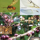
Gall (Fig. 8D–H). Similar in location, shape, and size to the galls of Coffeikokkos copeyensis Pujade-Villar & Melika. However, the galls of this new species are much more regularly spherical and its surface is not uniformly colored, but spotty. Diameter of gall measures 5 to 8 mm. They are formed, solitary or more frequently in groups, in stems of Quercus bumelioides Liebm. The surface of the gall is smooth and shiny; whitish, green or yellowish when fresh with red spots, becoming brown when mature. Monothalamic, with compact woody tissue internally containing the single larval cell (Fig. 8H). Similar spherical and spotty galls are also induced by the Nearctic Cynips (=Besbicus) mirabilis (Kinsey 1922), but this galls are larger, pubescent, formed in leaves and with an internal structure of irradiant filaments (Kinsey 1930).
- license
- cc-by-3.0
- copyright
- Enrique Medianero, José Luis Nieves-Aldrey
- bibliographic citation
- Medianero E, Nieves-Aldrey J (2013) Barucynips panamensis , a new genus and species of oak gallwasps (Hymenoptera, Cynipidae, Cynipini) from Panama, and description of one new species of Coffeikokkos ZooKeys 277: 25–46
- author
- Enrique Medianero
- author
- José Luis Nieves-Aldrey
Distribution
provided by Zookeys
Coffeikokkos korytkowskii was found between 2515–3045 m a.s.l. at Volcán Barú, Chiriqui, Panama.
- license
- cc-by-3.0
- copyright
- Enrique Medianero, José Luis Nieves-Aldrey
- bibliographic citation
- Medianero E, Nieves-Aldrey J (2013) Barucynips panamensis , a new genus and species of oak gallwasps (Hymenoptera, Cynipidae, Cynipini) from Panama, and description of one new species of Coffeikokkos ZooKeys 277: 25–46
- author
- Enrique Medianero
- author
- José Luis Nieves-Aldrey