-
-
Steiglitz, Victoria, Australia
-
Three Lakes, Wisconsin, United States
-

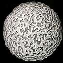
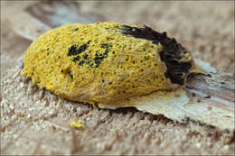
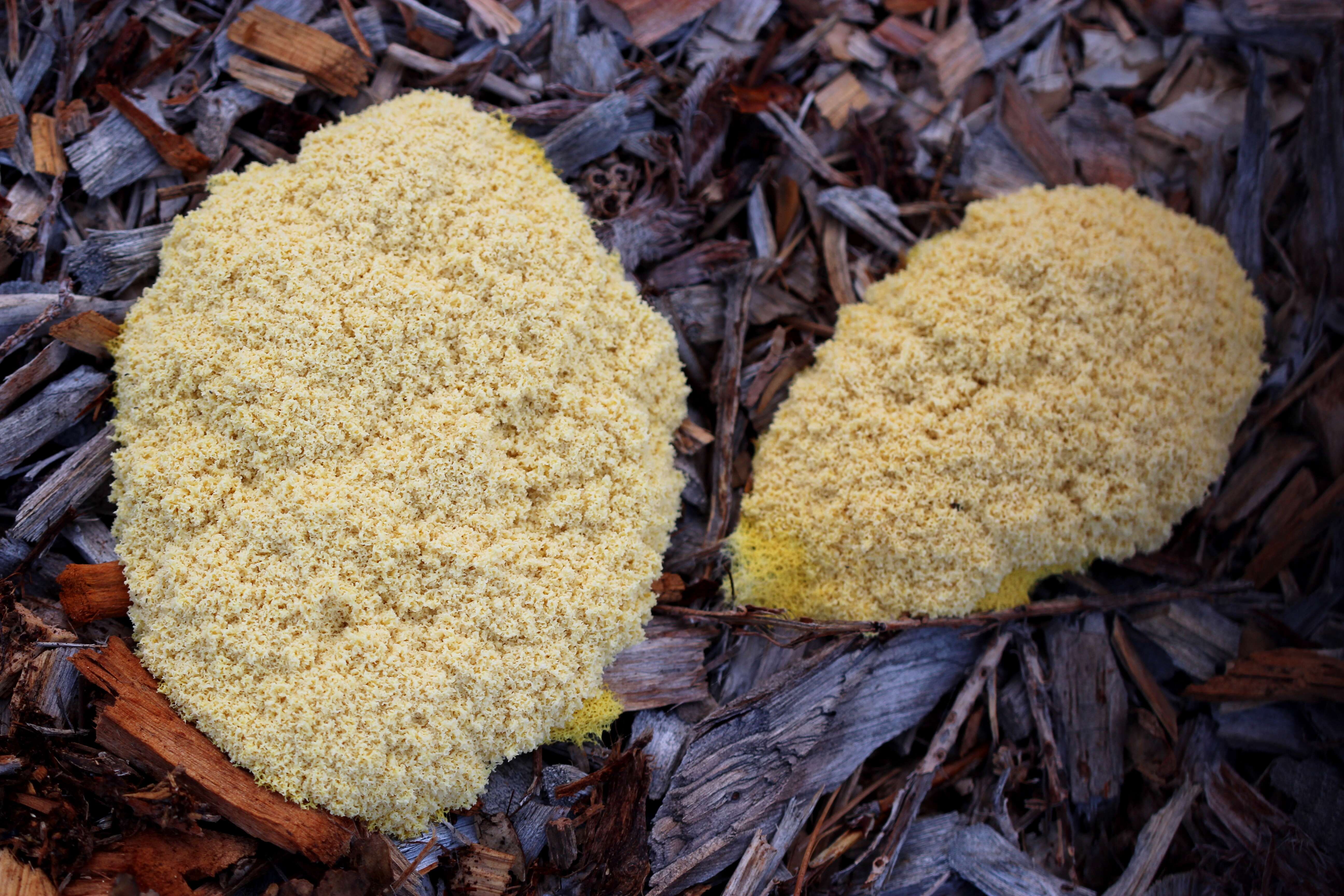
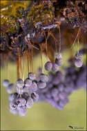
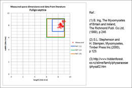
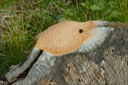
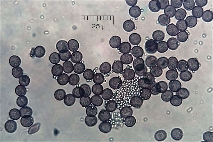
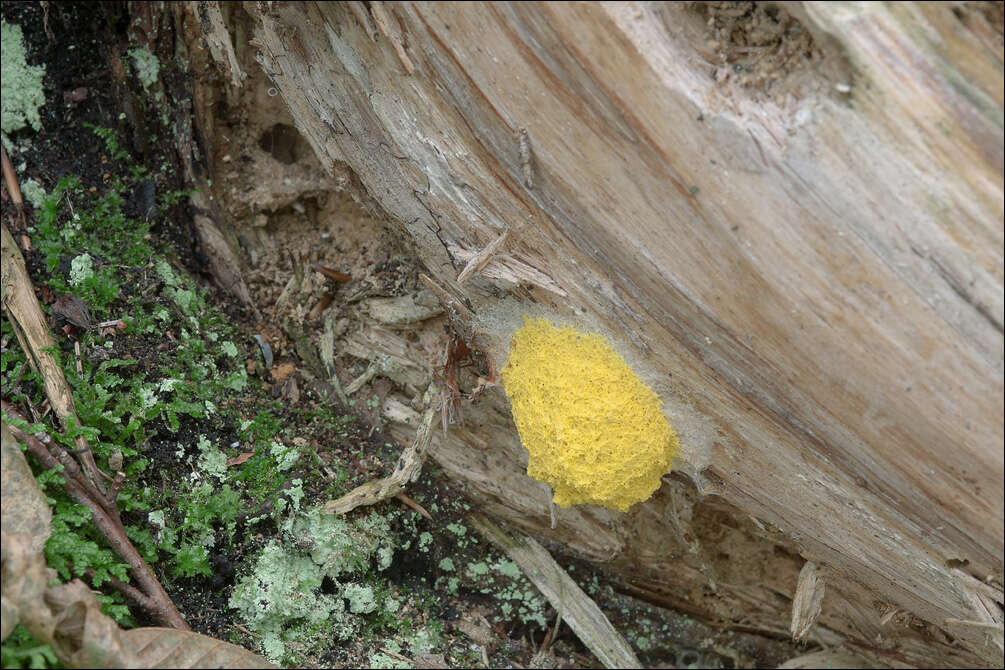
Fuligo septica (L.)Wigg., syn. Mucorsepticus L., Reticularia septica (L.) With., Aethalium septicum (L.) Fr., Fuligo varians Sommerf.Scrambled-egg slime, Dog vomit slime mold, Flowers of Tan, DE: Gelbe Lohblte, HexenbutterSlo.: reslov cvetThe aethalium picture taken on July 21. Dat.: July 21. 2014Code: Bot_815/2014_DSC2009 Lat.: 46.36114 Long.: 13.70122Habitat: old partly tree overgrown pasture, near mixed wood edge; moderately southeast inclined foot of an old overgrown scree slope; open, dry, sunny place; shallow, skeletal, calcareous ground, exposed to direct rain, average precipitations ~ 3.000 mm/year, average temperature 7-9 deg C, elevation 630 m (2.070 feet), alpine phytogeographical region. Substratum: a stump of Picea abies cut down three years ago.Place: Lower Trenta valley, between villages Soa and Trenta, upper part of 'Na Melu' place, East Julian Alps, Posoje, Slovenia EC.Comment: Myxomycetes are poorly known yet very interesting creatures. For decades they have been shuffled back and forth between the animal and plants kingdoms until recognized as separate creatures. They are not animals because they proliferate by spores. They are also not plants since they crumble around (an animal like ability) and fix themselves firmly to substrate only at the end of their life cycle. They don't produce their own food like plants but feed by 'hunting' (actually engulfing) bacteria and tiny bits of other organic matter, which is another animal like feature. The first stage in their development cycle, which is observable in the field, is called plasmodium. Earlier stages (from myxoflagellates, myxoamoebae, to zygote) are microscopic and can be observed only in labs. Plasmodium is a single giant living cell, a clump of protoplasm filled with thousands of cell nuclei, crawling around, eating bacteria and growing. Some of such plasmodia are the largest cells of living creatures known. In some species they can measure several meters across or weight up to 20 kg. That it much larger than ostrich's eggs, which are popularly considered as 'largest living cells'! Plasmodia could be found in the field, some are even brightly colored and easy to spot; however, it is almost impossible to determine to which species they belong. Plasmodium of Fuligo septica is commonly described like disgusting mucus, spilled scrambled eggs, dogs vomiting and other 'benevolent' portrayals.When the time is right or delicate environmental conditions required for growth worsen plasmodia eventually evolves (usually almost completely) into sporocarps of different forms. These are bodies producing spores and then vanishing. In genus Fuligo sporocarp is a cushion like aethalium sitting on a thin whitish, 'fibrous' layer called hypothallus (Fig.14.). These'cushions' are what one usually finds in the field. But, other Myxomycetes develop also many other forms of sporocarps full of beauty, delicacy and imagination. Aethalia of Fuligo septica are usually covered with a kind of crust called cortex, which is brittle and soon crumbles away. In humid conditions it may not fully develop (Ref.1). Inside a mature aethalium there is a mesh of thin tubes or fibers called capillitium and zillions of dark brown spores. Fuligo septica has characteristic nodes on capillitial tubes, which are clearly seen on Fig. 4M. In due course the aethalium decomposes almost entirely into spore mass (Fig.17., 18.), which are sooner or later blown or washed away (Fig.20. taken about three weeks after the first photo). Size and shape of spores and structure of their surface are important traits for species determination. Cushion-shaped aethalium measured approximately 14 x 5 cm and was about 3 cm thick. Spores are minutely warty and globose to subglobose. Dimensions: 8 [8,4 ; 8,7] 9,1 x 7,4 [8 ; 8,2] 8,7 microns; Q = [1 ; 1,07] 1,1; N = 25; C = 95%; Me = 8,5 x 8,1 microns; Qe = 1,05. Olympus CH20, NEA 100x/1.25, magnification 1.000 x, oil (spores), NEA 40x/0.65, magnification 400x (capillitium), NEA 10x/0.25, magnification 100x (hypothallus); in water, living material. AmScope MA500 digital camera. Spore sample taken on July 23. 2014.Herbarium: Mycotheca and lichen herbarium (LJU-Li) of Slovenian Forestry Institute, Vena pot 2, Ljubljana, Index Herbariorum LJFRef.:(1) B. Ing, The Myxomycetes of Britain and Ireland, The Richmond Publ. Co.Ltd, (1999), p 246(2) S.L.Stephenson and H.Stempen, Myxomycetes, Timber Press Inc.(2000), p 123(3)
www.hiddenforest.co.nz/slime/family/physaraceae/physa02.htm 6-9(4) M. Poulain, M. Meyer, J. Bozzonet, Les Myxomycetes, FMBDS (2011), Vol.1., p128; Vol.2., p168.
-
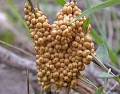
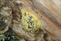
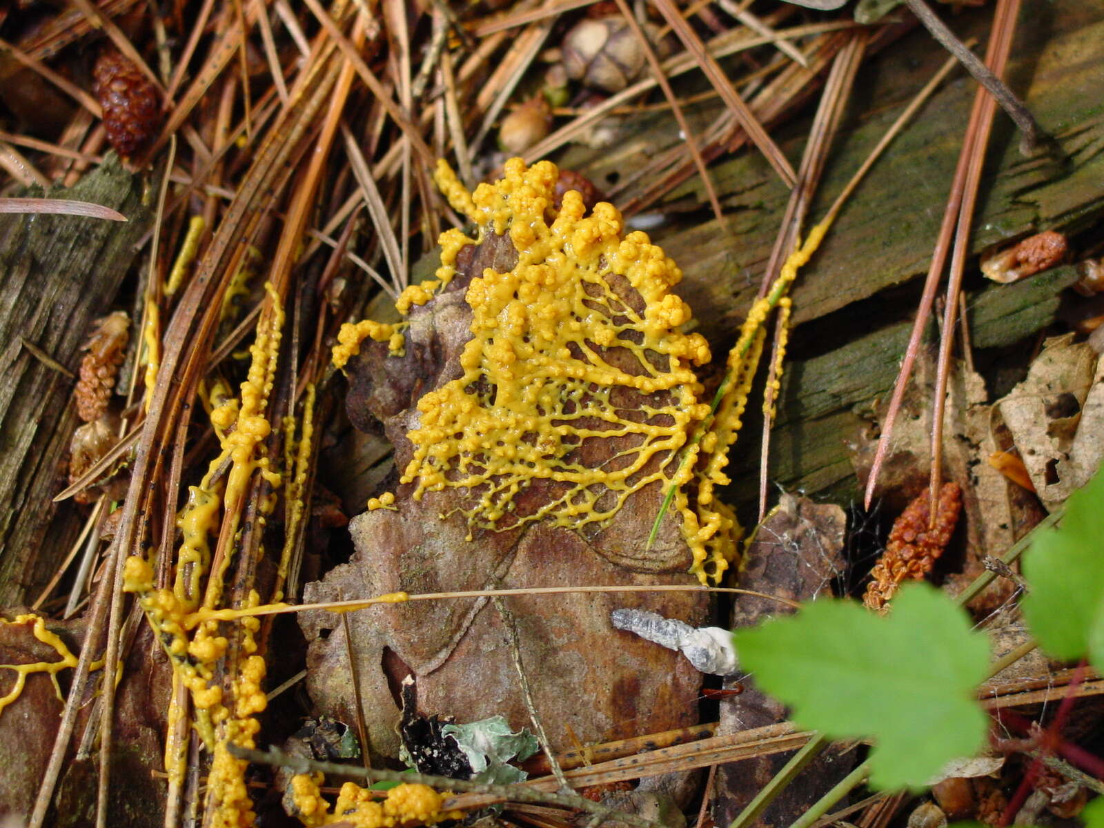
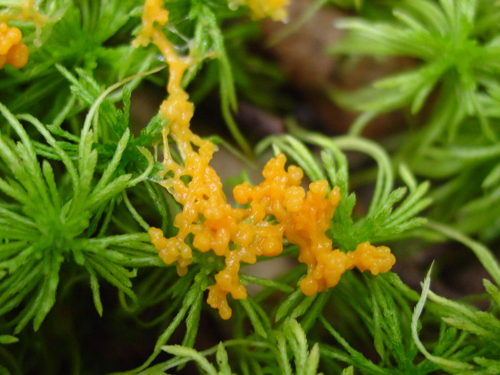
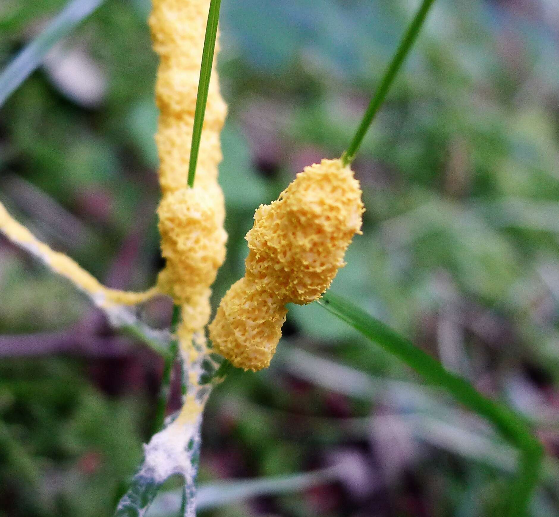
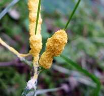
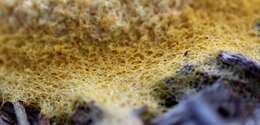
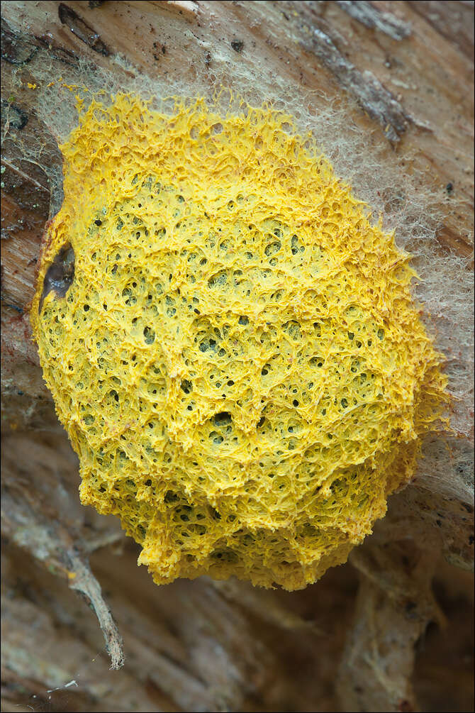
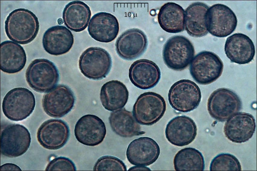
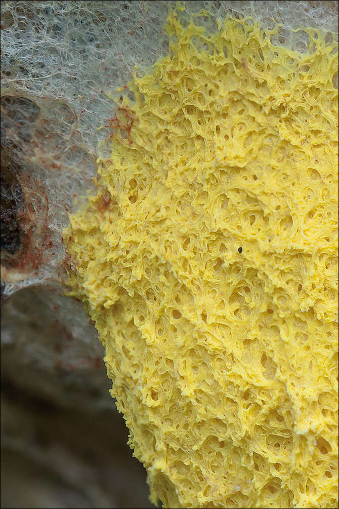
Fuligo septica var. flava (Pers.) Morgan, syn.: Mucor septicus L., Aethalium flavum (Pers) LinkFlowers of Tan, DE: Gelbe LohblteSlo.: reslov cvet, rumeni razliekDat.: Sept. 05. 2014Lat.: 46.35965 Long.: 13.70116Code: Bot_832/2014_DSC3621Habitat: mixed wood, Fagus sylvatica and Picea abies dominant, moderately southeast inclined mountain slope, shallow, skeletal calcareous ground, old overgrown slope and moraine scree with larger rocks and boulders, in shade, relatively warm place, partly protected from direct rain by tree canopies, average precipitation ~ 3.000 mm/year, average temperature 7 - 9 deg C, elevation 600 m (1.970 feet), alpine phytogeographical region. Substratum: debarked trunk of Picea abies lying on ground in its late disintegration stage.Place: Lower Trenta valley, between villages Soa and Trenta, next to the trail from Trenta 2b cottage to abandoned farmhouse 'Strgulc', East Julian Alps, Posoje, Slovenia EC.Comment: Fuligo septica is probably the most common and widely known Myxomicete. The latest monograph on Myxomycetes I have (Ref.:1) describes six varieties of this species, which differ mostly in cortex structure (single versus double layered) and color of different parts of sporocarp and plasmodium. Fuligo septica var. flava should have vivid yellow aethalia, yellow inner lime and yellow plasmodium. Two days before I took these pictures I had seen the plasmodium, which was in a form of vividly yellow colored patch of densely packed small half-spheres. The rest of traits of Fuligo septica var. flava also fit well to my observation.Spores minutely warty, globose to subglobose. Dimensions: 8 [8,4 ; 8,7] 9,1 x 7,4 [8 ; 8,2] 8,7 microns; Q = [1 ; 1,07] 1,1; N = 25; C = 95%; Me = 8,5 x 8,1 microns; Qe = 1,1. Olympus CH20, NEA 100x/1.25, magnification 1.000 x, oil (spores), NEA 40x/0.65, magnification 400x (capillitium, calcareous granules); in water; live material. AmScope MA500 digital camera.Herbarium: Mycotheca and lichen herbarium (LJU-Li) of Slovenian Forestry Institute, Vena pot 2, Ljubljana, Index Herbariorum LJFRef.:(1) M. Poulain, M. Meyer, J. Borronet, Les Myxomycetes, FMBDS (2011), Vol.1: p390, Vol.2: (2) B. Ing, The Myxomycetes of Britain and Ireland, The Richmond Publ. Co.Ltd, (1999), p 246. (3) S.L.Stephenson and H.Stempen, Myxomycetes, Timber Press Inc.(2000), p 123.
-
Zaragoza: Aragn (Espaa)Reino: ProtozoaFilo: MycetozoaClase: MyxomycetesOrden: PhysaralesFamilia: Physaraceae
-
Three Lakes, Wisconsin, United States
-
Brodick Castle, Arran. NS007381
-
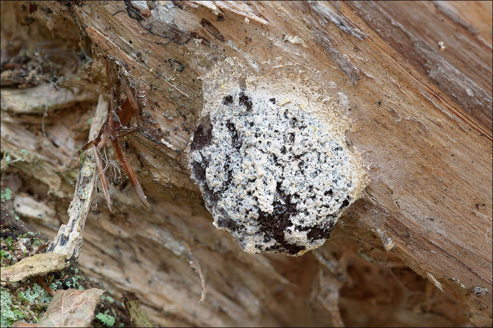
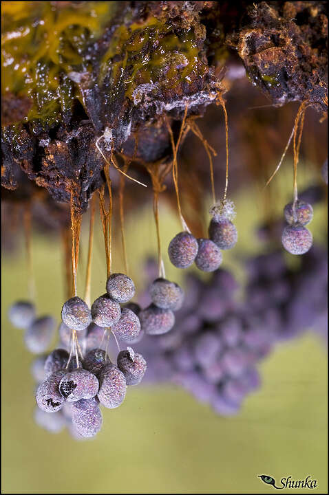
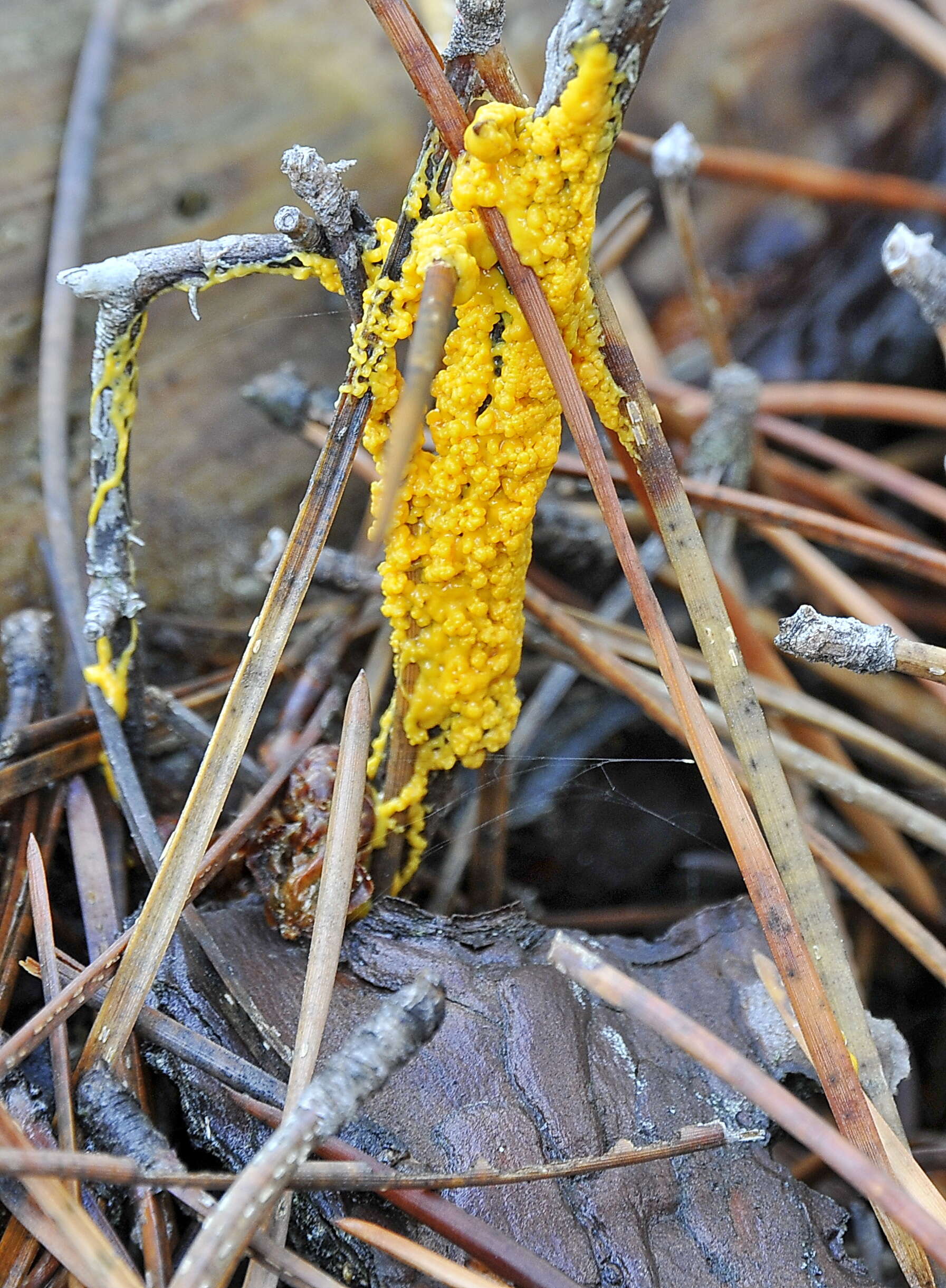
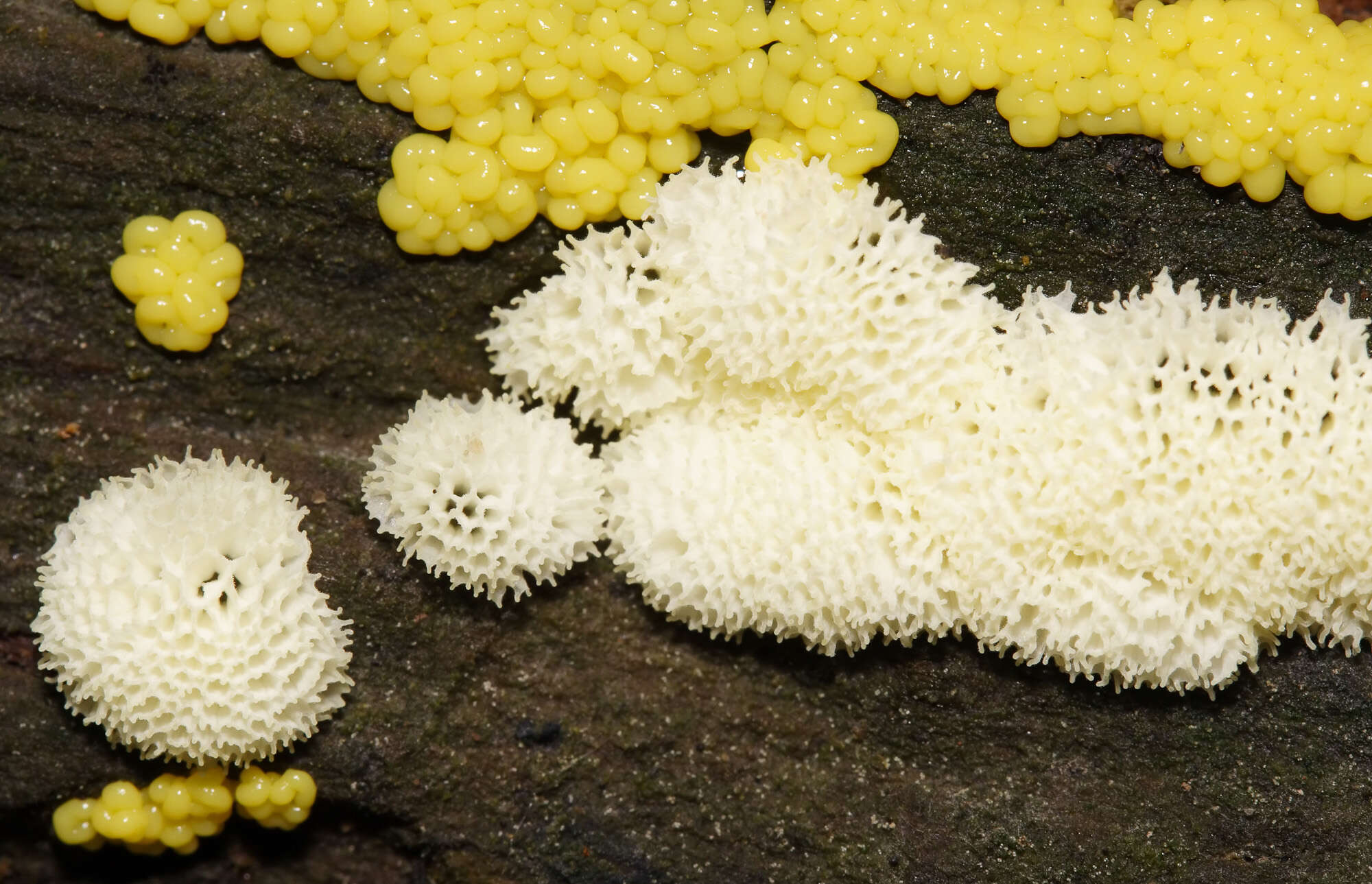
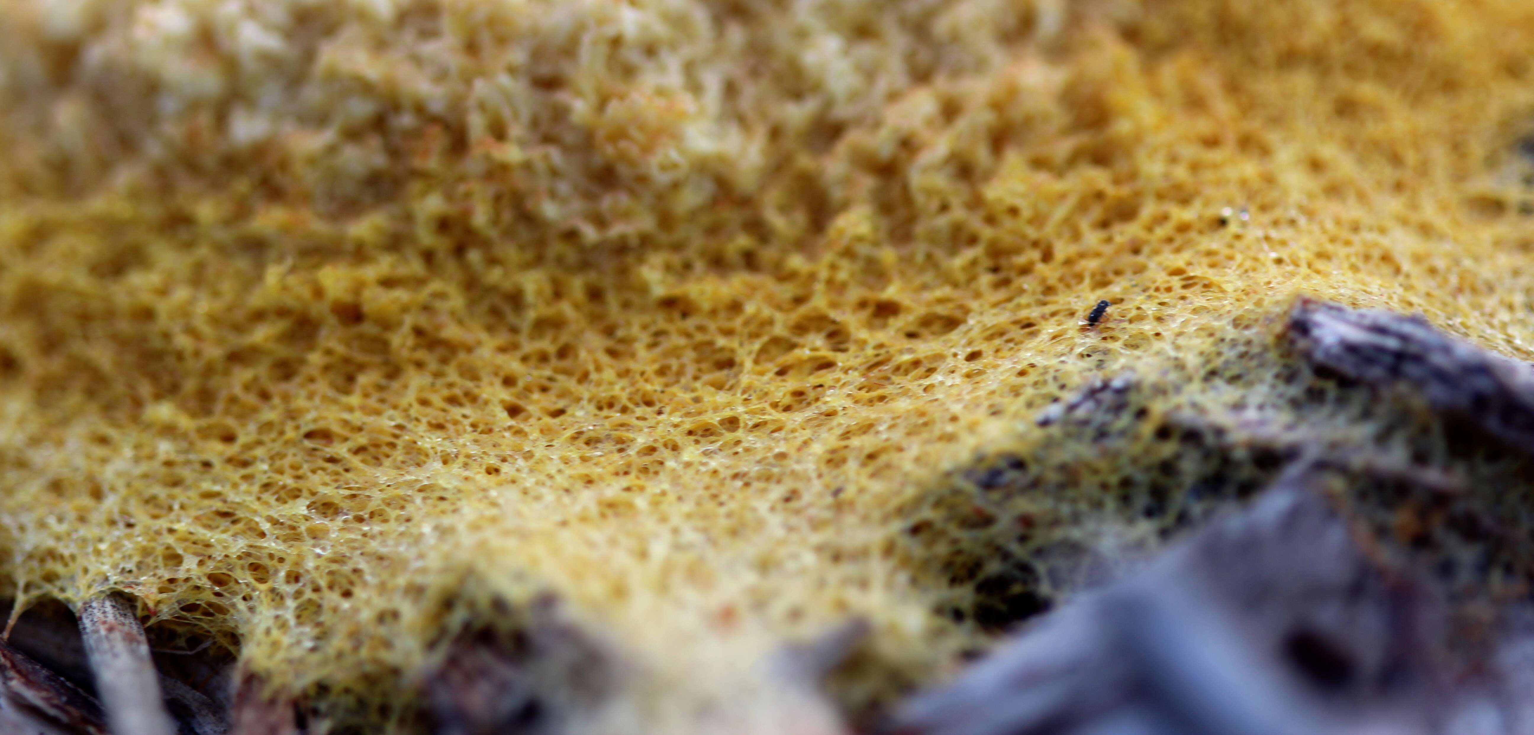
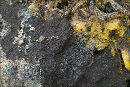
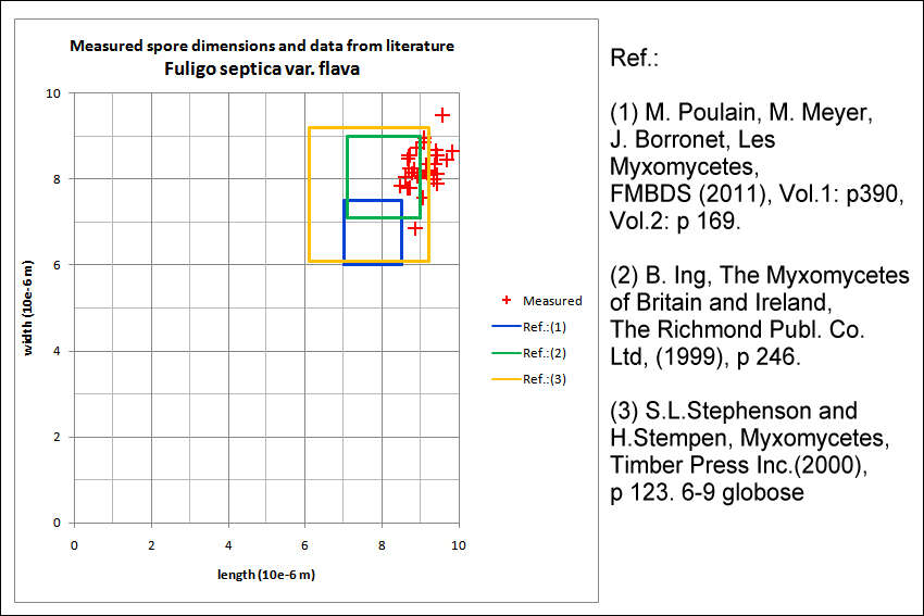
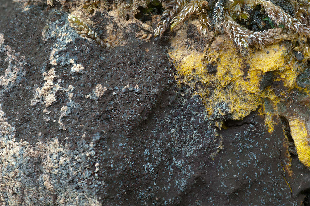
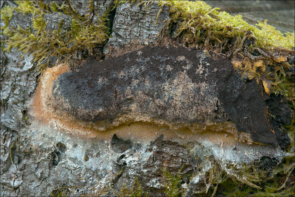
Fuligo septica var. flava (Pers.) Morgan, syn.: Mucor septicus L., Aethalium flavum (Pers) LinkFlowers of Tan, DE: Gelbe LohblteSlo.: reslov cvet, rumeni razliekDat.: Nov. 02. 2016Lat.: 46.35977 Long.: 13.70137Code: Bot_1025/2016_DSC6323Habitat: mixed wood, Fagus sylvatica and Picea abies dominant, moderately southeast inclined mountain slope, shallow, skeletal calcareous ground, old overgrown slope and moraine scree with larger rocks and boulders, in shade, relatively warm place, partly protected from direct rain by tree canopies, average precipitation ~ 3.000 mm/year, average temperature 7 - 9 deg C, elevation 600 m (1.970 feet), alpine phytogeographical region. Substratum: debarked roots of dead Picea abies, still standing.Place: Lower Trenta valley, between villages Soa and Trenta, next to the trail from Trenta 2b cottage to abandoned farmhouse 'Strgulc', East Julian Alps, Posoje, Slovenia EC.Comment: Fuligo septica is probably the most common and widely known Myxomicete. One of its varieties - Fuligo septica var. flava - has vivid yellow aethalia and yellow plasmodium (I haven't seen it this time). Yet, this typical color pertains only to mature but still fresh aethalia (pictures 10-13). With time cortex becomes darker, almost golden (picture 14), then black inside becomes visible (pictures 15, 16). In next phase cortex become whitish (pictures 17, 18) and finally it disappears and only blackish-reddish-brown spore mass is seen (pictures 19, 20). It is interesting that this find was found only a few meters away of the site where I photographed the same species two years ago (see Album Fuligo septica var. flava - I, Dec. 2014) (see MO observations #194527). In both observations spores are unusually large.Spores minutely warty, globose to subglobose. Dimensions: 8,5 [9,1 ; 9,4] 10 x 8 [8,6 ; 8,8] 9,4 microns; Q = [1 ; 1,08] 1,1; N = 35; C = 95%; Me = 9,2 x 8,7 microns; Qe = 1,1. Olympus CH20, NEA 100x/1.25, magnification 1.000 x, oil, in water; fresh material. AmScope MA500 digital camera.Herbarium: Mycotheca and lichen herbarium (LJU-Li) of Slovenian Forestry Institute, Vena pot 2, Ljubljana, Index Herbariorum LJFRef.:(1) M. Poulain, M. Meyer, J. Borronet, Les Myxomycetes, FMBDS (2011), Vol.1: p390, Vol.2: p 169.(2) B. Ing, The Myxomycetes of Britain and Ireland, The Richmond Publ. Co.Ltd, (1999), p 246. (3) S.L.Stephenson and H.Stempen, Myxomycetes, Timber Press Inc.(2000), p 123.
-
Ballan, Victoria, Australia
-

Fuligo septica var. flava (Pers.) Morgan, syn.: Mucor septicus L., Aethalium flavum (Pers) LinkFlowers of Tan, DE: Gelbe LohblteSlo.: reslov cvet, rumeni razliekDat.: Sept. 05. 2014Lat.: 46.35965 Long.: 13.70116Code: Bot_832/2014_DSC3621Habitat: mixed wood, Fagus sylvatica and Picea abies dominant, moderately southeast inclined mountain slope, shallow, skeletal calcareous ground, old overgrown slope and moraine scree with larger rocks and boulders, in shade, relatively warm place, partly protected from direct rain by tree canopies, average precipitation ~ 3.000 mm/year, average temperature 7 - 9 deg C, elevation 600 m (1.970 feet), alpine phytogeographical region. Substratum: debarked trunk of Picea abies lying on ground in its late disintegration stage.Place: Lower Trenta valley, between villages Soa and Trenta, next to the trail from Trenta 2b cottage to abandoned farmhouse 'Strgulc', East Julian Alps, Posoje, Slovenia EC.Comment: Fuligo septica is probably the most common and widely known Myxomicete. The latest monograph on Myxomycetes I have (Ref.:1) describes six varieties of this species, which differ mostly in cortex structure (single versus double layered) and color of different parts of sporocarp and plasmodium. Fuligo septica var. flava should have vivid yellow aethalia, yellow inner lime and yellow plasmodium. Two days before I took these pictures I had seen the plasmodium, which was in a form of vividly yellow colored patch of densely packed small half-spheres. The rest of traits of Fuligo septica var. flava also fit well to my observation.Spores minutely warty, globose to subglobose. Dimensions: 8 [8,4 ; 8,7] 9,1 x 7,4 [8 ; 8,2] 8,7 microns; Q = [1 ; 1,07] 1,1; N = 25; C = 95%; Me = 8,5 x 8,1 microns; Qe = 1,1. Olympus CH20, NEA 100x/1.25, magnification 1.000 x, oil (spores), NEA 40x/0.65, magnification 400x (capillitium, calcareous granules); in water; live material. AmScope MA500 digital camera.Herbarium: Mycotheca and lichen herbarium (LJU-Li) of Slovenian Forestry Institute, Vena pot 2, Ljubljana, Index Herbariorum LJFRef.:(1) M. Poulain, M. Meyer, J. Borronet, Les Myxomycetes, FMBDS (2011), Vol.1: p390, Vol.2: (2) B. Ing, The Myxomycetes of Britain and Ireland, The Richmond Publ. Co.Ltd, (1999), p 246. (3) S.L.Stephenson and H.Stempen, Myxomycetes, Timber Press Inc.(2000), p 123.
-
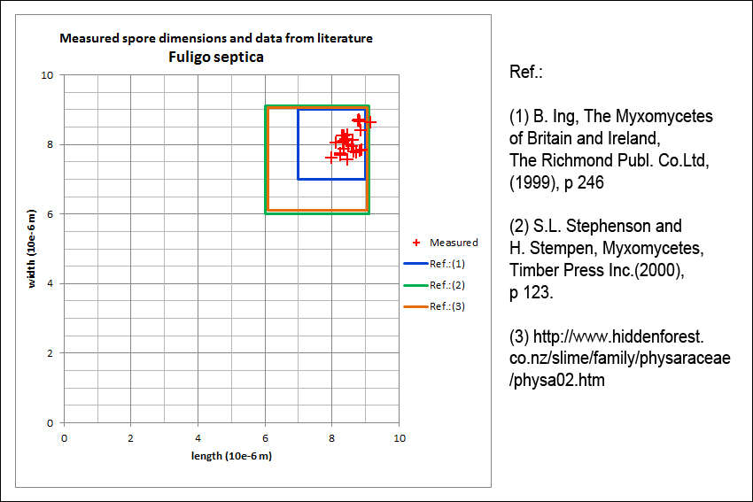
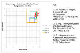
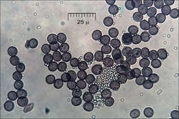
Fuligo septica (L.)Wigg., syn. Mucorsepticus L., Reticularia septica (L.) With., Aethalium septicum (L.) Fr., Fuligo varians Sommerf.Scrambled-egg slime, Dog vomit slime mold, Flowers of Tan, DE: Gelbe Lohblte, HexenbutterSlo.: reslov cvetSpore statistics and comparison with data from literature. (Fig. 2M).Dat.: July 21. 2014Code: Bot_815/2014_DSC2009 Lat.: 46.36114 Long.: 13.70122Habitat: old partly tree overgrown pasture, near mixed wood edge; moderately southeast inclined foot of an old overgrown scree slope; open, dry, sunny place; shallow, skeletal, calcareous ground, exposed to direct rain, average precipitations ~ 3.000 mm/year, average temperature 7-9 deg C, elevation 630 m (2.070 feet), alpine phytogeographical region. Substratum: a stump of Picea abies cut down three years ago.Place: Lower Trenta valley, between villages Soa and Trenta, upper part of 'Na Melu' place, East Julian Alps, Posoje, Slovenia EC.Comment: Myxomycetes are poorly known yet very interesting creatures. For decades they have been shuffled back and forth between the animal and plants kingdoms until recognized as separate creatures. They are not animals because they proliferate by spores. They are also not plants since they crumble around (an animal like ability) and fix themselves firmly to substrate only at the end of their life cycle. They don't produce their own food like plants but feed by 'hunting' (actually engulfing) bacteria and tiny bits of other organic matter, which is another animal like feature. The first stage in their development cycle, which is observable in the field, is called plasmodium. Earlier stages (from myxoflagellates, myxoamoebae, to zygote) are microscopic and can be observed only in labs. Plasmodium is a single giant living cell, a clump of protoplasm filled with thousands of cell nuclei, crawling around, eating bacteria and growing. Some of such plasmodia are the largest cells of living creatures known. In some species they can measure several meters across or weight up to 20 kg. That it much larger than ostrich's eggs, which are popularly considered as 'largest living cells'! Plasmodia could be found in the field, some are even brightly colored and easy to spot; however, it is almost impossible to determine to which species they belong. Plasmodium of Fuligo septica is commonly described like disgusting mucus, spilled scrambled eggs, dogs vomiting and other 'benevolent' portrayals.When the time is right or delicate environmental conditions required for growth worsen plasmodia eventually evolves (usually almost completely) into sporocarps of different forms. These are bodies producing spores and then vanishing. In genus Fuligo sporocarp is a cushion like aethalium sitting on a thin whitish, 'fibrous' layer called hypothallus (Fig.14.). These'cushions' are what one usually finds in the field. But, other Myxomycetes develop also many other forms of sporocarps full of beauty, delicacy and imagination. Aethalia of Fuligo septica are usually covered with a kind of crust called cortex, which is brittle and soon crumbles away. In humid conditions it may not fully develop (Ref.1). Inside a mature aethalium there is a mesh of thin tubes or fibers called capillitium and zillions of dark brown spores. Fuligo septica has characteristic nodes on capillitial tubes, which are clearly seen on Fig. 4M. In due course the aethalium decomposes almost entirely into spore mass (Fig.17., 18.), which are sooner or later blown or washed away (Fig.20. taken about three weeks after the first photo). Size and shape of spores and structure of their surface are important traits for species determination. Cushion-shaped aethalium measured approximately 14 x 5 cm and was about 3 cm thick. Spores are minutely warty and globose to subglobose. Dimensions: 8 [8,4 ; 8,7] 9,1 x 7,4 [8 ; 8,2] 8,7 microns; Q = [1 ; 1,07] 1,1; N = 25; C = 95%; Me = 8,5 x 8,1 microns; Qe = 1,05. Olympus CH20, NEA 100x/1.25, magnification 1.000 x, oil (spores), NEA 40x/0.65, magnification 400x (capillitium), NEA 10x/0.25, magnification 100x (hypothallus); in water, living material. AmScope MA500 digital camera. Spore sample taken on July 23. 2014.Herbarium: Mycotheca and lichen herbarium (LJU-Li) of Slovenian Forestry Institute, Vena pot 2, Ljubljana, Index Herbariorum LJFRef.:(1) B. Ing, The Myxomycetes of Britain and Ireland, The Richmond Publ. Co.Ltd, (1999), p 246(2) S.L.Stephenson and H.Stempen, Myxomycetes, Timber Press Inc.(2000), p 123(3)
www.hiddenforest.co.nz/slime/family/physaraceae/physa02.htm 6-9(4) M. Poulain, M. Meyer, J. Bozzonet, Les Myxomycetes, FMBDS (2011), Vol.1., p128; Vol.2., p168.
-

Fuligo septica var. flava (Pers.) Morgan, syn.: Mucor septicus L., Aethalium flavum (Pers) LinkFlowers of Tan, DE: Gelbe LohblteSlo.: reslov cvet, rumeni razliekDat.: Oct. 26. 2016Lat.: 46.35977 Long.: 13.70137Code: Bot_1022/2016_DSC6083Habitat: mixed wood, Fagus sylvatica and Picea abies dominant, moderately southeast inclined mountain slope, shallow, skeletal calcareous ground, old overgrown slope and moraine scree with larger rocks and boulders, in shade, relatively warm place, partly protected from direct rain by tree canopies, average precipitation ~ 3.000 mm/year, average temperature 7 - 9 deg C, elevation 600 m (1.970 feet), alpine phytogeographical region. Substratum: debarked roots of dead Picea abies, still standing.Place: Lower Trenta valley, between villages Soa and Trenta, next to the trail from Trenta 2b cottage to abandoned farmhouse 'Strgulc', East Julian Alps, Posoje, Slovenia EC.Comment: Fuligo septica is probably the most common and widely known Myxomicete. One of its varieties - Fuligo septica var. flava - has vivid yellow aethalia and yellow plasmodium (I haven't seen it this time). Yet, this typical color pertains only to mature but still fresh aethalia (pictures 10-13). With time cortex becomes darker, almost golden (picture 14), then black inside becomes visible (pictures 15, 16). In next phase cortex become whitish (pictures 17, 18) and finally it disappears and only blackish-reddish-brown spore mass is seen (pictures 19, 20). It is interesting that this find was found only a few meters away of the site where I photographed the same species two years ago (see Album Fuligo septica var. flava - I, Dec. 2014) (see MO observations #194527). In both observations spores are unusually large.Spores minutely warty, globose to subglobose. Dimensions: 8,5 [9,1 ; 9,4] 10 x 8 [8,6 ; 8,8] 9,4 microns; Q = [1 ; 1,08] 1,1; N = 35; C = 95%; Me = 9,2 x 8,7 microns; Qe = 1,1. Olympus CH20, NEA 100x/1.25, magnification 1.000 x, oil, in water; fresh material. AmScope MA500 digital camera.Herbarium: Mycotheca and lichen herbarium (LJU-Li) of Slovenian Forestry Institute, Vena pot 2, Ljubljana, Index Herbariorum LJFRef.:(1) M. Poulain, M. Meyer, J. Borronet, Les Myxomycetes, FMBDS (2011), Vol.1: p390, Vol.2: p 169.(2) B. Ing, The Myxomycetes of Britain and Ireland, The Richmond Publ. Co.Ltd, (1999), p 246. (3) S.L.Stephenson and H.Stempen, Myxomycetes, Timber Press Inc.(2000), p 123.
-
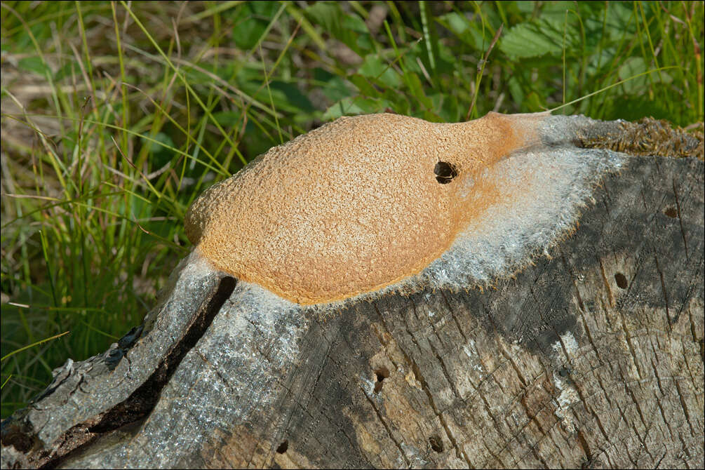
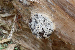
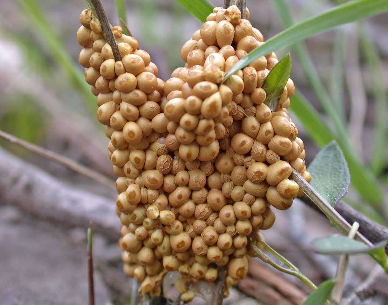
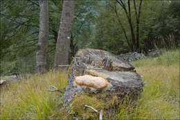
Fuligo septica (L.) Wigg., syn. Mucorsepticus L., Reticularia septica (L.) With., Aethalium septicum (L.) Fr., Fuligo varians Sommerf.Scrambled-egg slime, Dog vomit slime mold, Flowers of Tan, DE: Gelbe Lohblte, HexenbutterSlo.: reslov cvetDat.: July 18. 2014Lat.: 46.36113 Long.: 13.70122Code: Bot_814/2014_DSC1863 Habitat: old partly overgrown pasture, near mixed wood edge, moderately southeast inclined foot of a mountain; open, dry, sunny place; shallow, skeletal, calcareous ground, old overgrown scree slope; exposed to direct rain, average precipitations ~ 3.000 mm/year, average temperature 7-9 deg C, elevation 630 m (2.070 feet), alpine phytogeographical region. Substratum: stump of Picea abies cut down three years ago.Place: Lower Trenta valley, between villages Soa and Trenta, upper part of 'Na melu' place, East Julian Alps, Posoje, Slovenia ECComments: It is interesting to follow how aethalium was developing and decaying over almost one month period. 'Fibrous' layer called hypothallus is shown on picture 3. Cushion-shaped aethalium measured approximately 12 x 4 cm and was about 2 cm thick (when first photographed). I found six such aethalia this day on three stumps in only a few meters distance.Herbarium: Mycotheca and lichen herbarium (LJU-Li) of Slovenian Forestry Institute, Vena pot 2, Ljubljana, Index Herbariorum LJFRef.:(1) B. Ing, The Myxomycetes of Britain and Ireland, The Richmond Publ. Co.Ltd, (1999), p 246.(2) S.L.Stephenson and H.Stempen, Myxomycetes, Timber Press Inc.(2000), p 123.(3)
www.hiddenforest.co.nz/slime/family/physaraceae/physa02.htm
-
Fuligo septica var. flava (Pers.) Morgan, syn.: Mucor septicus L., Aethalium flavum (Pers) LinkFlowers of Tan, DE: Gelbe LohblteSlo.: reslov cvet, rumeni razliekDat.: Sept. 05. 2014Lat.: 46.35965 Long.: 13.70116Code: Bot_832/2014_DSC3621Habitat: mixed wood, Fagus sylvatica and Picea abies dominant, moderately southeast inclined mountain slope, shallow, skeletal calcareous ground, old overgrown slope and moraine scree with larger rocks and boulders, in shade, relatively warm place, partly protected from direct rain by tree canopies, average precipitation ~ 3.000 mm/year, average temperature 7 - 9 deg C, elevation 600 m (1.970 feet), alpine phytogeographical region. Substratum: debarked trunk of Picea abies lying on ground in its late disintegration stage.Place: Lower Trenta valley, between villages Soa and Trenta, next to the trail from Trenta 2b cottage to abandoned farmhouse 'Strgulc', East Julian Alps, Posoje, Slovenia EC.Comment: Fuligo septica is probably the most common and widely known Myxomicete. The latest monograph on Myxomycetes I have (Ref.:1) describes six varieties of this species, which differ mostly in cortex structure (single versus double layered) and color of different parts of sporocarp and plasmodium. Fuligo septica var. flava should have vivid yellow aethalia, yellow inner lime and yellow plasmodium. Two days before I took these pictures I had seen the plasmodium, which was in a form of vividly yellow colored patch of densely packed small half-spheres. The rest of traits of Fuligo septica var. flava also fit well to my observation.Spores minutely warty, globose to subglobose. Dimensions: 8 [8,4 ; 8,7] 9,1 x 7,4 [8 ; 8,2] 8,7 microns; Q = [1 ; 1,07] 1,1; N = 25; C = 95%; Me = 8,5 x 8,1 microns; Qe = 1,1. Olympus CH20, NEA 100x/1.25, magnification 1.000 x, oil (spores), NEA 40x/0.65, magnification 400x (capillitium, calcareous granules); in water; live material. AmScope MA500 digital camera.Herbarium: Mycotheca and lichen herbarium (LJU-Li) of Slovenian Forestry Institute, Vena pot 2, Ljubljana, Index Herbariorum LJFRef.:(1) M. Poulain, M. Meyer, J. Borronet, Les Myxomycetes, FMBDS (2011), Vol.1: p390, Vol.2: (2) B. Ing, The Myxomycetes of Britain and Ireland, The Richmond Publ. Co.Ltd, (1999), p 246. (3) S.L.Stephenson and H.Stempen, Myxomycetes, Timber Press Inc.(2000), p 123.
-
Fuligo septica (L.)Wigg., syn. Mucorsepticus L., Reticularia septica (L.) With., Aethalium septicum (L.) Fr., Fuligo varians Sommerf.Scrambled-egg slime, Dog vomit slime mold, Flowers of Tan, DE: Gelbe Lohblte, HexenbutterSlo.: reslov cvetThe aethalium after two days. Dat.: July 23. 2014Code: Bot_816/2014_DSC2074Lat.: 46.36114 Long.: 13.70122Habitat: old partly tree overgrown pasture, near mixed wood edge; moderately southeast inclined foot of an old overgrown scree slope; open, dry, sunny place; shallow, skeletal, calcareous ground, exposed to direct rain, average precipitations ~ 3.000 mm/year, average temperature 7-9 deg C, elevation 630 m (2.070 feet), alpine phytogeographical region. Substratum: a stump of Picea abies cut down three years ago.Place: Lower Trenta valley, between villages Soa and Trenta, upper part of 'Na Melu' place, East Julian Alps, Posoje, Slovenia EC.Comment: Myxomycetes are poorly known yet very interesting creatures. For decades they have been shuffled back and forth between the animal and plants kingdoms until recognized as separate creatures. They are not animals because they proliferate by spores. They are also not plants since they crumble around (an animal like ability) and fix themselves firmly to substrate only at the end of their life cycle. They don't produce their own food like plants but feed by 'hunting' (actually engulfing) bacteria and tiny bits of other organic matter, which is another animal like feature. The first stage in their development cycle, which is observable in the field, is called plasmodium. Earlier stages (from myxoflagellates, myxoamoebae, to zygote) are microscopic and can be observed only in labs. Plasmodium is a single giant living cell, a clump of protoplasm filled with thousands of cell nuclei, crawling around, eating bacteria and growing. Some of such plasmodia are the largest cells of living creatures known. In some species they can measure several meters across or weight up to 20 kg. That it much larger than ostrich's eggs, which are popularly considered as 'largest living cells'! Plasmodia could be found in the field, some are even brightly colored and easy to spot; however, it is almost impossible to determine to which species they belong. Plasmodium of Fuligo septica is commonly described like disgusting mucus, spilled scrambled eggs, dogs vomiting and other 'benevolent' portrayals.When the time is right or delicate environmental conditions required for growth worsen plasmodia eventually evolves (usually almost completely) into sporocarps of different forms. These are bodies producing spores and then vanishing. In genus Fuligo sporocarp is a cushion like aethalium sitting on a thin whitish, 'fibrous' layer called hypothallus (Fig.14.). These'cushions' are what one usually finds in the field. But, other Myxomycetes develop also many other forms of sporocarps full of beauty, delicacy and imagination. Aethalia of Fuligo septica are usually covered with a kind of crust called cortex, which is brittle and soon crumbles away. In humid conditions it may not fully develop (Ref.1). Inside a mature aethalium there is a mesh of thin tubes or fibers called capillitium and zillions of dark brown spores. Fuligo septica has characteristic nodes on capillitial tubes, which are clearly seen on Fig. 4M. In due course the aethalium decomposes almost entirely into spore mass (Fig.17., 18.), which are sooner or later blown or washed away (Fig.20. taken about three weeks after the first photo). Size and shape of spores and structure of their surface are important traits for species determination. Cushion-shaped aethalium measured approximately 14 x 5 cm and was about 3 cm thick. Spores are minutely warty and globose to subglobose. Dimensions: 8 [8,4 ; 8,7] 9,1 x 7,4 [8 ; 8,2] 8,7 microns; Q = [1 ; 1,07] 1,1; N = 25; C = 95%; Me = 8,5 x 8,1 microns; Qe = 1,05. Olympus CH20, NEA 100x/1.25, magnification 1.000 x, oil (spores), NEA 40x/0.65, magnification 400x (capillitium), NEA 10x/0.25, magnification 100x (hypothallus); in water, living material. AmScope MA500 digital camera. Spore sample taken on July 23. 2014.Herbarium: Mycotheca and lichen herbarium (LJU-Li) of Slovenian Forestry Institute, Vena pot 2, Ljubljana, Index Herbariorum LJFRef.:(1) B. Ing, The Myxomycetes of Britain and Ireland, The Richmond Publ. Co.Ltd, (1999), p 246(2) S.L.Stephenson and H.Stempen, Myxomycetes, Timber Press Inc.(2000), p 123(3)
www.hiddenforest.co.nz/slime/family/physaraceae/physa02.htm 6-9(4) M. Poulain, M. Meyer, J. Bozzonet, Les Myxomycetes, FMBDS (2011), Vol.1., p128; Vol.2., p168.
-
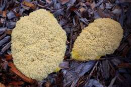
Fuligo septica var. flava (Pers.) Morgan, syn.: Mucor septicus L., Aethalium flavum (Pers) LinkFlowers of Tan, DE: Gelbe LohblteSlo.: reslov cvet, rumeni razliekDat.: Oct. 18. 2016Lat.: 46.35977 Long.: 13.70137Code: Bot_1020/2016_DSC5862Habitat: mixed wood, Fagus sylvatica and Picea abies dominant, moderately southeast inclined mountain slope, shallow, skeletal calcareous ground, old overgrown slope and moraine scree with larger rocks and boulders, in shade, relatively warm place, partly protected from direct rain by tree canopies, average precipitation ~ 3.000 mm/year, average temperature 7 - 9 deg C, elevation 600 m (1.970 feet), alpine phytogeographical region. Substratum: debarked roots of dead Picea abies, still standing.Place: Lower Trenta valley, between villages Soa and Trenta, next to the trail from Trenta 2b cottage to abandoned farmhouse 'Strgulc', East Julian Alps, Posoje, Slovenia EC.Comment: Fuligo septica is probably the most common and widely known Myxomicete. One of its varieties - Fuligo septica var. flava - has vivid yellow aethalia and yellow plasmodium (I haven't seen it this time). Yet, this typical color pertains only to mature but still fresh aethalia (pictures 10-13). With time cortex becomes darker, almost golden (picture 14), then black inside becomes visible (pictures 15, 16). In next phase cortex become whitish (pictures 17, 18) and finally it disappears and only blackish-reddish-brown spore mass is seen (pictures 19, 20). It is interesting that this find was found only a few meters away of the site where I photographed the same species two years ago (see Album Fuligo septica var. flava - I, Dec. 2014) (see MO observations #194527). In both observations spores are unusually large.Spores minutely warty, globose to subglobose. Dimensions: 8,5 [9,1 ; 9,4] 10 x 8 [8,6 ; 8,8] 9,4 microns; Q = [1 ; 1,08] 1,1; N = 35; C = 95%; Me = 9,2 x 8,7 microns; Qe = 1,1. Olympus CH20, NEA 100x/1.25, magnification 1.000 x, oil, in water; fresh material. AmScope MA500 digital camera.Herbarium: Mycotheca and lichen herbarium (LJU-Li) of Slovenian Forestry Institute, Vena pot 2, Ljubljana, Index Herbariorum LJFRef.:(1) M. Poulain, M. Meyer, J. Borronet, Les Myxomycetes, FMBDS (2011), Vol.1: p390, Vol.2: p 169.(2) B. Ing, The Myxomycetes of Britain and Ireland, The Richmond Publ. Co.Ltd, (1999), p 246. (3) S.L.Stephenson and H.Stempen, Myxomycetes, Timber Press Inc.(2000), p 123.
-
Castel Fusano, Lazio, Italy
-
Fuligo septica var. flava (Pers.) Morgan, syn.: Mucor septicus L., Aethalium flavum (Pers) LinkFlowers of Tan, DE: Gelbe LohblteSlo.: reslov cvet, rumeni razliekDat.: Sept. 05. 2014Lat.: 46.35965 Long.: 13.70116Code: Bot_832/2014_DSC3621Habitat: mixed wood, Fagus sylvatica and Picea abies dominant, moderately southeast inclined mountain slope, shallow, skeletal calcareous ground, old overgrown slope and moraine scree with larger rocks and boulders, in shade, relatively warm place, partly protected from direct rain by tree canopies, average precipitation ~ 3.000 mm/year, average temperature 7 - 9 deg C, elevation 600 m (1.970 feet), alpine phytogeographical region. Substratum: debarked trunk of Picea abies lying on ground in its late disintegration stage.Place: Lower Trenta valley, between villages Soa and Trenta, next to the trail from Trenta 2b cottage to abandoned farmhouse 'Strgulc', East Julian Alps, Posoje, Slovenia EC.Comment: Fuligo septica is probably the most common and widely known Myxomicete. The latest monograph on Myxomycetes I have (Ref.:1) describes six varieties of this species, which differ mostly in cortex structure (single versus double layered) and color of different parts of sporocarp and plasmodium. Fuligo septica var. flava should have vivid yellow aethalia, yellow inner lime and yellow plasmodium. Two days before I took these pictures I had seen the plasmodium, which was in a form of vividly yellow colored patch of densely packed small half-spheres. The rest of traits of Fuligo septica var. flava also fit well to my observation.Spores minutely warty, globose to subglobose. Dimensions: 8 [8,4 ; 8,7] 9,1 x 7,4 [8 ; 8,2] 8,7 microns; Q = [1 ; 1,07] 1,1; N = 25; C = 95%; Me = 8,5 x 8,1 microns; Qe = 1,1. Olympus CH20, NEA 100x/1.25, magnification 1.000 x, oil (spores), NEA 40x/0.65, magnification 400x (capillitium, calcareous granules); in water; live material. AmScope MA500 digital camera.Herbarium: Mycotheca and lichen herbarium (LJU-Li) of Slovenian Forestry Institute, Vena pot 2, Ljubljana, Index Herbariorum LJFRef.:(1) M. Poulain, M. Meyer, J. Borronet, Les Myxomycetes, FMBDS (2011), Vol.1: p390, Vol.2: (2) B. Ing, The Myxomycetes of Britain and Ireland, The Richmond Publ. Co.Ltd, (1999), p 246. (3) S.L.Stephenson and H.Stempen, Myxomycetes, Timber Press Inc.(2000), p 123.
-
Castel Fusano, Lazio, Italy
-
Fuligo septica var. flava (Pers.) Morgan, syn.: Mucor septicus L., Aethalium flavum (Pers) LinkFlowers of Tan, DE: Gelbe LohblteSlo.: reslov cvet, rumeni razliekDat.: Oct. 17. 2016Lat.: 46.35977 Long.: 13.70137Code:Bot_1019/2016_DSC5774Habitat: mixed wood, Fagus sylvatica and Picea abies dominant, moderately southeast inclined mountain slope, shallow, skeletal calcareous ground, old overgrown slope and moraine scree with larger rocks and boulders, in shade, relatively warm place, partly protected from direct rain by tree canopies, average precipitation ~ 3.000 mm/year, average temperature 7 - 9 deg C, elevation 600 m (1.970 feet), alpine phytogeographical region. Substratum: debarked roots of dead Picea abies, still standing.Place: Lower Trenta valley, between villages Soa and Trenta, next to the trail from Trenta 2b cottage to abandoned farmhouse 'Strgulc', East Julian Alps, Posoje, Slovenia EC.Comment: Fuligo septica is probably the most common and widely known Myxomicete. One of its varieties - Fuligo septica var. flava - has vivid yellow aethalia and yellow plasmodium (I haven't seen it this time). Yet, this typical color pertains only to mature but still fresh aethalia (pictures 10-13). With time cortex becomes darker, almost golden (picture 14), then black inside becomes visible (pictures 15, 16). In next phase cortex become whitish (pictures 17, 18) and finally it disappears and only blackish-reddish-brown spore mass is seen (pictures 19, 20). It is interesting that this find was found only a few meters away of the site where I photographed the same species two years ago (see Album Fuligo septica var. flava - I, Dec. 2014) (see MO observations #194527). In both observations spores are unusually large.Spores minutely warty, globose to subglobose. Dimensions: 8,5 [9,1 ; 9,4] 10 x 8 [8,6 ; 8,8] 9,4 microns; Q = [1 ; 1,08] 1,1; N = 35; C = 95%; Me = 9,2 x 8,7 microns; Qe = 1,1. Olympus CH20, NEA 100x/1.25, magnification 1.000 x, oil, in water; fresh material. AmScope MA500 digital camera.Herbarium: Mycotheca and lichen herbarium (LJU-Li) of Slovenian Forestry Institute, Vena pot 2, Ljubljana, Index Herbariorum LJFRef.:(1) M. Poulain, M. Meyer, J. Borronet, Les Myxomycetes, FMBDS (2011), Vol.1: p390, Vol.2: p 169.(2) B. Ing, The Myxomycetes of Britain and Ireland, The Richmond Publ. Co.Ltd, (1999), p 246. (3) S.L.Stephenson and H.Stempen, Myxomycetes, Timber Press Inc.(2000), p 123.
-
Ballan, Victoria, Australia
-
Fuligo septica var. flava (Pers.) Morgan, syn.: Mucor septicus L., Aethalium flavum (Pers) LinkFlowers of Tan, DE: Gelbe LohblteSlo.: reslov cvet, rumeni razliekDat.: Oct. 17. 2016Lat.: 46.35977 Long.: 13.70137Code:Bot_1019/2016_DSC5774Habitat: mixed wood, Fagus sylvatica and Picea abies dominant, moderately southeast inclined mountain slope, shallow, skeletal calcareous ground, old overgrown slope and moraine scree with larger rocks and boulders, in shade, relatively warm place, partly protected from direct rain by tree canopies, average precipitation ~ 3.000 mm/year, average temperature 7 - 9 deg C, elevation 600 m (1.970 feet), alpine phytogeographical region. Substratum: debarked roots of dead Picea abies, still standing.Place: Lower Trenta valley, between villages Soa and Trenta, next to the trail from Trenta 2b cottage to abandoned farmhouse 'Strgulc', East Julian Alps, Posoje, Slovenia EC.Comment: Fuligo septica is probably the most common and widely known Myxomicete. One of its varieties - Fuligo septica var. flava - has vivid yellow aethalia and yellow plasmodium (I haven't seen it this time). Yet, this typical color pertains only to mature but still fresh aethalia (pictures 10-13). With time cortex becomes darker, almost golden (picture 14), then black inside becomes visible (pictures 15, 16). In next phase cortex become whitish (pictures 17, 18) and finally it disappears and only blackish-reddish-brown spore mass is seen (pictures 19, 20). It is interesting that this find was found only a few meters away of the site where I photographed the same species two years ago (see Album Fuligo septica var. flava - I, Dec. 2014) (see MO observations #194527). In both observations spores are unusually large.Spores minutely warty, globose to subglobose. Dimensions: 8,5 [9,1 ; 9,4] 10 x 8 [8,6 ; 8,8] 9,4 microns; Q = [1 ; 1,08] 1,1; N = 35; C = 95%; Me = 9,2 x 8,7 microns; Qe = 1,1. Olympus CH20, NEA 100x/1.25, magnification 1.000 x, oil, in water; fresh material. AmScope MA500 digital camera.Herbarium: Mycotheca and lichen herbarium (LJU-Li) of Slovenian Forestry Institute, Vena pot 2, Ljubljana, Index Herbariorum LJFRef.:(1) M. Poulain, M. Meyer, J. Borronet, Les Myxomycetes, FMBDS (2011), Vol.1: p390, Vol.2: p 169.(2) B. Ing, The Myxomycetes of Britain and Ireland, The Richmond Publ. Co.Ltd, (1999), p 246. (3) S.L.Stephenson and H.Stempen, Myxomycetes, Timber Press Inc.(2000), p 123.
-
Fuligo septica (L.)Wigg., syn. Mucorsepticus L., Reticularia septica (L.) With., Aethalium septicum (L.) Fr., Fuligo varians Sommerf.Scrambled-egg slime, Dog vomit slime mold, Flowers of Tan, DE: Gelbe Lohblte, HexenbutterSlo.: reslov cvetThe aethalium after two days. Dat.: July 23. 2014Code: Bot_816/2014_DSC2074Lat.: 46.36114 Long.: 13.70122Habitat: old partly tree overgrown pasture, near mixed wood edge; moderately southeast inclined foot of an old overgrown scree slope; open, dry, sunny place; shallow, skeletal, calcareous ground, exposed to direct rain, average precipitations ~ 3.000 mm/year, average temperature 7-9 deg C, elevation 630 m (2.070 feet), alpine phytogeographical region. Substratum: a stump of Picea abies cut down three years ago.Place: Lower Trenta valley, between villages Soa and Trenta, upper part of 'Na Melu' place, East Julian Alps, Posoje, Slovenia EC.Comment: Myxomycetes are poorly known yet very interesting creatures. For decades they have been shuffled back and forth between the animal and plants kingdoms until recognized as separate creatures. They are not animals because they proliferate by spores. They are also not plants since they crumble around (an animal like ability) and fix themselves firmly to substrate only at the end of their life cycle. They don't produce their own food like plants but feed by 'hunting' (actually engulfing) bacteria and tiny bits of other organic matter, which is another animal like feature. The first stage in their development cycle, which is observable in the field, is called plasmodium. Earlier stages (from myxoflagellates, myxoamoebae, to zygote) are microscopic and can be observed only in labs. Plasmodium is a single giant living cell, a clump of protoplasm filled with thousands of cell nuclei, crawling around, eating bacteria and growing. Some of such plasmodia are the largest cells of living creatures known. In some species they can measure several meters across or weight up to 20 kg. That it much larger than ostrich's eggs, which are popularly considered as 'largest living cells'! Plasmodia could be found in the field, some are even brightly colored and easy to spot; however, it is almost impossible to determine to which species they belong. Plasmodium of Fuligo septica is commonly described like disgusting mucus, spilled scrambled eggs, dogs vomiting and other 'benevolent' portrayals.When the time is right or delicate environmental conditions required for growth worsen plasmodia eventually evolves (usually almost completely) into sporocarps of different forms. These are bodies producing spores and then vanishing. In genus Fuligo sporocarp is a cushion like aethalium sitting on a thin whitish, 'fibrous' layer called hypothallus (Fig.14.). These'cushions' are what one usually finds in the field. But, other Myxomycetes develop also many other forms of sporocarps full of beauty, delicacy and imagination. Aethalia of Fuligo septica are usually covered with a kind of crust called cortex, which is brittle and soon crumbles away. In humid conditions it may not fully develop (Ref.1). Inside a mature aethalium there is a mesh of thin tubes or fibers called capillitium and zillions of dark brown spores. Fuligo septica has characteristic nodes on capillitial tubes, which are clearly seen on Fig. 4M. In due course the aethalium decomposes almost entirely into spore mass (Fig.17., 18.), which are sooner or later blown or washed away (Fig.20. taken about three weeks after the first photo). Size and shape of spores and structure of their surface are important traits for species determination. Cushion-shaped aethalium measured approximately 14 x 5 cm and was about 3 cm thick. Spores are minutely warty and globose to subglobose. Dimensions: 8 [8,4 ; 8,7] 9,1 x 7,4 [8 ; 8,2] 8,7 microns; Q = [1 ; 1,07] 1,1; N = 25; C = 95%; Me = 8,5 x 8,1 microns; Qe = 1,05. Olympus CH20, NEA 100x/1.25, magnification 1.000 x, oil (spores), NEA 40x/0.65, magnification 400x (capillitium), NEA 10x/0.25, magnification 100x (hypothallus); in water, living material. AmScope MA500 digital camera. Spore sample taken on July 23. 2014.Herbarium: Mycotheca and lichen herbarium (LJU-Li) of Slovenian Forestry Institute, Vena pot 2, Ljubljana, Index Herbariorum LJFRef.:(1) B. Ing, The Myxomycetes of Britain and Ireland, The Richmond Publ. Co.Ltd, (1999), p 246(2) S.L.Stephenson and H.Stempen, Myxomycetes, Timber Press Inc.(2000), p 123(3)
www.hiddenforest.co.nz/slime/family/physaraceae/physa02.htm 6-9(4) M. Poulain, M. Meyer, J. Bozzonet, Les Myxomycetes, FMBDS (2011), Vol.1., p128; Vol.2., p168.