-
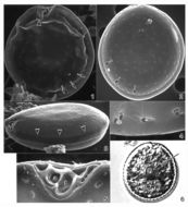
Fig 1: Schematic drawing of Prorocentrum lima
-
Exuviaella lima.
-
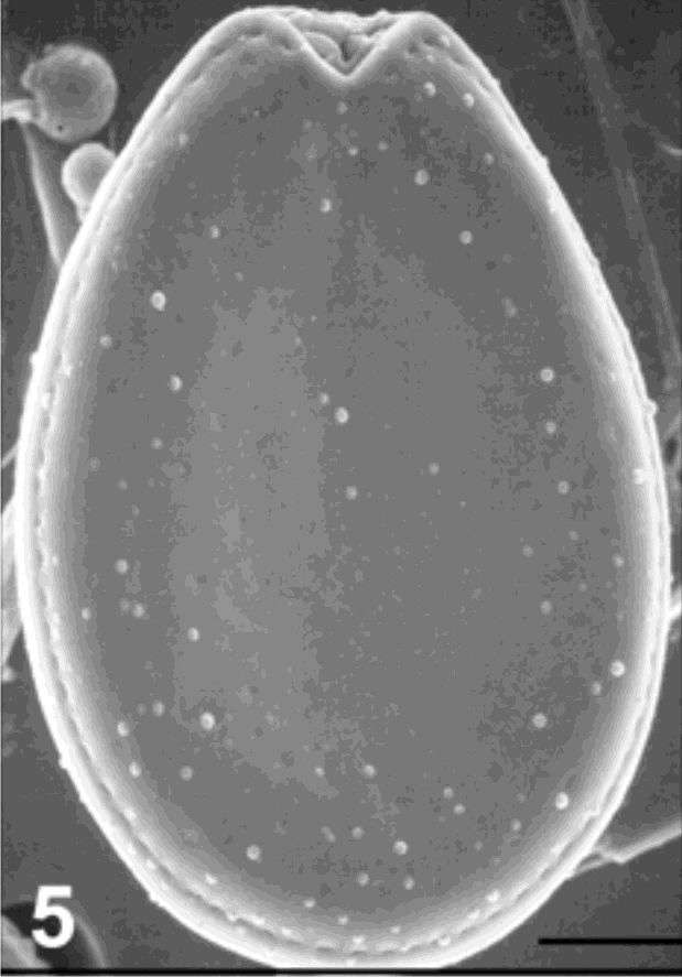
Fig 5:Prorocentrum lima SEM image of cell in right valve view
-
Fig 1: Prorocentrum emarginatum Schematic diagram (ventral view) redrawn from Tomas et al. 1997.
-
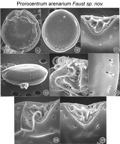
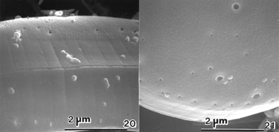
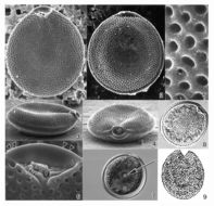
Figs. 14-19. Prorocentrum arenarium sp. nov. FIG.14. Cells in right valve view are round to oval. Cell distorted due to preparation. The periflagellar area is a broad, V-shaped depression. The longitudinal short flagellum is visible (arrowhead). Marginal poroids (arrows). FIG.15. The valve surface in left valve view is smooth and scattered with valve and marginal poroids (arrows). FIG. 6. The oblique ventral view is ellipsoid. The intercalary band is smooth. The marginal poroids are evenly spaced (arrows). FIG.17. The periflagellar area is triangular and unornamented. It has a large flagellar pore (F) and one smaller auxiliary pore (A). The apical platelets appear vertical when viewed from the anterior end of the cell. FIG. 18. Both flagella emerge from the flagellar pore (F): longitudinal flagellum (arrowheads), transverse flagellum (arrows). FIG. 9. At higher magnification, valve and marginal poroids are kidney-shaped to oblong (arrowheads) and are similar in size.FIGS. 20-21. Prorocentrum arenarium sp. nov. with peduncle-like structures. FIG. 20. The periflagellar area exhibits a short flagellum 11µm long (arrows), and a short, curved tubular structure 2-3 µm long (arrowheads), both emerging from the flagellar pore (F). The width of the curved tubular structure and flagellum are the same. FIG. 21. Both tubular structures emerge from the flagellar pore (F); one is straight, 2 µm long (arrow), and the other curved, 3µm long (arrowheads). The auxiliary pore (A) is to the left of the flagellar pore.
EMu: HOLOTYPE SEM NEGATIVE #133024; SEM stub # 133; Field # 556-92; Accession # 407166; Catalog # 97; Figure # 14.
-
Fig 2: Prorocentrum emarginatum Schematic diagram (Pore arrangement) from Faust 1990.
-
Fig 1: Prorocentrum micans Schematic diagram (ventral view) redrawn from Tomas et al. 1997.
-
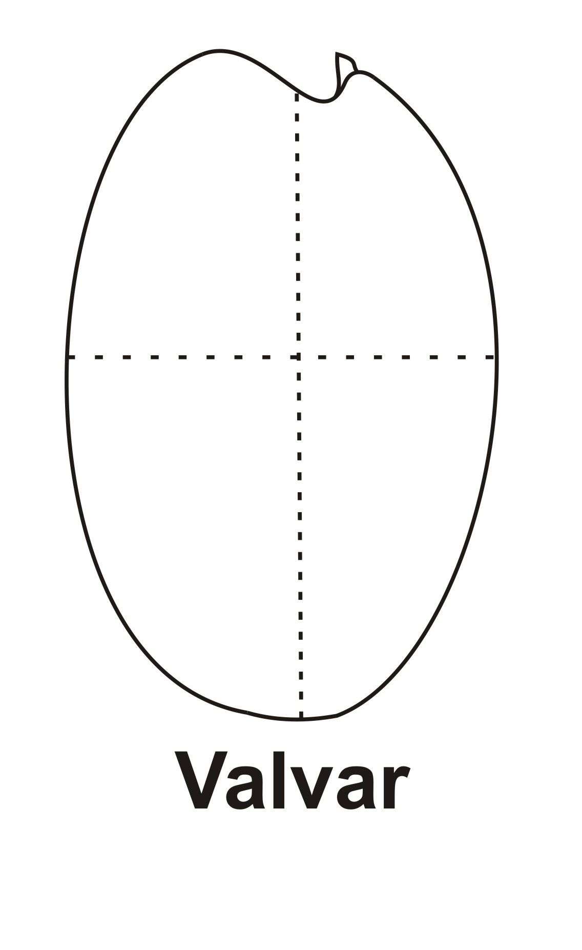
Fig 1: Prorocentrum rhathymum Schematic diagram (valvar view) redrawn (and edited) from Cortés-Altamirano & Sierra-Beltran 2003.
-
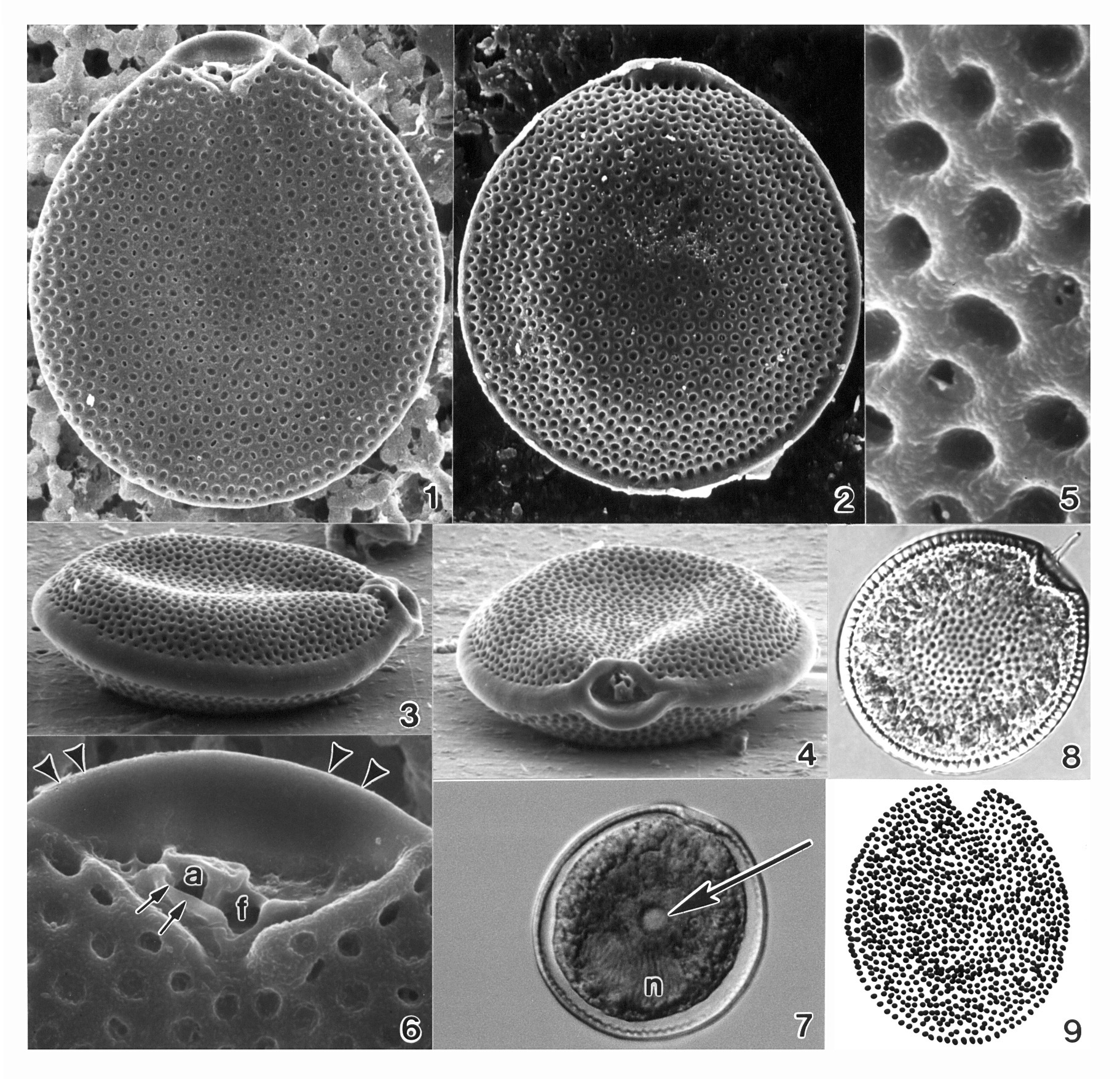
Plate 37. Prorocentrum arenarium. Figs. 1-5. SEM. Fig. 1. Right valve: cells round to ovoid. Periflagellar area is a broad, V-shaped depression. Short longitudinal flagellum visible (arrowhead). Marginal poroids present (arrows). Fig. 2. Left valve: surface smooth, with scattered valve and marginal poroids (arrows). Fig. 3. Lateral view: intercalary band smooth; marginal poroids evenly spaced (arrowheads). Fig.4. Marginal poroids oblong to kidney-shaped. Fig. 5. Periflagellar area: triangular and unornamented with large flagellar pore (f) and smaller auxiliary pore (a). Fig. 6. LM. Right valve: posterior nucleus (n) and prominent central pyrenoid (arrow).
-
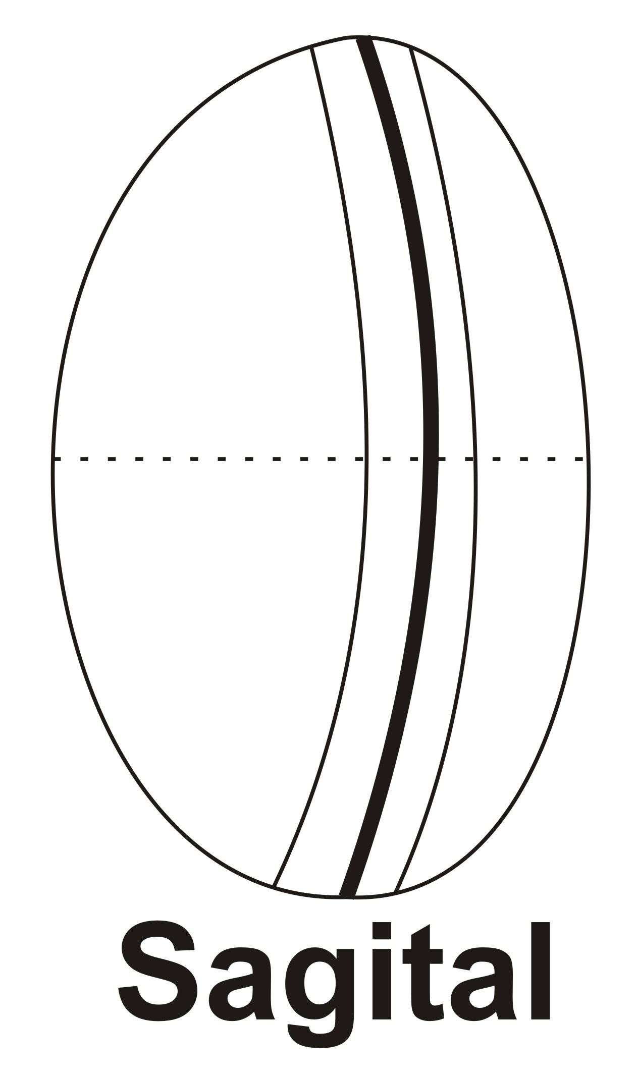
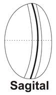
Fig 2: Prorocentrum rhathymum Schematic diagram (sagital view) redrawn from Cortés-Altamirano & Sierra-Beltran 2003.
-
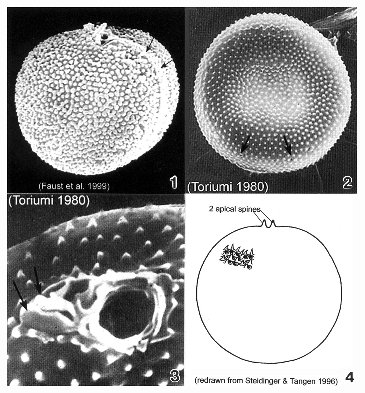
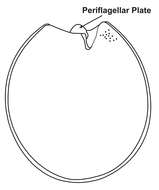
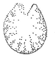
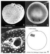
Plate 38. Prorocentrum balticum. Figs. 1-3. SEM. Fig. 1. Valve view: cell round to spherical, covered with many tiny spines. Apical spine apparent. Intercalary band broad, transversely striated (arrows). Fig. 2. Surface with scattered rimmed pores (arrows). Fig. 3. Periflagellar region: two different sized pores and two small apical projections (arrows). Fig. 4. Line drawing.
-
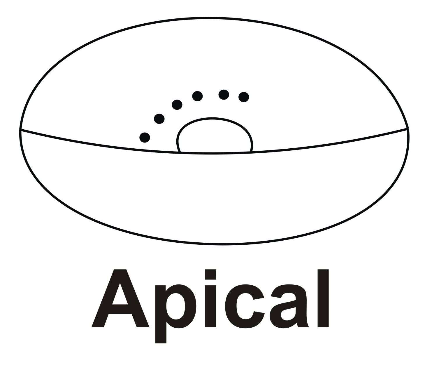
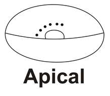
Fig 3: Prorocentrum rhathymum Schematic diagram (apical view) redrawn (and edited) from Cortés-Altamirano & Sierra-Beltran 2003.
-

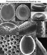
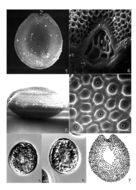
Figs. 1-10. Prorocentrum belizeanum sp. nov. Fig.1. Valve surface is areolated. Fig.2. Cells are round to oval in valve view. Fig.3. In side view, cells are ellipsoid, the apical area exhibits a rounded lip, and both left and right valves are excavated. Fig.4. Periflagellar area is a wide, V-shaped depression located in the right valve. It has a flagellar and auxiliary pore, equal in size. Fig.5. Auxiliary pore (A) is surrounded by a curved apical collar (arrowheads) and is adjacent to the flagellar pore (F). The left valve margin exhibits a wide, rounded ridge (arrow). The two flagella are not shown. Fig.6. Intercalary band is horizontally striated. Fig.7. Areolae are round to ovoid with a smooth margin. Some areolae have trichocyst pores (arrowheads). Fig.8 . The center of the inside valve surface is smooth. The arrangement of round trichocyst pores is shown. Fig. 9. Inside valve surface shown at higher magnification. Trichocyst pores (arrowheads) are arranged in an array along the intercalary band. Fig.10. Inside of the intercalary band is constructed of evenly spaced rectangular sections separated by shallow grooves. Scale bars =10 µm.
EMu: Holotype SEM negative # 104052; SEM stub #104; Field # n.a; Accession # 407164; Catalog # 34; Figure # 1.
-
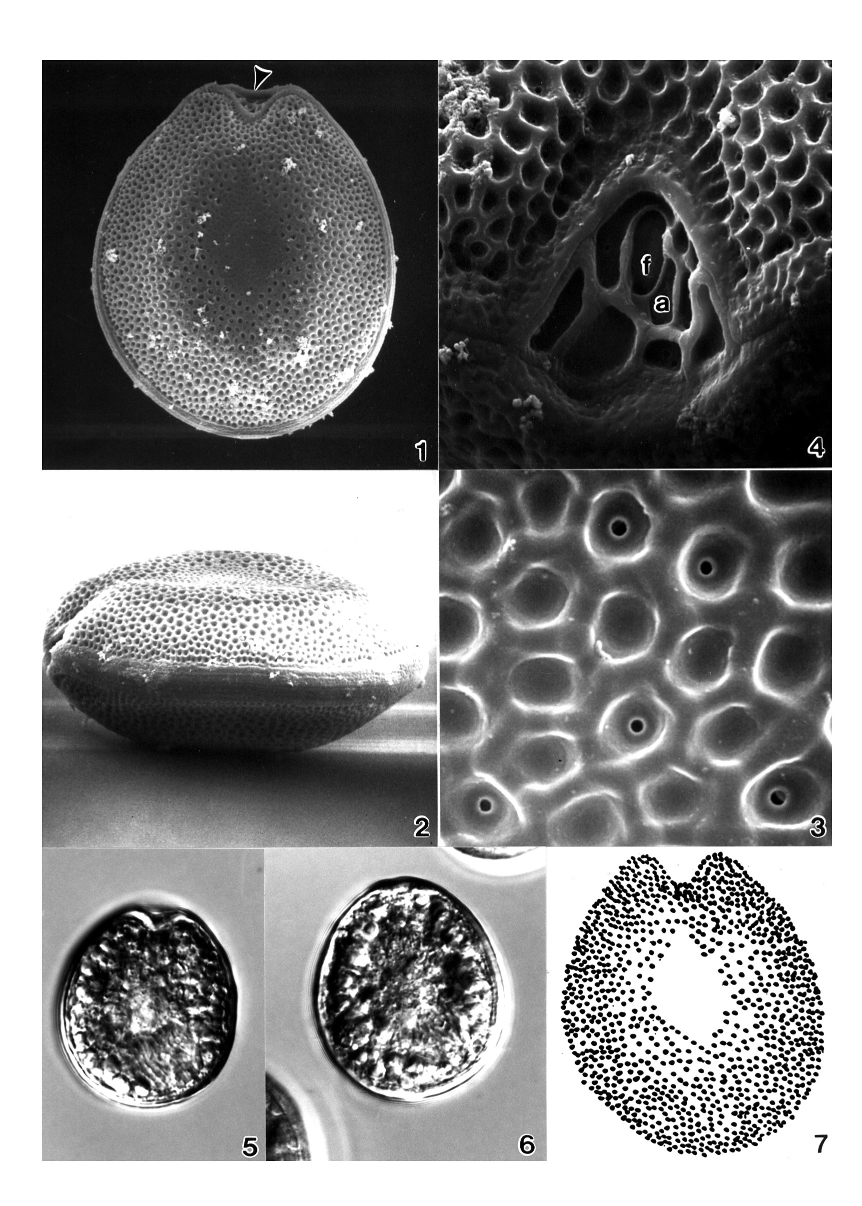
Plate 39. Prorocentrum belizeanum. Figs. 1-6. SEM. Fig. 1. Right valve: cell round to oval; surface heavily areolated. Fig. 2. Left valve: anterior margin with flared curved apical collar. Marginal areolae visible. Fig. 3. Lateral view: valve center concave; intercalary band smooth and wide. Fig. 4. Apical view: apical area with rounded lip; both valves excavated. Fig. 5. Areolae round to ovoid with smooth margins; some with pores. Fig. 6. Periflagellar area: auxiliary pore (a) surrounded by curved periflagellar collar (arrows); adjacent to flagellar pore (f). Left valve with flared apical collar (arrowheads). Fig. 7. Left valve: central pyrenoid (arrow) and posterior nucleus (n). Fig. 8. LM: right valve; flagella present. Fig. 9. Line drawing: areolae arrangement (after Faust 1993a).
-

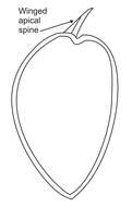
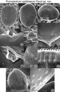
Figs. 17-27. Prorocentrum caribbaeum sp. nov. FIG.17. Cell shape is oval with a rounded anterior and a pointed posterior end. FIG.18. Valve surface is smooth with minute depressions. Radially arranged trichocyst pores are present on each valve. The two flagella are not shown. FIG.19. Cells are ovate to convex in side view. The periflagellar area is located in the right valve and is highly ornate. FIG.20. In valve view, the periflagellar area has a rectangular orientation and is composed of a curved apical collar (on the left) and a smaller protuberant apical plate (on the right). FIG.21. Curved apical collar (arrow) is the largest platelet situated adjacent to the auxiliary pore (A). The apical plate (arrowhead) is located next to the flagellar pore (F) and is separated by a rectangular platelet from the auxiliary pore. FIG.22. The trichocyst pores (arrow) are round with smooth edges and are similar in size. They are situated in furrowed depressions. Small, round pores (arrowhead) are also present, unevenly distributed on the valve surface. FIG.23. The posterior end is pointed and laced with trichocyst pores and small, round pores unique to this species. FIG.24.Ejected trichocysts are on valve surface. FIG.25. The intercalary band is transversally striated and sinous. FIG.6.Inner valve surface is smooth. The location of round trichocyst pores is illustrated, and a distinct striated intercalary band is present (arrowheads). FIG. 7. The inner face of the intercalary band is highly ornate and lacelike. Scale bars = 8 µm.
EMu: Holotype SEM negative # 103001; SEM stub #103; Field # 358-90 Accession # 407164; Catalog # 49; Figure # 17.
-
Plate 40. Prorocentrum concavum. Figs. 1-4. SEM. Fig. 1. Right valve. Cell ovate and heavily areolate. Valve center devoid of areolae. Left valve with anterior apical ridge (arrowhead). Fig. 2. Lateral view. Valve center concave and flattened. Intercalary band granulated and horizontally striated. Fig. 3. Valve areolae round to oval with smooth edges; some with small central pores. Fig. 4. Periflagellar area a V-shaped depression. Two pores: small auxiliary pore (a); large flagellar pore (f). Figs. 5-6. LM (M.A. Faust). Fig. 5. Right valve. Fig. 6. Left valve. Fig. 7. Line drawing: areolae arrangement. (Figs. 1-4,7 after Faust 1990b)
-

FIGS. 11-16. Prorocentrum elegans sp. nov. FIG 11. Cells are oval in valve view; the cell surface is smooth with few valve pores. The two flagella are not present. FIG.12. Left valve margin at the anterior end is flat or inclined. FIG.13. Cells are ovate in side view. The periflagellar area is large relative to the cell size. It has a flagellar (F) and auxiliary (A) pore and an angled flagellar plate (arrowhead adjacent to the auxiliary pore). Large pores (arrow) and small pores (arrowheads) are present on the valves. The small pores are better illustrated at higher magnification in Figure 15. FIG.14. Periflagellar area is detached from the right valve. It has a smooth inner surface, and discrete platelets are unequal in size. FIG.15. The intercalary band is transversely striated, and the inner surface appears ribbed. Small pores (arrowheads) are situated in an array along the intercalary band. FIG.16. The two apical pores are separated by a ridge in this naked cell. Scale bars = 5 µm.
Note:Holotype SEM negative # 86075A; SEM stub #86; Field # n.a; Accession # 407164; Catalog # 44; Figure # 11.
-
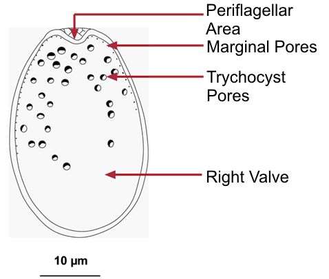
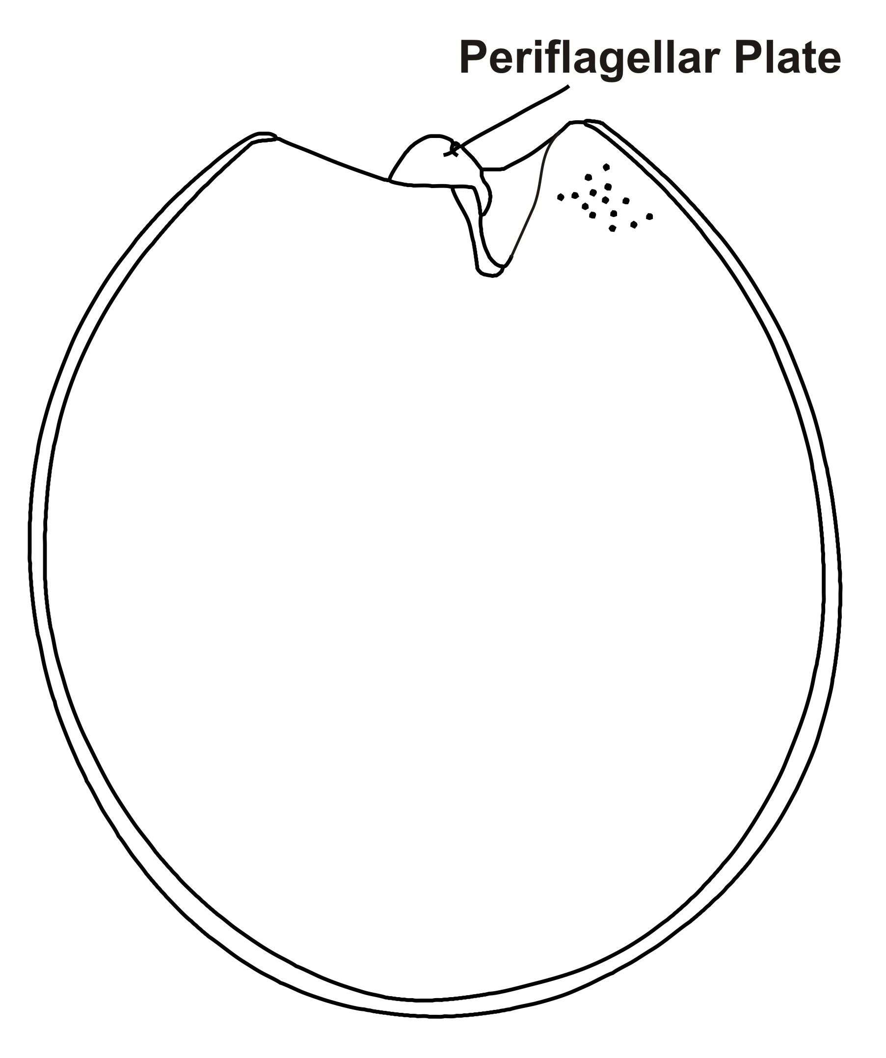
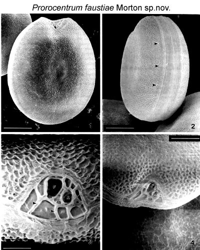
Figs. 1-4. Fig.1.Valve surface of Prorocentrum faustiae. Scale bar represents 5µm. Fig. 2. Intercalary band of Prorocentrum faustiae. Scale bar represents 5 um. Arrow heads point to the evenly spaced marginal pores. Fig. 3. Periflagellar area of Prorocentrum faustiae displays a large flagellar pore (F) and smaller auxiliary pore (A). Scale bar represents 1 µm. Arrow heads point to sutures which separate each periflagellar plate. Fig. 4. Periflagellar area in valve view. Scale bar represents 3 µm.
Note: Isolated from Heron Island, Australia (23.25° S, 151.55° E).
-
Plate 41. Prorocentrum faustiae. Figs. 1-4. SEM. Fig. 1. Right valve. Cells broadly ovate to rotundate with slightly concave center. Valve surface rugose. Periflagellar area situated apically. Fig. 2. Left valve: apical region slightly excavated. Fig. 3. Intercalary band wide and transversely striated. Small marginal pores evenly spaced along cell perifery (arrows). Fig. 4. Periflagellar area: apical view. Broad V-shaped depression; larger flagellar pore (f) adjacent to smaller auxiliary pore (a). (All figures donated by S.L. Morton)
-
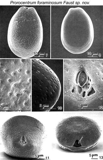
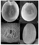
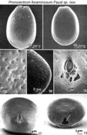
Figs. 7-13. Prorocentrum foraminosum sp. nov. Fig.7. Right valve view showing the apical area, which has a narrow and shallow depression. The surface is covered with a larger number of pores but the centre is devoid of pores. Fig.8. The left valve has a flat anterior end. Left and right valves are similar in surface appearance. Fig.9. The surface is covered with scattered valve pores. Fig.10. The pores are round, equal in size and situated in shallow depressions with an opening in the centre. Fig.11. The intercalary band is smooth, cells have no marginal pores and cell shape is convex. Fig.12. The periflagellar area is narrowly triangular. The platelets appear vertical when viewed from the anterior end of the cell. There is one flagellar pore (F), one auxiliary pore (A) and 8 platelets of unequal size and shape. Fig. 13. Below the periflagellar area the flagellar opening (arrow) is observed in the cytoplasm.
EMu: HOLOTYPE SEM NEGATIVE # 23042; SEM STUB # 23; FIELD # 78-87; ACCESSION # 407159; CATALOG # 66: FIGURE # 7.
-

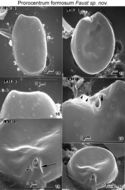
Figs. 14-19. Prorocentrum formosum sp. nov. Fig. 14. The cell is ovate in left valve view. The surface is smooth and the anterior end flat. Fig. 15 The periflagellar area is located in the right valve, forming a broad, shallow depression. Fig. 16. The valve surface has large trichocyst pores (0.2 µm in diameter) distributed in a distinct pattern, and small pores (< 0.1 µm in diameter) around the cell periphery. Fig. 17. The cell surface is concave in side view. The surface of the apical area is smooth and has one flagellar pore. Fig. 18. The apical area at higher magnification showing a prominent curved apical collar (arrow) and an angled plate (arrowhead) adjacent to the flagellar pore (F) which is ornate with a flange. Fig. 19. The curved apical collar is the largest apical plate and has a height of 0.8 µm (arrowheads). It is situated opposite the angled plate (arrow).
Prorocentrum formosum holotype plate
EMu: Holotype SEM negative # 115099; SEM Stub # 115; Field # Lair muck #3 Accession # 407165; Catalog # 73; Figure # 5.
-
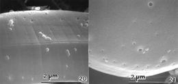
Figs. 20-21. Prorocentrum formosum sp. nov.Fig. 20. The intercalary band is transversely striated and wider in dividing cells. Fig. 21. Trichocyst pores are distributed in a characteristic pattern. They are round to oblong in shape with smooth edges, and equal size. The small pores are circular with smooth edges and distributed near the valve periphery.
EMu: Holotype SEM negative # 115099; SEM Stub # 115; Field # Lair muck #3 Accession # 407165; Catalog # 73; Figure # 5.
-
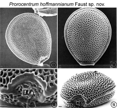
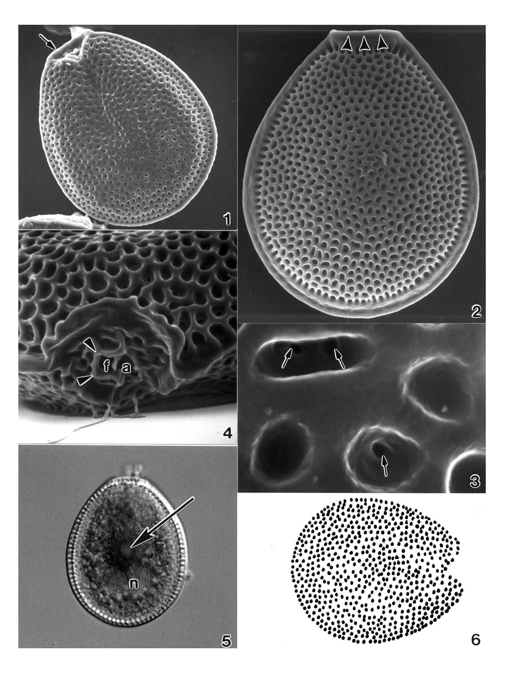
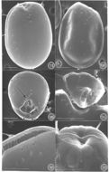
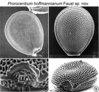
Figs. 13-16. . Prorocentrum hoffmannianum sp. nov. FIG.13. The Valve surface is areolated and slightly concave. FIG. 14. The body is ovoid in valve view with the maximum width behind the middle region. The body is narrow at the anterior end. FIG. 15. The cell is round in side view and convex in the middle of the valve. The flagellar pore area is attached to the right valve; surrounded by a distinct flared ridge that is V-shaped, triangular with a complex arrangement of flagellar platelets unequal in size. The intercalary band is smooth. FIG. 16. The areolae are round to ovoid with a smooth margin. Areolae are perforated by oval openings. The flagellar pore (f) (arrow) is surrounded by a flared apical collar (arrow) and is adjacent to an auxiliary pore (a). Scale bars = 200 µm.
EMu: SEM NEGATIVES # 27013; SEM STUB # 27; FIELD # 132-88; ACCESSION # 407160; CATALOG # 22; FIGURE # 13.
-
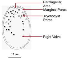
Plate 42. Prorocentrum hoffmannianum. Figs. 1-4. SEM. Fig. 1. Right valve: cell ovoid, tapering slightly apically. Valve surface areolated, slightly concave. Curved apical collar (arrow). Fig. 2. Left valve: distinct flared apical collar bordering periflagellar area (arrowheads). Marginal areolae large. Intercalary band smooth. Fig. 3. Areolae round to ovoid with smooth margins. Some with small pores (arrows). Fig. 4. Periflagellar area: flagellar pore (f) surrounded by flared periflagellar collar (arrowheads), adjacent to auxiliary pore (a); pores equal in size. Fig. 5. LM. Left valve: central pyrenoid (arrow); posterior nucleus (n). Intercalary band appears striated (M.A. Faust). Fig. 6. Line drawing: areolae arrangement. (Figs. 1-4,6 after Faust 1990b)