Ecology
provided by NMNH Marine Dinoflagellates
P. faustiae is a benthic species epiphytic on macroalgae (Morton 1998).
- bibliographic citation
- Faust, Maria A. and Rose A. Gulledge. Identifying Harmful Marine Dinoflagellates. Smithsonian Contributions from the United States National Herbarium, volume 42: 1-144 (including 48 plates, 1 figure and 1 table).
Etymology
provided by NMNH Marine Dinoflagellates
The species 'faustiae' is named in honor of Dr. Maria Faust, Smithsonian Institution, for her advancements in the taxonomy of non-planktonic dinoflagellates (Morton 1998).
- bibliographic citation
- Faust, Maria A. and Rose A. Gulledge. Identifying Harmful Marine Dinoflagellates. Smithsonian Contributions from the United States National Herbarium, volume 42: 1-144 (including 48 plates, 1 figure and 1 table).
Habitat and Locality
provided by NMNH Marine Dinoflagellates
Populations of P. faustiae are associated with macroalgae from Heron Island, Australia (Morton 1998).
- bibliographic citation
- Faust, Maria A. and Rose A. Gulledge. Identifying Harmful Marine Dinoflagellates. Smithsonian Contributions from the United States National Herbarium, volume 42: 1-144 (including 48 plates, 1 figure and 1 table).
Morphology and Structure
provided by NMNH Marine Dinoflagellates
P. faustiae is a photosynthetic species containing numerous golden-brown chloroplasts and a centrally located pyrenoid (Figs. 1, 2). A large kidney-shaped nucleus is situated posteriorly (Morton 1998).
- bibliographic citation
- Faust, Maria A. and Rose A. Gulledge. Identifying Harmful Marine Dinoflagellates. Smithsonian Contributions from the United States National Herbarium, volume 42: 1-144 (including 48 plates, 1 figure and 1 table).
Nomenclatural Types
provided by NMNH Marine Dinoflagellates
Holotype: Prorocentrum faustiae Morton, 1998: 567, figs. 1-4
Type Locality: Coral Sea: Heron Island, Australia
- bibliographic citation
- Faust, Maria A. and Rose A. Gulledge. Identifying Harmful Marine Dinoflagellates. Smithsonian Contributions from the United States National Herbarium, volume 42: 1-144 (including 48 plates, 1 figure and 1 table).
Reproduction
provided by NMNH Marine Dinoflagellates
P. faustiae reproduces asexually by binary fission.
- bibliographic citation
- Faust, Maria A. and Rose A. Gulledge. Identifying Harmful Marine Dinoflagellates. Smithsonian Contributions from the United States National Herbarium, volume 42: 1-144 (including 48 plates, 1 figure and 1 table).
Species Comparison
provided by NMNH Marine Dinoflagellates
Prorocentrum faustiae is similar in shape and size to P. hoffmannianum (45-55 µm long and 40-45 µm wide); however, the former lacks thecal areolae, which are very abundant on the latter. P. faustiae lacks a distinct ridge along the valve perifery which distinguishes this species from P. maculosum (Morton 1998).
- bibliographic citation
- Faust, Maria A. and Rose A. Gulledge. Identifying Harmful Marine Dinoflagellates. Smithsonian Contributions from the United States National Herbarium, volume 42: 1-144 (including 48 plates, 1 figure and 1 table).
Species Overview
provided by NMNH Marine Dinoflagellates
P. faustiae is an armoured, marine, benthic dinoflagellate species. This species is associated with macroalge from the Australian Barrier Reef.
- bibliographic citation
- Faust, Maria A. and Rose A. Gulledge. Identifying Harmful Marine Dinoflagellates. Smithsonian Contributions from the United States National Herbarium, volume 42: 1-144 (including 48 plates, 1 figure and 1 table).
Taxonomic Description
provided by NMNH Marine Dinoflagellates
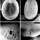
P. faustiae is a bivalvate species often observed in valve view. Cells are broadly ovate to rotundate with a rugose appearance (Figs. 1-3). Valve centers are concave (Figs. 1-3). Cells are 43-49 µm long and 38-42 µm wide. Small pores are (0.1 µm in diameter), probably containing trichocysts, are dense on the valve surface and along the valve perifery (Figs. 1-3). The intercalary band is transversely striated (Fig. 3) (Morton 1998).
The periflagellar area is a wide triangular, V-shaped region located apically on the right valve (Figs. 1, 4). Sixteen apical platelets make up the periflagellar area. Included also are two pores: a large flagellar pore, and a smaller auxiliary pore (Fig. 4)(Morton 1998).
- bibliographic citation
- Faust, Maria A. and Rose A. Gulledge. Identifying Harmful Marine Dinoflagellates. Smithsonian Contributions from the United States National Herbarium, volume 42: 1-144 (including 48 plates, 1 figure and 1 table).
Toxicity
provided by NMNH Marine Dinoflagellates
P. faustiae is a diarrhetic shellfish poison (DSP) toxin-producing species producing okadaic acid (OA) and Dinophysistoxin-1 (DTX1) (Morton 1998).
- bibliographic citation
- Faust, Maria A. and Rose A. Gulledge. Identifying Harmful Marine Dinoflagellates. Smithsonian Contributions from the United States National Herbarium, volume 42: 1-144 (including 48 plates, 1 figure and 1 table).