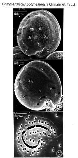
Figs. 5-7. Scanning electron micrographs of Gamblerdiscus polynesiensis (TB-92), sp. nov. Figs. 5, 6. Cells are round to ellipsoid. Cell surface is smooth with small scattered pores. Fig. 5. Epithecal view. The PO plate is oriented ventrally. Fig. 6. Hypothecal view. The Ip plate, broad and pentagonal, occupies 60% of the of hypotheca width. Postcingular plates 2'", 3'" and 4'" are narrow. The cingulum, deep, is ascending into a deep sulcal hollow. Fig. 7. The Po plate is triangular with fish-hook-shaped apical opening surrounded by 44 pores.
EMu: Holotype SEM negative # 242010; SEM stub # 242; Field # TB-92; Catalog # 1522; Figure #5.