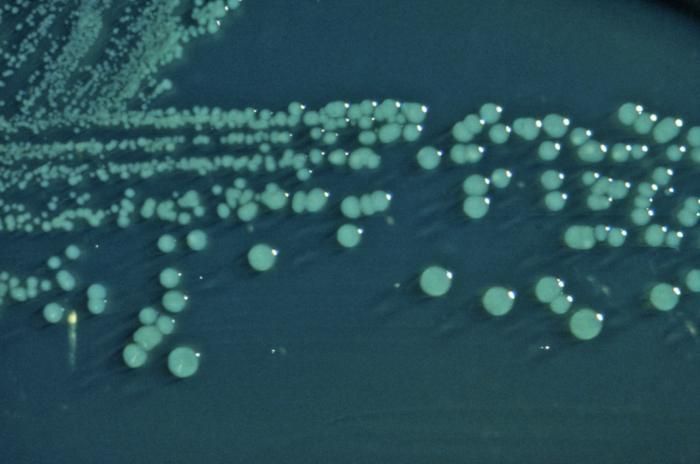
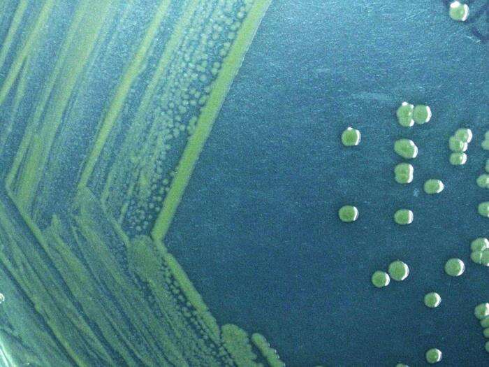
This photograph depicts the colonial morphology displayed by Shigella boydii bacteria cultivated on a Hektoen enteric (HE) agar surface; colonies of S. boydii bacteria grown on HE agar display a raised, green, and moist appearance.Created: 1976
This photograph depicts the colonial morphology displayed by Shigella boydii bacteria cultivated on a Hektoen enteric (HE) agar surface; colonies of S. boydii bacteria grown on HE agar display a raised, green, and moist appearance.Created: 1976
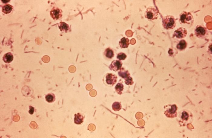
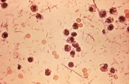
This photomicrograph revealed stool exudates in a patient with shigellosis, which is also known as Shigella dysentery, or Bacterial dysentery.Created: 1980
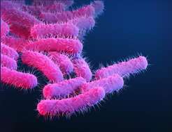
Description: English: This is a medical illustration of drug-resistant, Shigella sp. bacteria, presented in the Centers for Disease Control and Prevention (CDC) publication entitled, Antibiotic Resistance Threats in the United States, 2019 (AR Threats Report). See the link below for more on the topic of antimicrobial resistance (AR). Date: 2019. Source: https://phil.cdc.gov/Details.aspx?pid=23252. Author: Medical Illustrator: Stephanie Rossow.
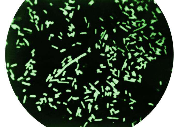
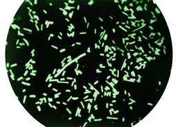
Description: English: Dark field microscopy revealing Shigella dysenteriae bacteria. Date: 1964. Source: https://phil.cdc.gov/Details.aspx?pid=22229. Author: Armed Forces Institute of Pathology, AFIP.
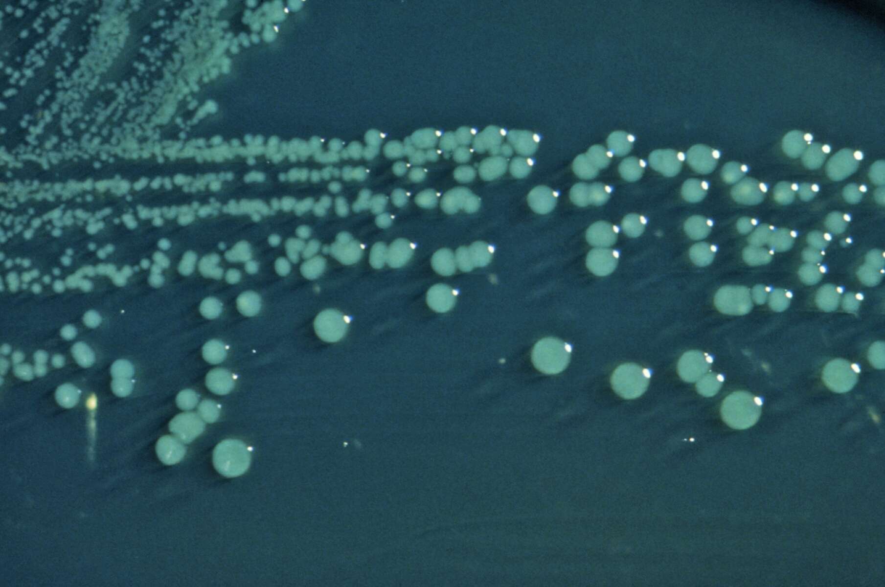
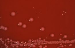
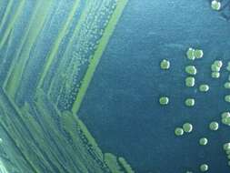
Description: English: Gram negative Shigella sonnei bacteria which spent 48 hours cultured on Hektoen enteric agar (HEK). Date: 2014. Source: https://phil.cdc.gov/Details.aspx?pid=17186. Author: Todd Parker, Ph.D., Assoc Director for Laboratory Science, Div of Preparedness and Emerging Infections at CDC.


