-
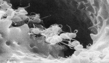
After gametogenesis, there is very little left of the parent cell. In this image, remnant cytoplasm in the empty test (which has been decalcified to make it more transparent) is being scavenged by a ciliate (arrow). Image courtesy of Susan T. Goldstein, University of Georgia. This image first appeared in J. Foram Res. 23:213-220, and is used with permission.
-
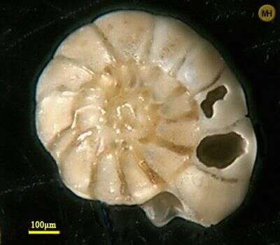
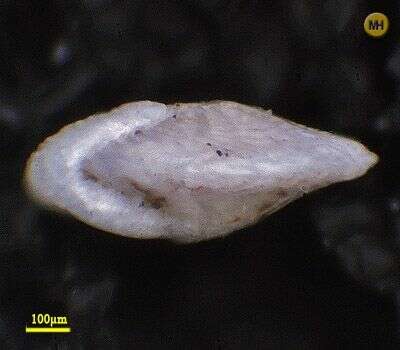
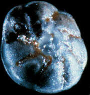
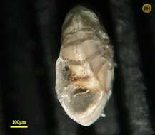
Test is 1 mm. across (long dimension). Image courtesy of David B. Scott, Dalhousie University. This image was originally published in
Palaeologica Electronica, vol. 3, issue 2, and is used with the kind permission of that journal and the Paleontological Association.
-
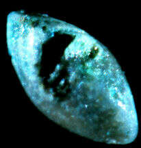
A closeup of biflagellated foraminiferal gametes leaving the aperture of the parent gamont. The gametes are about 2 um in diameter. Image courtesy of Susan T. Goldstein, University of Georgia. This image first appeared in J. Foram. Res. 23:213-220 and is used with permission.
-
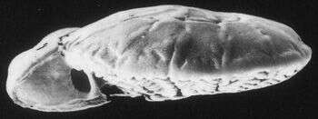
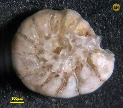
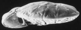
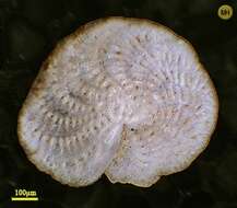
Image of the holotype, which is 1.1 mm. across. Image courtesy of David B. Scott, Dalhousie University. This image was originally published in
Palaeologica Electronica, vol. 3, issue 2, and is used with the kind permission of that journal and the Paleontological Association.
-
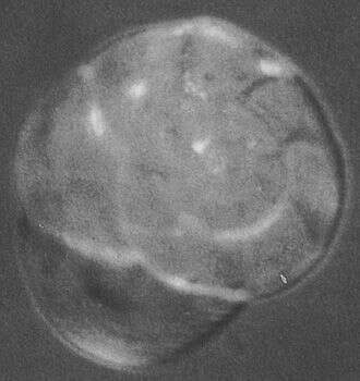
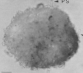
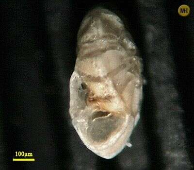
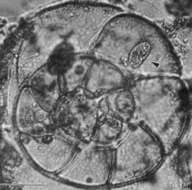
A light micrograph of a "vegetative" gamont; i.e., on that has not yet begun the process of producing gametes. The test is approximately 250 um in diameter. Image courtesy of Susan T. Goldstein, University of Georgia. This image first appeared in J. Foram. Res. 23:213-220 and is used with permission.
-
Discription to come
-
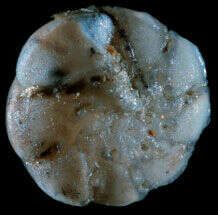
A decalcified test after gamete release. The test is mostly empty; the dark spheres are the remnants of the cytoplasm. Image courtesy of Susan T. Goldstein, University of Georgia. This image first appeared in J. Foram. Res. 23:213-220 and is used with permission.
-
Discription to come
-
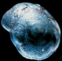
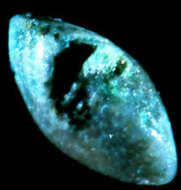
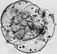
During gametogenesis, some forams draw a mass of sediment and detritus around their tests; this is called a "reproductive cyst". It is shed just before gamete release. Image courtesy of Susan T. Goldstein, University of Georgia. This image first appeared in J. Foram. Res. 23:213-220 and is used with permission.
-
Discription to come
-
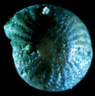
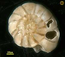
This individual was harvested near Rimini, Italy. The aperture (on the left side) is quite prominent in this image, as are the different textures on the upper (spiral) and lower (umbilical) faces of the test. Image courtesy of Stefan Revets. This image first appeared in Hansen and Revets, J. Foram. Res. 22:166-180 (1992) and is used with permission.
-
Discription to come
-
found at Cherai beach Nov 2007 near Kochi/India 9° 58' N, 76° 17' E
-
ound at Cherai beach Nov 2007 near Kochi/India 9° 58' N, 76° 17' E
-
ound at Cherai beach Nov 2007 near Kochi/India 9° 58' N, 76° 17' E
-
Sample collected at Hamble Estuary, Hampshire, England. Image courtesy of Elisabeth Alve, University of Oslo. Originally published in the Journal of Foraminiferal Research 31:1; used with permission.
-
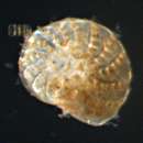
Smoegen/Skagerrak/Sweden, 58.35 N, 11.22 E found at beach
-
Smoegen/Skagerrak/Sweden, 58.35 N, 11.22 E found at beach.
-
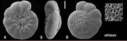
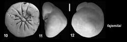
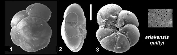
From: Hayward, B.W.; Holzmann, M.; Pawlowski, J.; Parker, J.H.; Kaushik, T.; Toyofuku, M.S.; Tsuchiya, M. 2021. Molecular and morphological taxonomy of living Ammonia and related taxa (Foraminifera) and their biogeography. Micropaleontology 67: 109-313. Plate 28, figs. 4-6. Type locality: Ghana, Accra, outer part of Densu Estuary, sample GH21 (5° 30' 53.6" N, 0°17' 59.7" W), 0.8 m water depth, salinity at time of sampling (7 March 2017) 34.7 psu and temperature 29° C, shelly mud.
-
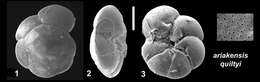
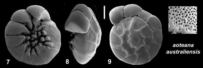
From: Hayward, B.W.; Holzmann, M.; Pawlowski, J.; Parker, J.H.; Kaushik, T.; Toyofuku, M.S.; Tsuchiya, M. 2021. Molecular and morphological taxonomy of living Ammonia and related taxa (Foraminifera) and their biogeography. Micropaleontology 67: 109-313. Plate 28, Figs 10-12. Type locality: Intertidal mudflats of Rohini village, Rajapuri creek, Raigad, Maharashtra, India (northeast Arabian Sea). The figure of location and locality is described in Kaushik et al. (2019).
-
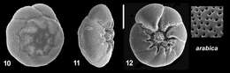
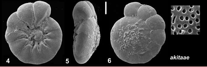
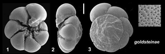
From: Hayward, B.W.; Holzmann, M.; Pawlowski, J.; Parker, J.H.; Kaushik, T.; Toyofuku, M.S.; Tsuchiya, M. 2021. Molecular and morphological taxonomy of living Ammonia and related taxa (Foraminifera) and their biogeography. Micropaleontology 67: 109-313.Pl. 28, figs 7-9. Type locality and age: Australia, New South Wales, Port Hacking, low tide Zostera sea grass, Recent, collected by BWH in 2001, AU17644.
-
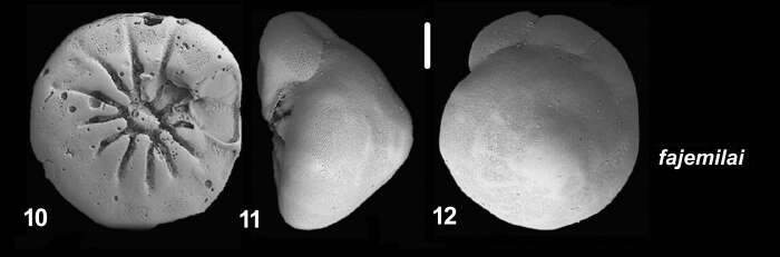
From: Hayward, B.W.; Holzmann, M.; Pawlowski, J.; Parker, J.H.; Kaushik, T.; Toyofuku, M.S.; Tsuchiya, M. 2021. Molecular and morphological taxonomy of living Ammonia and related taxa (Foraminifera) and their biogeography. Micropaleontology 67: 109-313. Plate 29, Figs. 1-3. Type locality and age: Fiji, Suva,Walu Bay, mid-high tide mud, Recent (AU15350), collector Bruce Hayward 1999.
-
in: Hayward, B.W.; Holzmann, M.; Pawlowski, J.; Parker, J.H.; Kaushik, T.; Toyofuku, M.S.; Tsuchiya, M. 2021. Molecular and morphological taxonomy of living Ammonia and related taxa (Foraminifera) and their biogeography. Micropaleontology 67: 109-313. Plate 29, figs. 10-12. Type locality: South Atlantic, Gabon, Pointe Denise, 1.5 m depth, Recent (Langer et al. 2016).
-
in: Hayward, B.W.; Holzmann, M.; Pawlowski, J.; Parker, J.H.; Kaushik, T.; Toyofuku, M.S.; Tsuchiya, M. 2021. Molecular and morphological taxonomy of living Ammonia and related taxa (Foraminifera) and their biogeography. Micropaleontology 67: 109-313. Plate 30, figs. 1-3. Type locality and age: New Caledonia, Noumea, Baie de Magenta, near Tjibaou Cultural Centre, AU17669, collected by Margaret Morley, July 2001, Recent.