-
Maria Cleide de Mendonça, Eduardo A. Abrantes, Ana Carolina R. Neves
Zookeys
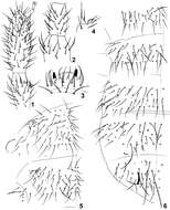
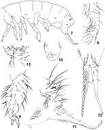
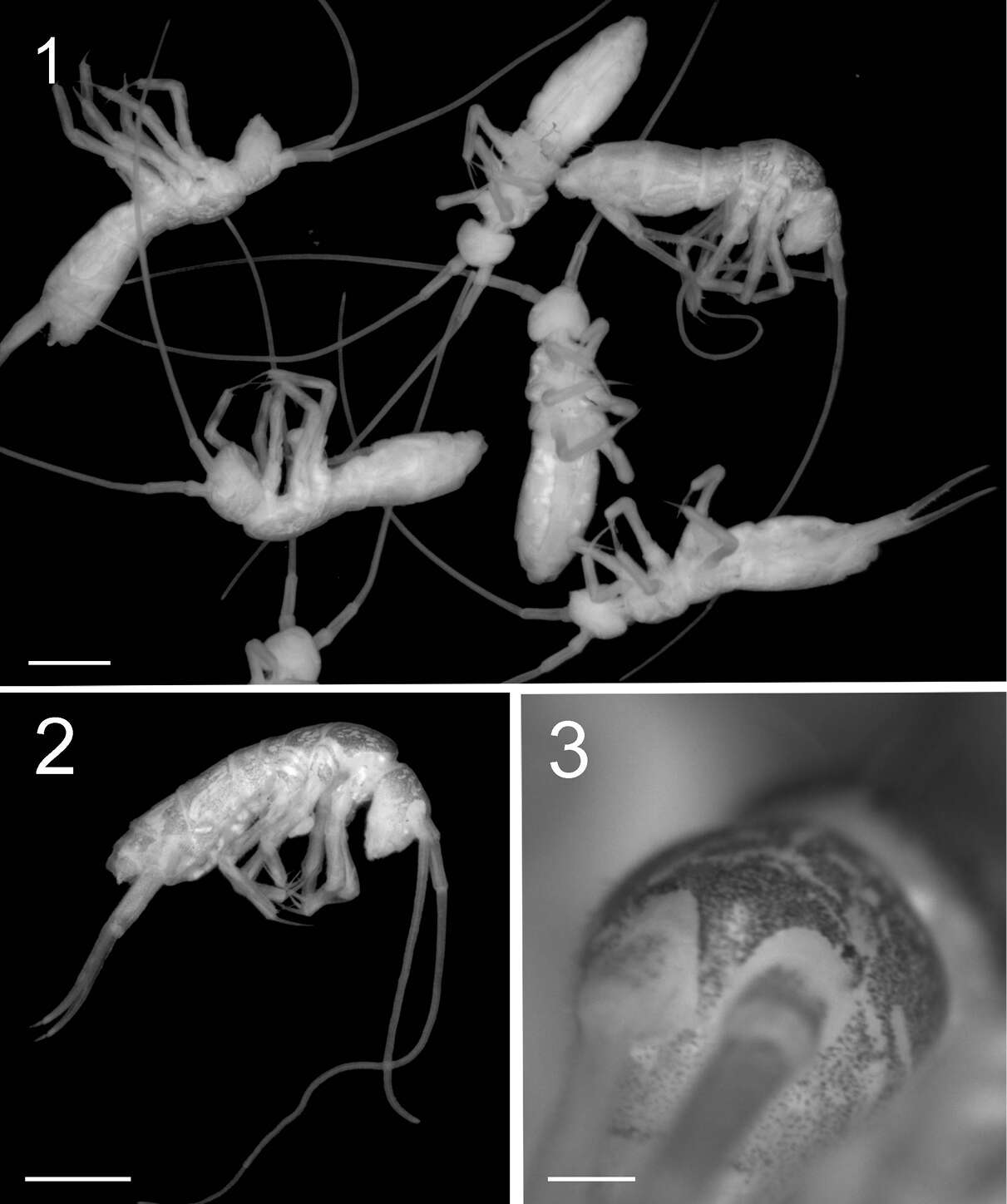
Figure 1–6.Isotomiella macedoi sp.n. 1 Ant III-IV Dorsal view, detail of the apical microsensillum 2 Ant I-II Dorsal view 3 Labral and prelabral chaetae 4 Outer lobe of maxilla 5 Dorsal chaetotaxy of Th II-III 6 Dorsal chaetotaxy of Abd I-VI.
-
Felipe N. Soto-Adames, Steven J. Taylor
Zookeys
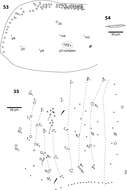
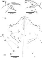
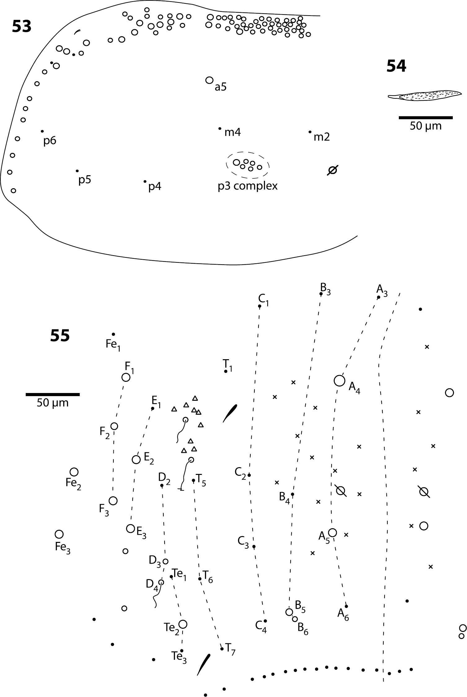
Figures 53–55.Trogolaphysa jataca 53 Mesothorax chaetotaxy 54 Second abdominal segment seta p5 55 Complete chaetotaxy of fourth abdominal segment.
-
Xiang-Qun Yuan, Zhi-Xiang Pan
Zookeys
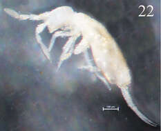
Figure 22.Habitus of Sinella triseta sp. n.
-
Sopark Jantarit, Chutamas Satasook, Louis Deharveng
Zookeys
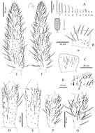
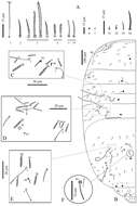
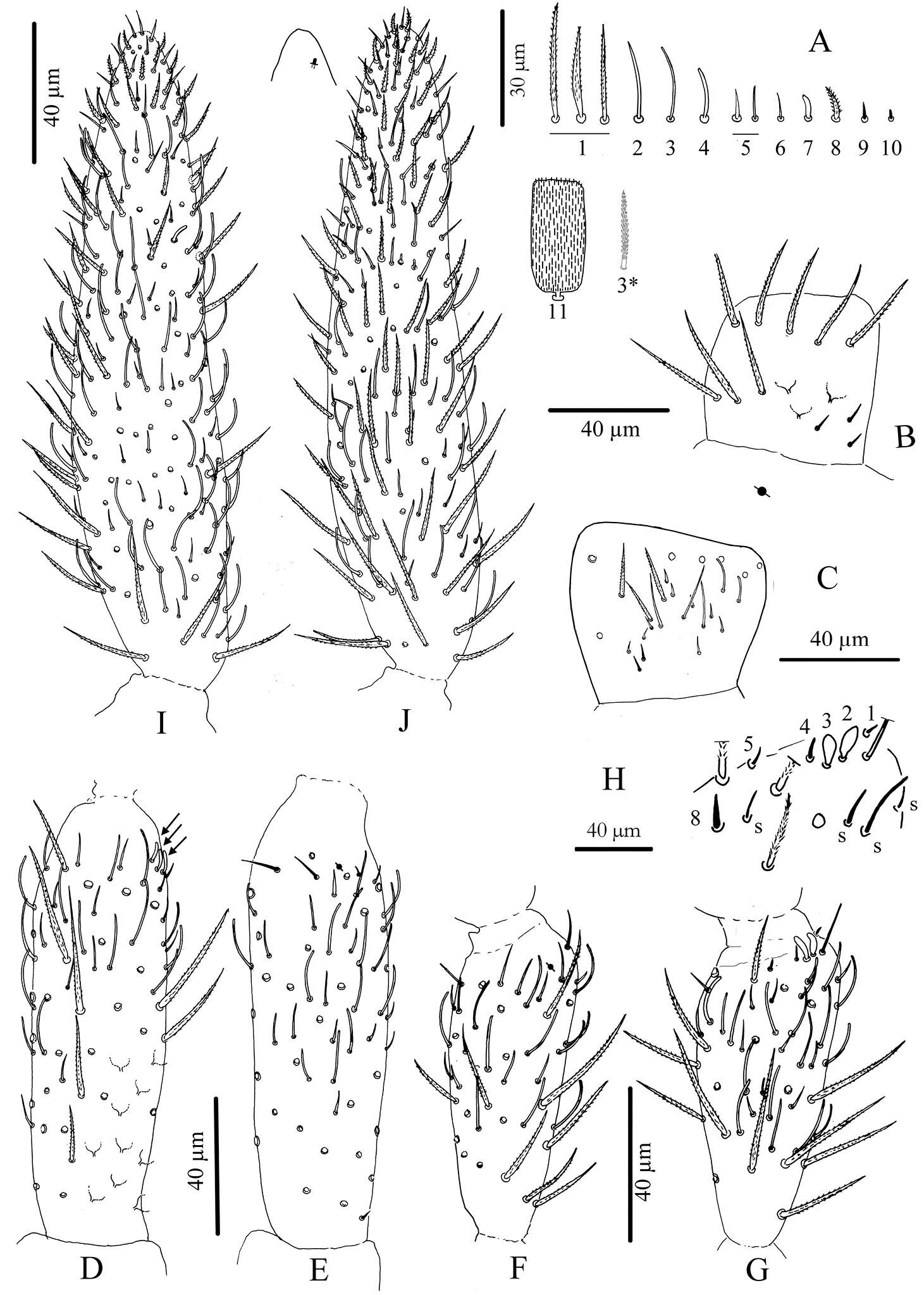
Figure 3.Cyphoderus songkhlaensis sp. n. continued A chaetae of antenna drawn from optical microscope, except 3* derived from SEM image B dorsal side of right Ant.I C ventral side of right Ant.I D dorsal side of right Ant.II; the apical swollen sens of type-7 are indicated by arrows E ventral side of right Ant.II with apical pseudopore F ventral side of right Ant.III with apical pseudopore G dorsal side of right Ant.III H distal organite of Ant.III I ventral side of Ant.IV J dorsal side of Ant.IV with separate view of the subapical organite (left).
-
Daoyuan Yu, Feng Zhang, Louis Deharveng
Zookeys
Figure 1.Tomocerus postantennalis sp. n. Appearance in alcohol. Scale bar: 1000 μm.
-
Marko Lukić, Céline Houssin, Louis Deharveng
Zookeys
Figures 1–3. Tritomurus veles sp. n. (optical stereomicroscope). 1, 2 Habitus (scale 1 mm) 3 Head (scale 0.2 mm).
-
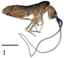
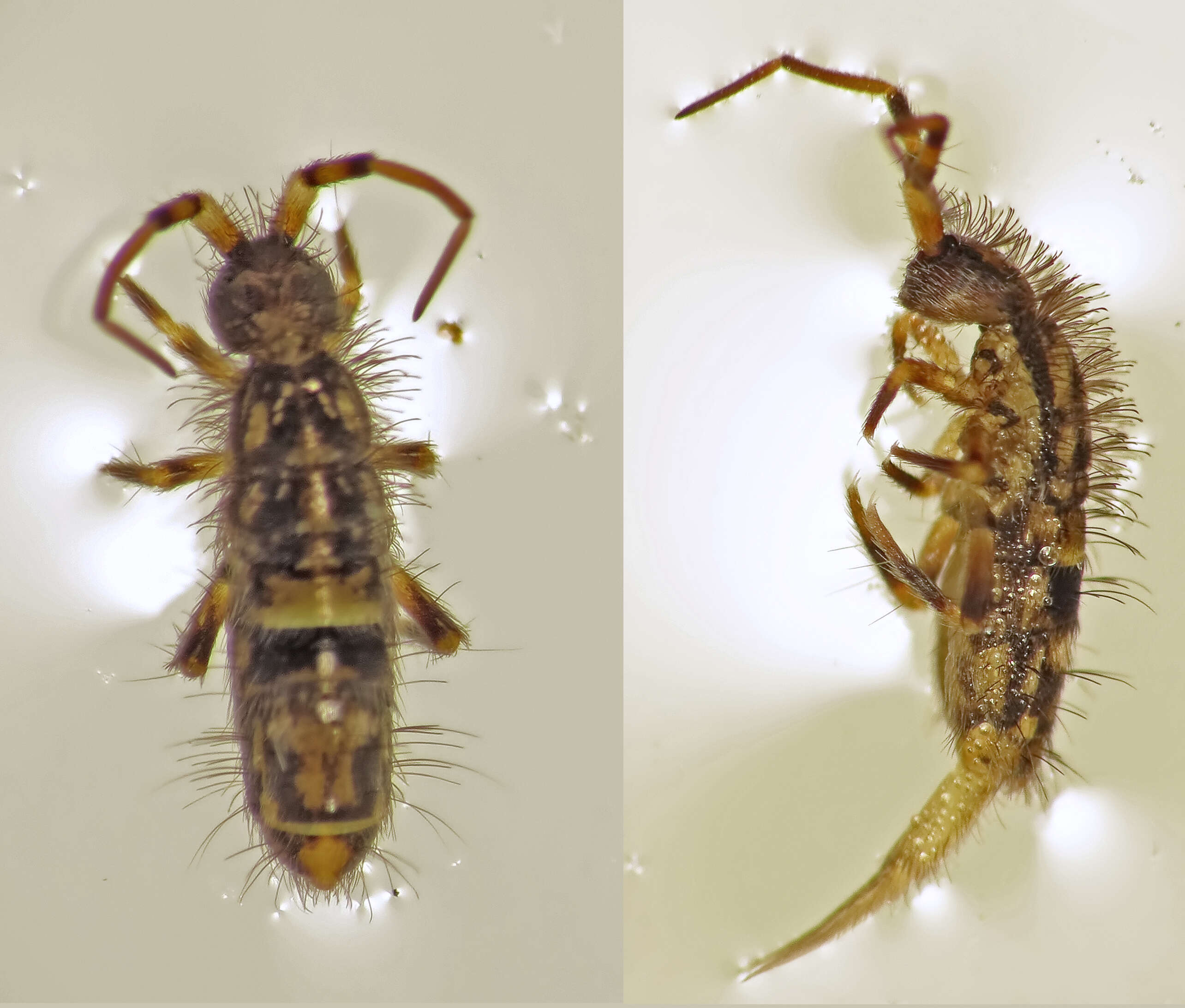
possibly: Genus: Pogonognathellus ???Family: Tomoceridae SCHFFER, 1896[det. Manuel Valdueza, 2012, based on this photo]Subclass: Collembola LUBBOCK, 1870 (Springschwnze, Springtails)Class: Entognatha Subphylum: HexapodaPhylum: Arthropoda2011-07-24_vic. Regensburg, Bavaria, GermanyIMG_3928
-
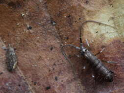
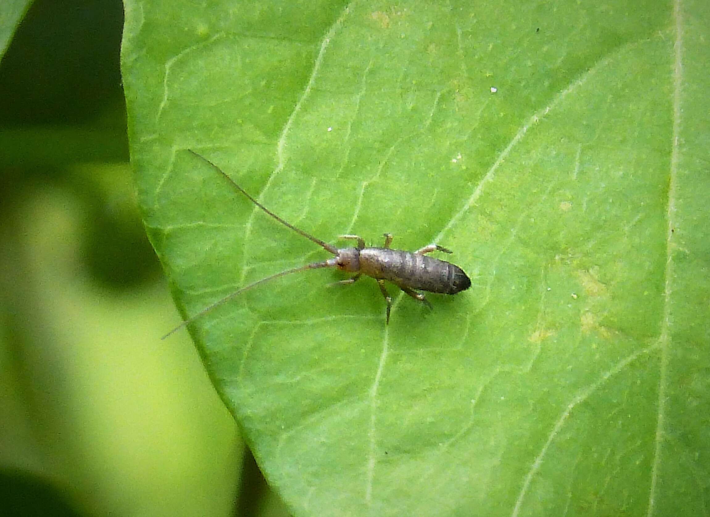
Ipswich, England, United Kingdom
-
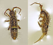
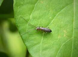
WWT Knapp Reserve, Worcs. SO748520
-
Maria Cleide de Mendonça, Eduardo A. Abrantes, Ana Carolina R. Neves
Zookeys
Figure 7–13.Isotomiella macedoi sp.n. 7 Sensillary pattern of the body 8 Subcoxa and femur of leg III 9 Tibiotarsus and unguis of leg III 10 Lateral view of ventral tube 11 Lateral view of abd. V-VI, subcoxa furcal, furca and tenaculum 12 Furca 13 Male genital plate.
-
Felipe N. Soto-Adames, Steven J. Taylor
Zookeys
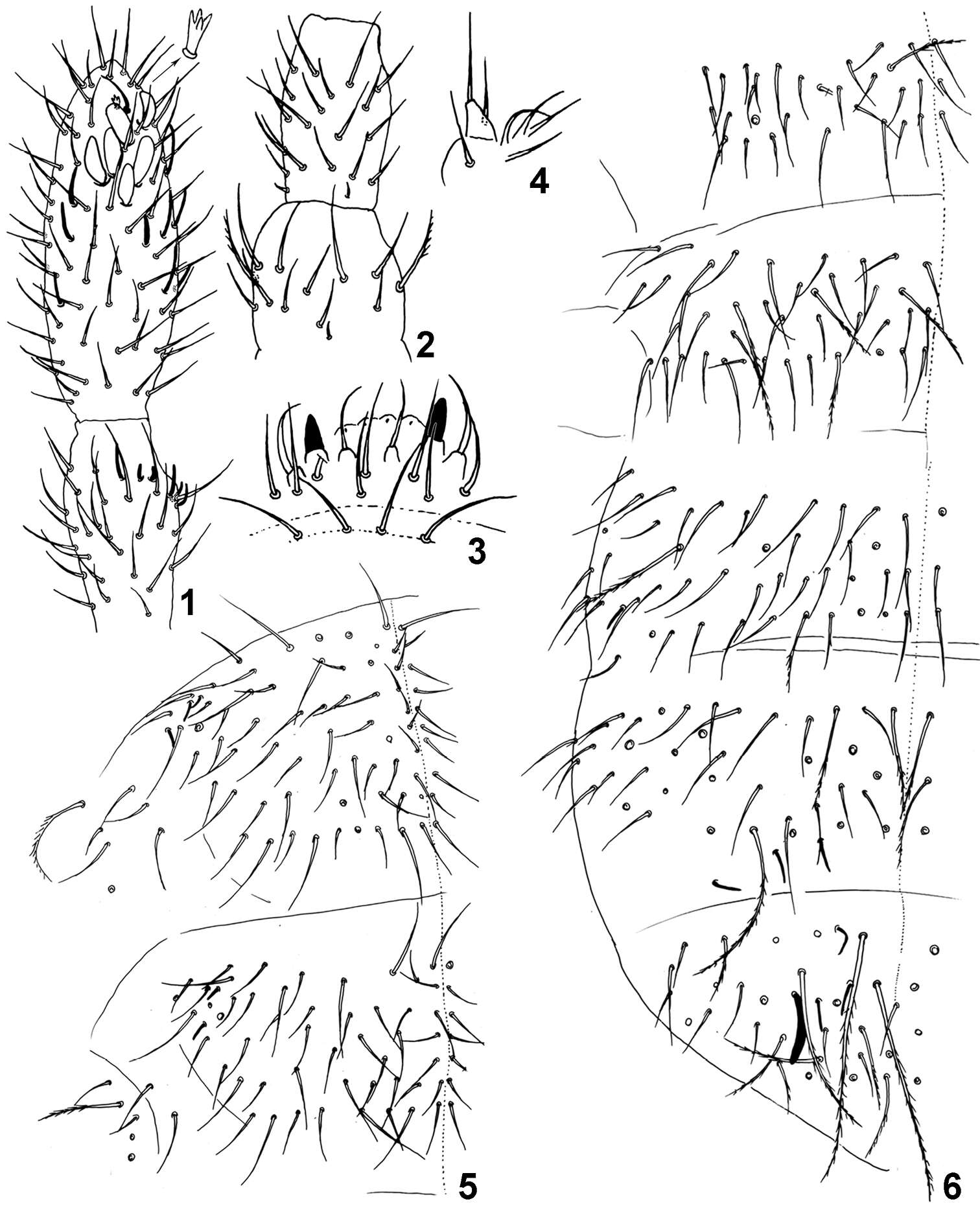
Figures 50–52.Trogolaphysa belizeana (50, 51) and Trogolaphysa jataca (52) 50 Prothoracic claw 51 Metathoracic claw 52 Dorsal chaetotaxy of head.
-
Xiang-Qun Yuan, Zhi-Xiang Pan
Zookeys
Figures 36–40.Sinella triseta sp. n. 36 dorsal chaetotaxy of Abd. IV–V 37 anterior face of VT 38 posterior face and lateral flap of VT 39 manubrial plaque 40 apical dentes and mucro.
-
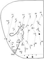
Sopark Jantarit, Chutamas Satasook, Louis Deharveng
Zookeys
Figure 4.Cyphoderus songkhlaensis sp. n. continued A chaetae of tergites drawn from optical microscope, except 5* derived from SEM image B chaetotaxy of tergites with types of S-chaetae S1 to S4 C trichobothrial complexes of Abd.II D trichobothrial complexes of Abd.III E anterior trichobothrial complexes of Abd.IV F tandem of chaetae on Abd.IV; the smallest is a short type-5 mes and the largest a S4 sens.
-
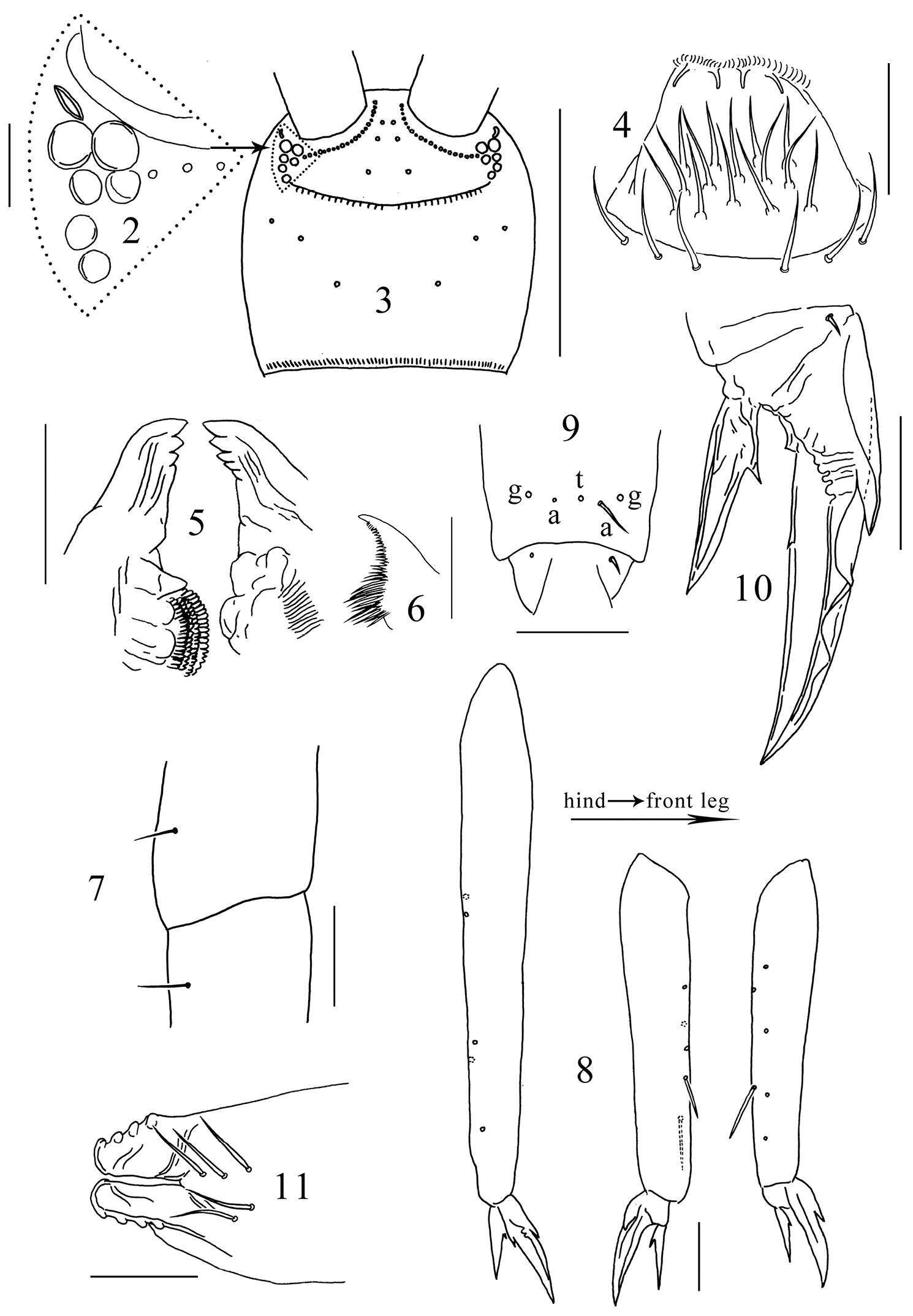
Daoyuan Yu, Feng Zhang, Louis Deharveng
Zookeys
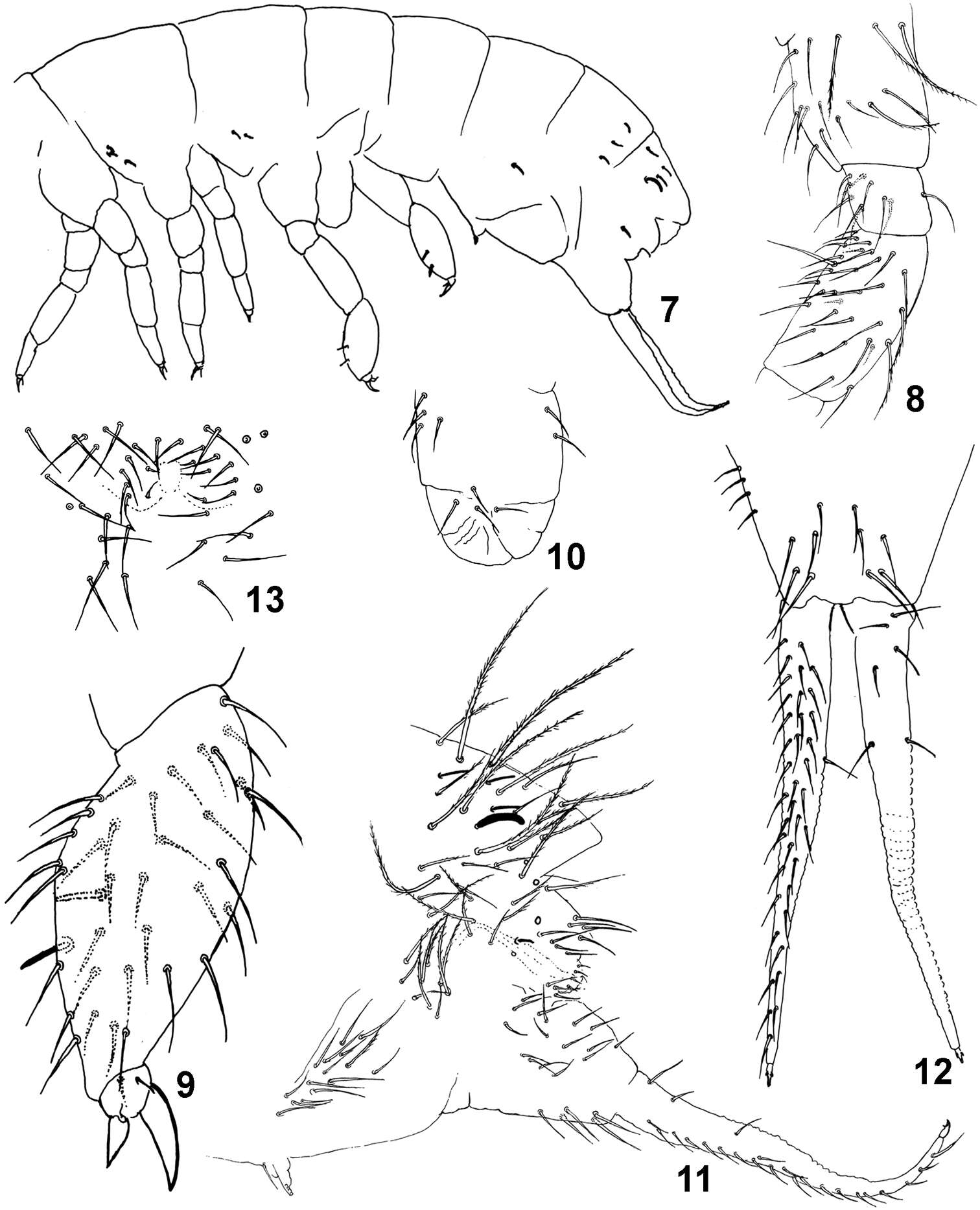
Figures 2–11.Tomocerus postantennalis sp. n. 2 PAO and ocelli 3 cephalic dorsal chaetotaxy 4 labrum 5 mandible 6 maxillary lamella five 7 trochanteral-femoral organ 8 tibiotarsus 9 anterior view of distal tibiotarsal chaetae (t: tenent hair, a: accessory chaetae, g: guard chaetae) 10 claw 11 tenaculum. Scale bars: 2, 7, 9, 10, 11 = 50 μm; 3 = 500 μm; 4, 5, 8 = 100 μm; 6= 20 μm.
-
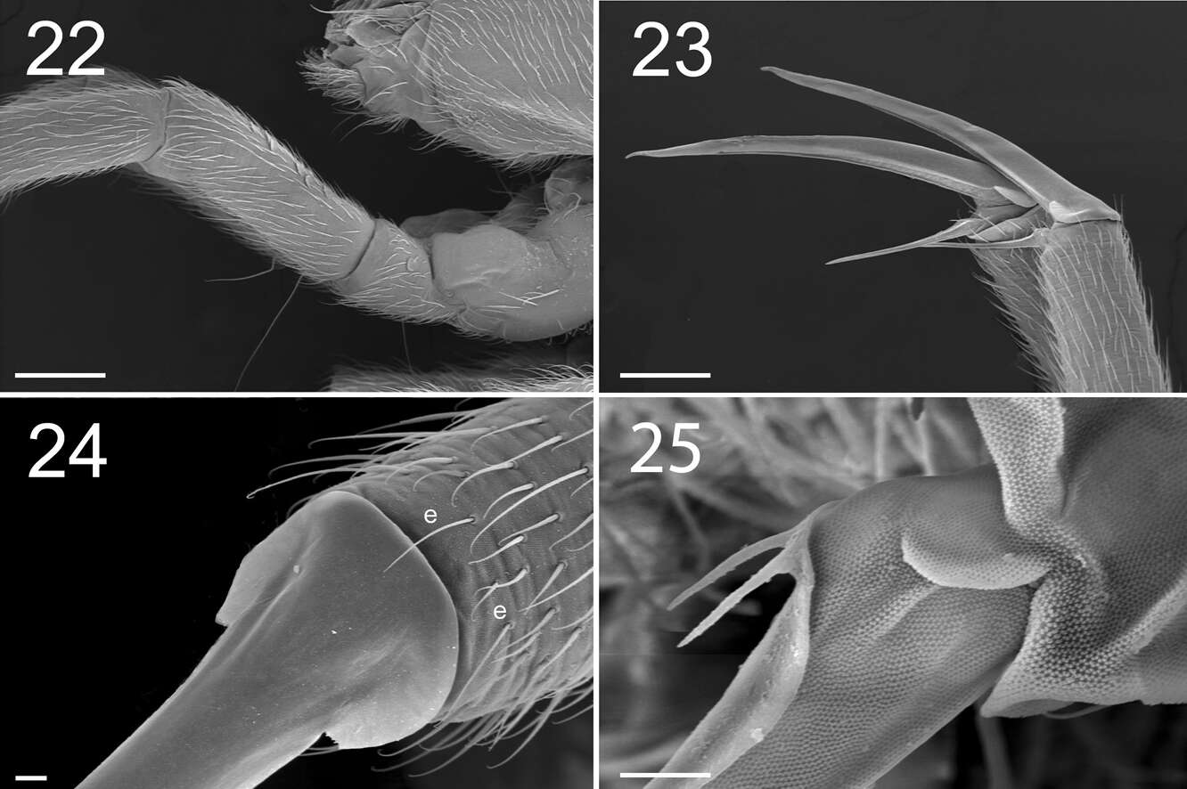
Marko Lukić, Céline Houssin, Louis Deharveng
Zookeys
Figures 22–25. Tritomurus veles sp. n. (SEM). 22 Leg I, with ventro-basal macrochaetae of femur and ventral macrochaetae of trochanter (scale 100 μm); the second visible macrochaetae of femur belongs to other leg 23 Claws of legs I (scale 100 µm) 24 Claw of leg I, basal part in dorsal view (scale 10µm); e, thin distal tenent hairs 25 Bifurcate empodial appendage of leg II (scale 10 μm).
-
WWT Knapp Reserve, Worcs. SO748520
-
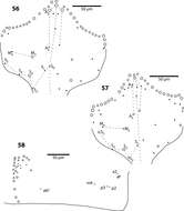
Maria Cleide de Mendonça, Eduardo A. Abrantes, Ana Carolina R. Neves
Zookeys
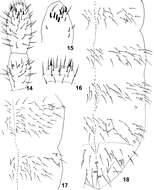
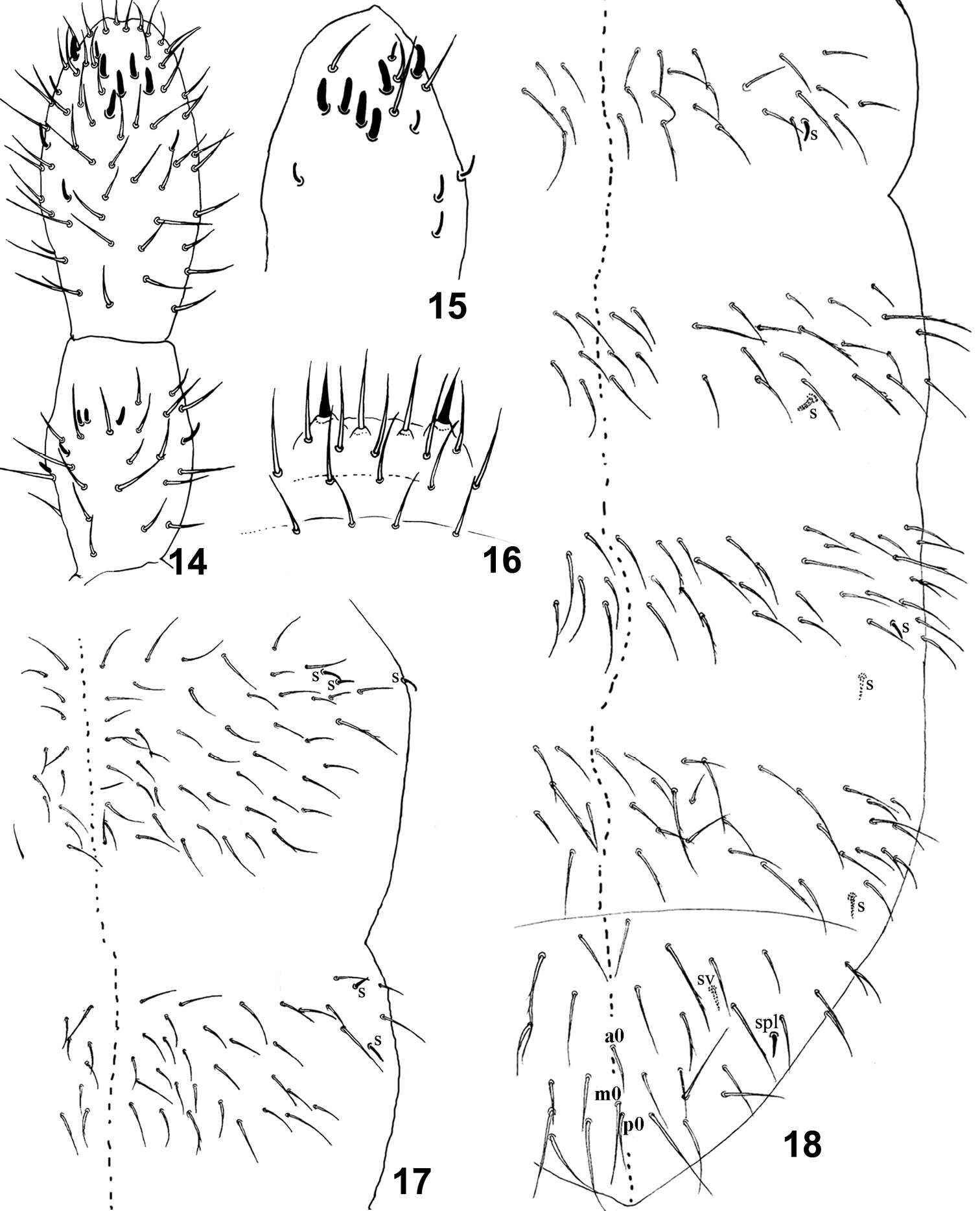
Figure 14–18.Isotomiella uaisp.n. 14 Ant III-IV Dorsal view 15 Sensillary pattern of Ant IV 16 Labral chaetae 17 Dorsal chaetotaxy of Th II-III 18 Dorsal chaetotaxy of Abd I-VI.
-
Felipe N. Soto-Adames, Steven J. Taylor
Zookeys
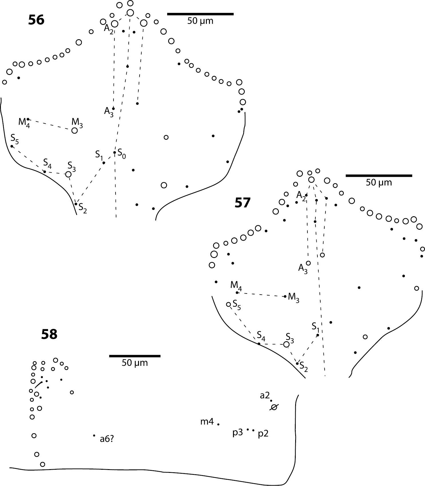
Figures 56–58.Trogolaphysa geminata (56) and Trogolaphysa riopedrensis (57, 58) 56 Head dorsal chaetotaxy 57 Head dorsal chaetotaxy 58 Metathorax chaetotaxy.
-
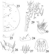
Xiang-Qun Yuan, Zhi-Xiang Pan
Zookeys
Figures 23–29.Sinella triseta sp. n. 23 dorsal cephalic chaetotaxy 24 basal chaetae of Ant. I 25 basal chaetae of Ant. II 26 Ant. III organ 27 clypeus 28 labrum 29 labial base 30 labial palp.
-
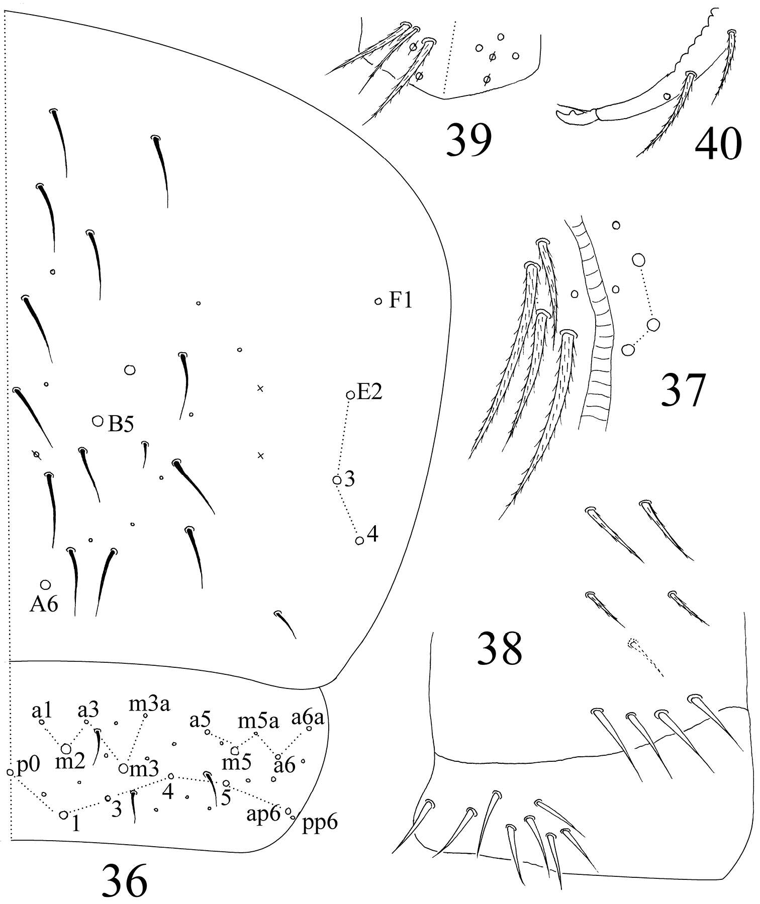
Sopark Jantarit, Chutamas Satasook, Louis Deharveng
Zookeys
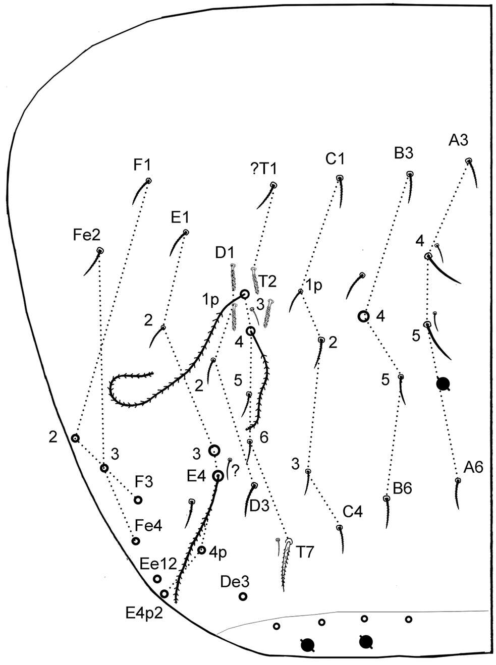
Figure 6.Cyphoderus songkhlaensis sp. n. continued, Szeptycki’s notation of tergal chaetae on Abd.IV (Szeptycki 1979).
-
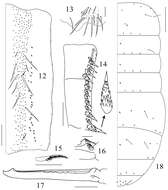
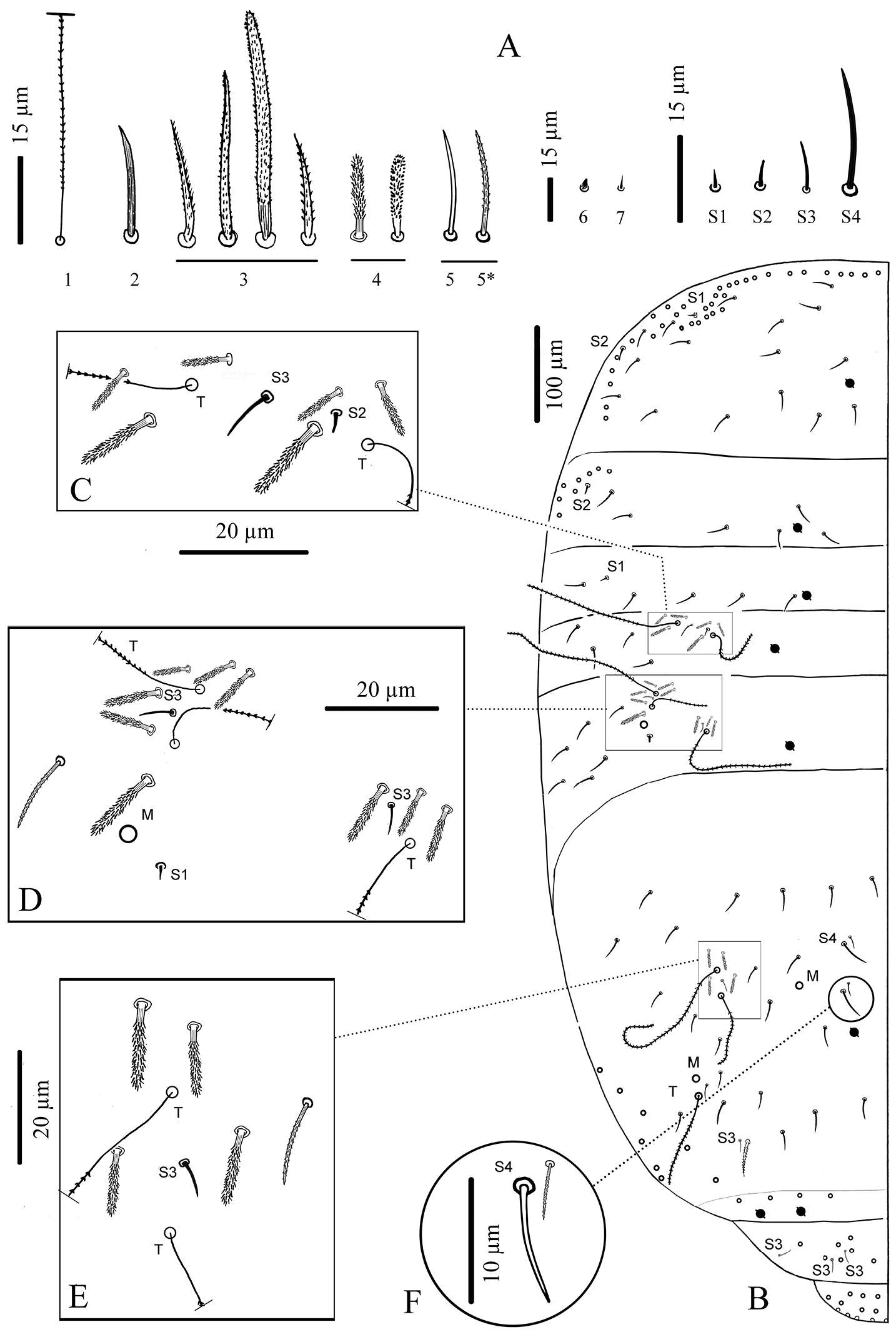
Daoyuan Yu, Feng Zhang, Louis Deharveng
Zookeys
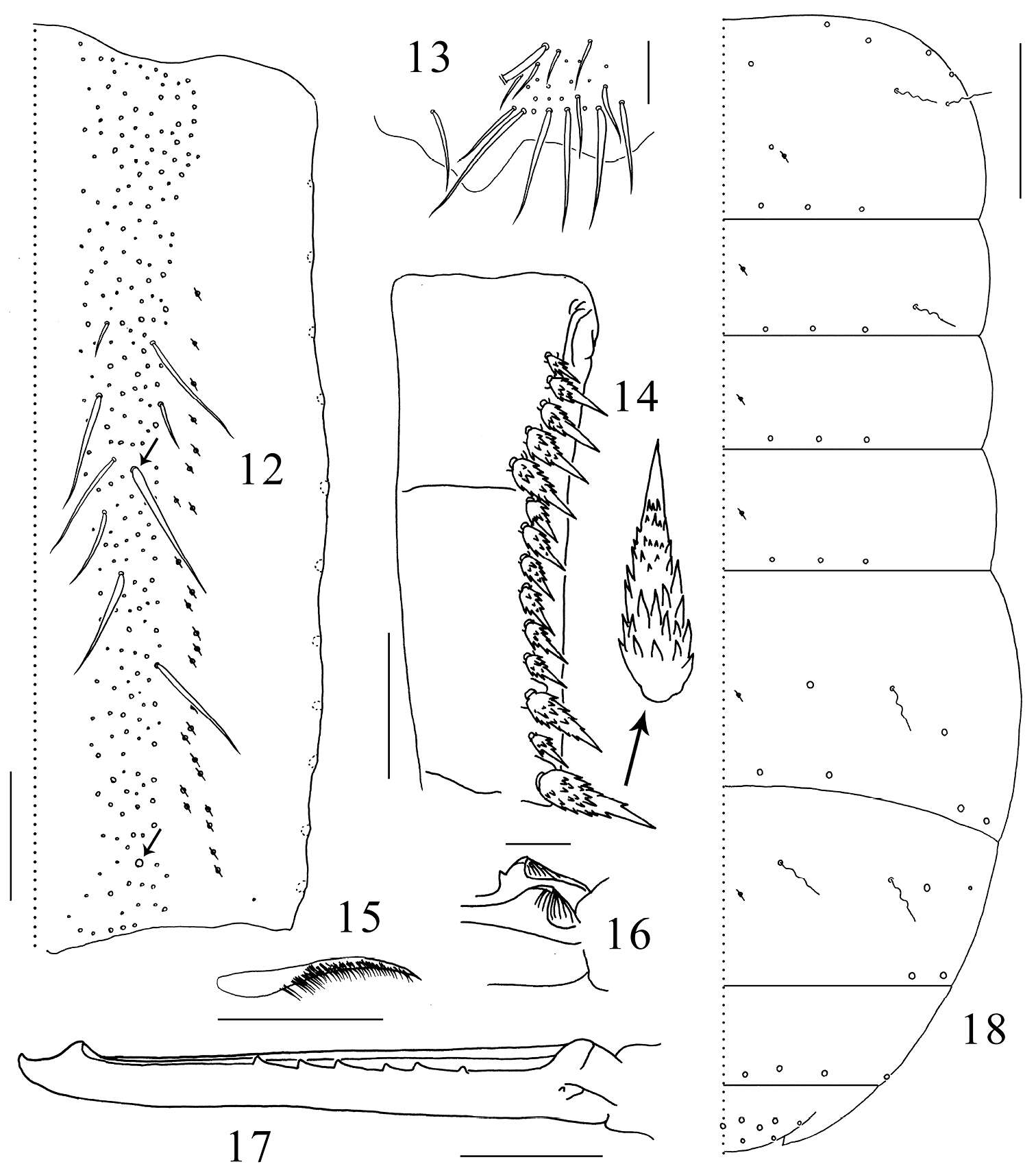
Figures 12–18.Tomocerus postantennalis sp. n. 12 dorsal face of manubrium (right side; prominent chaetae arrowed) 13 disto-dorsal chaetae on manubrium (left side) 14 dental spines (left side) 15 feathered chaeta on dens 16 basal teeth of left mucro 17 right mucro 18 body chaetotaxy. Scale bars: 12, 14 = 100μm; 13, 17 = 50 μm; 15, 16 = 21 μm; 18 = 400 μm. Large circles: macrochaetae; small circles: mesochaetae; wavy lines: bothriotricha; circles with a slash: pseudopores.
-
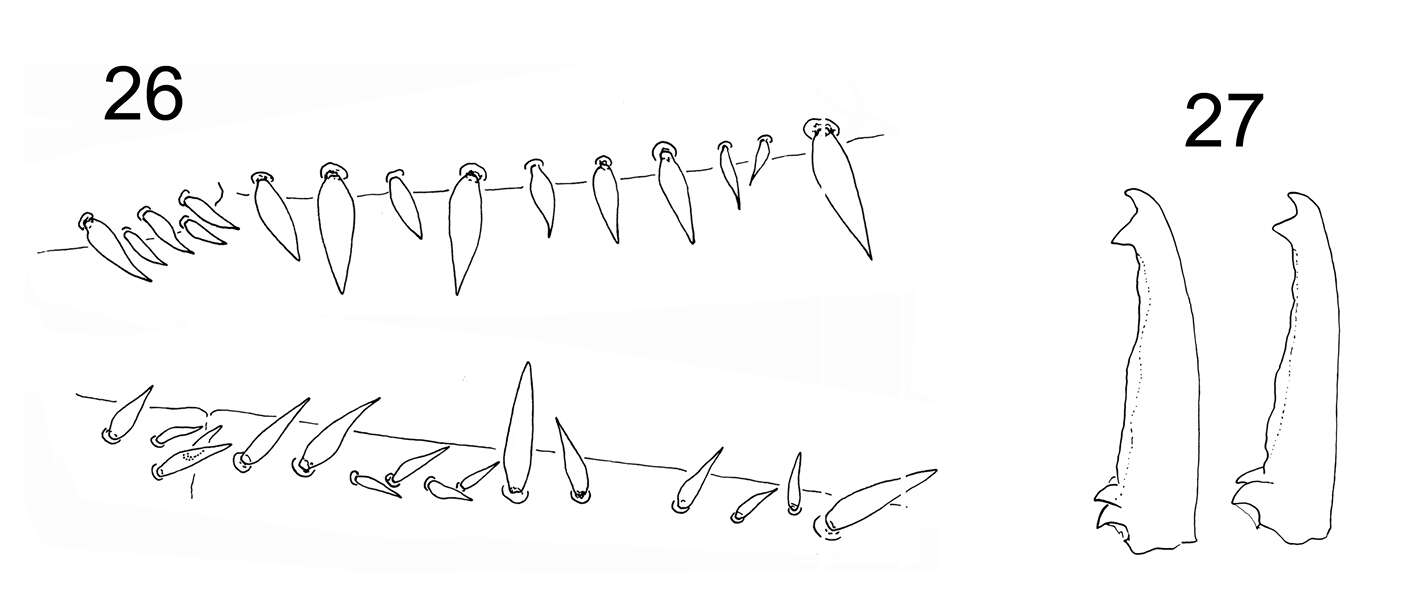
Marko Lukić, Céline Houssin, Louis Deharveng
Zookeys
Figures 26–27. Tritomurus veles sp. n. 26 Dental spines formula in a female specimen: 4/2,4,1,4,1 (lower, right dens) and 5/1,1,1,1, 2,1,2,1 (upper, left dens) 27 Mucro in two different specimens.
-
Maria Cleide de Mendonça, Eduardo A. Abrantes, Ana Carolina R. Neves
Zookeys
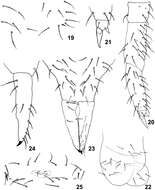
Figure 19–25.Isotomiella uai sp.n. 19 Detail of chaetotaxy of Abd II 20 Leg III 21 Unguis of leg III 22 Ventral tube 23 Furca 24 Lateral view of dens and mucro 25 Female genital opening.
-
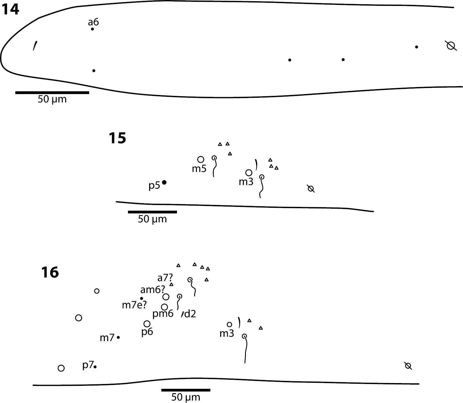
Felipe N. Soto-Adames, Steven J. Taylor
Zookeys
Figures 14–16.Trogolaphysa giordanoae sp. n. Dorsal chaetotaxy of abdominal segments 1–3, triangles are fan-shaped setae, circles are macrochaetae, filled are circles ciliate microchaeta14 First abdominal segment 15 Second abdominal segment 16 Third abdominal segment.