-
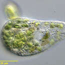
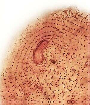
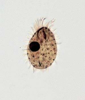
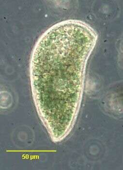
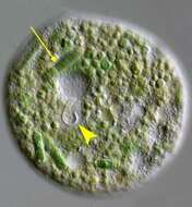
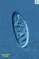
Portrait of Platyophrya sphagni, a colpodid ciliate. Cells are flask shaped with an indistinct subapical oral aperture. The cells are very flexible and squirm about rather slowly along the substratum. When swimming they tend to rotate around their long axis. The right lateral surface is more densely ciliated than the left (best seen anteriorly here). The cytoplasm contains both large ingested algae and smaller symbiotic zoochlorellae which distinguish this species from other members of the genus. The central macronucleus and micronucleus are seen in this image. Many small extrusomes are also seen in this image. From freshwater pond near Boise, Idaho. DIC optics.
-
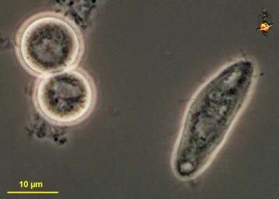
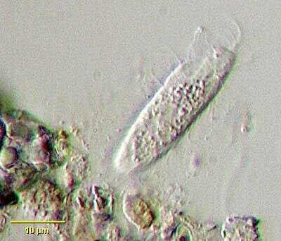
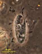
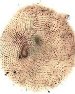
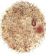
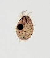
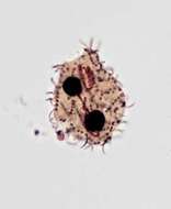
Kuklikophrya ougandae (DRAGESCO, 1972) FOISSNER, 1993. Collected from an organically enriched temporary freshwater pool near Boise, Idaho. May 2006.Brightfield.
-
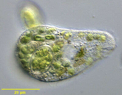
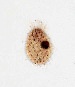
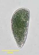
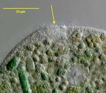
Ventral view of Platyophrya sphagni, a colpodid ciliate. Cells are flask shaped with an indistinct subapical oral aperture (well seen in this image). The cells are very flexible and squirm about rather slowly along the substratum. When swimming they tend to rotate around their long axis. The oral aperture has a right paraoral membrane, and is bordered on the left by a row of adoral organelles and an outer "postoral pseudomembrane". The right lateral surface is more densely ciliated than the left. The cytoplasm contains both large ingested algae and smaller symbiotic zoochlorellae which distinguish this species from other members of the genus. Many small extrusomes are also seen in this image. From freshwater pond near Boise, Idaho. DIC optics.
-
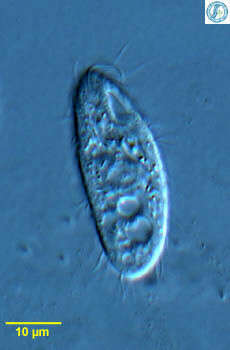
Ventral view of Kuklikophrya ougandae (DRAGESCO, 1972) FOISSNER, 1993 squashed and distorted by pressure from the coverglass. The arrowhead indicates the oral apparatus. The arrow points to a segment of ingested filamentous cyanobacterium. Collected from an organically enriched temporary freshwater pool near Boise, Idaho. May 2006.DIC.
-
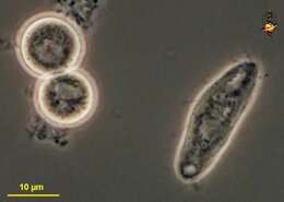
Cyrtolophosis (sir-till-owe-foe-sis) ciliate usually located in a flimsy mucus sheath, recessed mouth with small arc of projecting cilia. Eats bacteria. Phase contrast.
-
Detail view of the oral basket of Kuklikophrya ougandae (DRAGESCO, 1972) FOISSNER, 1993 squashed and distorted by pressure from the coverglass. The arrow points to the mematodesmata of the funnel-shaped cytopharynx.. Collected from an organically enriched temporary freshwater pool near Boise, Idaho. May 2006.DIC.
-
Cyrtolophosis (sir-till-owe-foe-sis) ciliate usually located in a flimsy mucus sheath, recessed mouth with small arc of projecting cilia. Eats bacteria. Phase contrast.
-
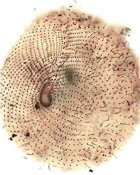
Ventral infraciliature of Kuklikophrya ougandae (DRAGESCO, 1972) FOISSNER, 1993 squashed and distorted by pressure from the coverglass and by fixation. The yellow arrow indicates the posterior end of the oral apparatus. The green arrow points to the left termination of the paraoral membrane.The adoral membranelles are arrayed in an oblique line from this termination to the left anterior apex of the cell (yellow bracket).Stained by the silver carbonate technique (see Foissner, W. Europ. J. Protistol., 27:313-330;1991). Collected from an organically enriched temporary freshwater pool near Boise, Idaho. May 2006.Brightfield.
-
Cyrtolophosis (sir-toe-low-foe-sis), usually found in mucus sheaths, with only the anterior end projecting. Typically with a quiff of anterior cilia, and the mouth located just subapically. Cysts also form. Phase contrast micrograph.
-
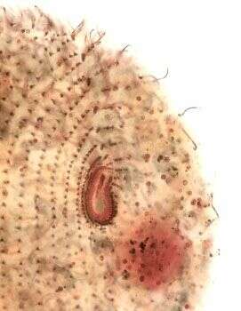
Ventral infraciliature of Kuklikophrya ougandae (DRAGESCO, 1972) FOISSNER, 1993. Stained by the silver carbonate technique (see Foissner, W. Europ. J. Protistol., 27:313-330;1991). Collected from an organically enriched temporary freshwater pool near Boise, Idaho. May 2006.Brightfield.
-
Cyrtolophosis (sir-toe-low-foe-sis), usually found in mucus sheaths, with only the anterior end projecting. Typically with a quiff of anterior cilia, and the mouth located just subapically. Cysts also form. Phase contrast micrograph.
-
Ventral infraciliature of Kuklikophrya ougandae (DRAGESCO, 1972) FOISSNER, 1993. Stained by the silver carbonate technique (see Foissner, W. Europ. J. Protistol., 27:313-330;1991). Collected from an organically enriched temporary freshwater pool near Boise, Idaho. May 2006.Brightfield.
-
This is a ciliate which consumesbacteria, the mouth being located to the upper right of this cell. Usually found in a mucus tube. From Lake Donghu, China. Differential interference contrast micrograph.
-
Oral infraciliature of Kuklikophrya ougandae (DRAGESCO, 1972) FOISSNER, 1993 The adoral membranelles are arrayed in an oblique line from the termination of the left part of the paraoral membrane to the left anterior apex of the cell.Stained by the silver carbonate technique (see Foissner, W. Europ. J. Protistol., 27:313-330;1991). Collected from an organically enriched temporary freshwater pool near Boise, Idaho. May 2006.Brightfield.
-
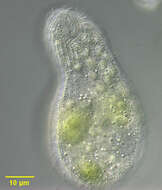
Cyrtolophosis, a colorless colpodid ciliate most often found in a mucous sheath (seen in this image) possibly secreted by mucocysts. Cells often flee the mucous sheath and swim free when transferred to a microscope slide. There is a characteristic prominent anterior ciliary tuft and well developed 'adoral ciliary organelles" (similar in appearance to an undulating membrane) along the left margin of the buccal cavity (both are seen in this image). From temporary rainwater pool in grass field near Boise, Idaho. Oblique illumination
-
Oral infraciliature of Kuklikophrya ougandae (DRAGESCO, 1972) FOISSNER, 1993. The adoral membranelles are arrayed in an oblique line from the termination of the left part of the paraoral membrane to the left anterior apex of the cell. Stained by the silver carbonate technique (see Foissner, W. Europ. J. Protistol., 27:313-330;1991). Collected from an organically enriched temporary freshwater pool near Boise, Idaho. May 2006.Brightfield.
-
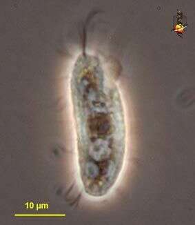
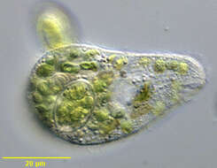
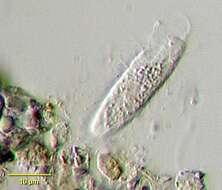
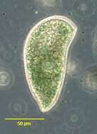
Portrait of Cyrtolophosis mucicola (Stokes,1885). An individual is seen here just fleeing its mucus dwelling tube (to viewer's left of organism). The tube is apparently formed from the contents of mucocysts.Collected from a freshwater pond near Boise, Idaho. July 2005.Phase contrast.
-
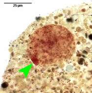
Nuclear apparatus of Kuklikophrya ougandae (DRAGESCO, 1972) FOISSNER, 1993. The more densely stained micronucleus (arrowhead) is attached to the macronucleus and may lie within its nuclear membrane. A similar arrangement is seen in the genus Rostrophrya.Stained by the silver carbonate technique (see Foissner, W. Europ. J. Protistol., 27:313-330;1991). Collected from an organically enriched temporary freshwater pool near Boise, Idaho. May 2006.Brightfield.
-
Ventral infraciliature of Cyrtolophosis mucicola (Stokes,1885). The paraoral membrane is seen to the viewer's right and the adoral membranelles to the left.A short "oblique kinety" is present between the anterior end of the paraoral membrane and the most anterior adoral membranelle. The sparse (~10) slightly spiralled somatic kineties are visible.Collected from a freshwater pond near Boise, Idaho.June 2005). Stained by the silver carbonate technique (see Foissner, W. Europ. J. Protistol., 27:313-330;1991). Brightfield.
-
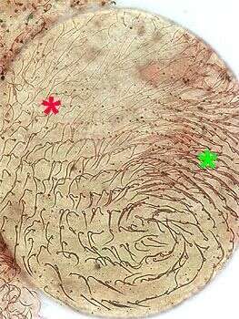
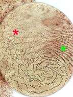
Dorsal somatic ciliature of Kuklikophrya ougandae (DRAGESCO, 1972) FOISSNER, 1993. The cilia are paired in the right anterior portion of the cell (green asterisk) but only the posterior basal body of the dikinetids in other parts of the body are ciliated (red asterisk).Stained by the silver carbonate technique (see Foissner, W. Europ. J. Protistol., 27:313-330;1991). Collected from an organically enriched temporary freshwater pool near Boise, Idaho. May 2006.Brightfield.
-
Dorsal infraciliature of Cyrtolophosis mucicola (Stokes, 1885).The sparse (~10) slightly spiralled somatic kineties are seen here.Collected from a freshwater pond near Boise, Idaho.June 2005). Stained by the silver carbonate technique (see Foissner, W. Europ. J. Protistol., 27:313-330;1991). Brightfield.
-
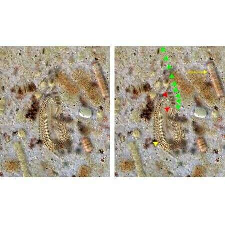
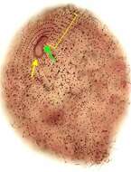
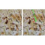
Oral infraciliature of Kuklikophrya ougandae (DRAGESCO, 1972) FOISSNER, 1993.The paraoral membrane is continuous except for a short open area at the left (between red arrowheads) anterior portion at which point the proximal adoral membranelles arise. The adoral membranelles are arrayed in an oblique line from the termination of the left part of the paraoral membrane to the left anterior apex of the cell (green arrowheads). Several postoral somatic kineties bend sharply around the posterior end of the paraoral membrane to parallel the right side of the oral opening (yellow arrowhead). A segment of ingested cyanobacteria is indicated by the yellow arrow.Stained by the silver carbonate technique (see Foissner, W. Europ. J. Protistol., 27:313-330;1991). Collected from an organically enriched temporary freshwater pool near Boise, Idaho. May 2006.DIC.
-
Ventral infraciliature of Cyrtolophosis mucicola (Stokes,1885)in middle division. The paraoral membrane is seen to the viewer's right and the adoral membranelles to the left.A very short "oblique kinety" is present between the anterior end of the paraoral membrane and the most anterior adoral membranelle. The sparse (~10) slightly spiralled somatic kineties are visible.The oral apparatus of the forming posterior daughter cell (opisthe) is obscured by its densley stained macronucleus. Collected from a freshwater pond near Boise, Idaho.June 2005). Stained by the silver carbonate technique (see Foissner, W. Europ. J. Protistol., 27:313-330;1991). Brightfield.
-
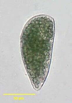
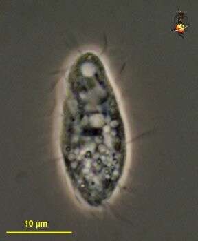
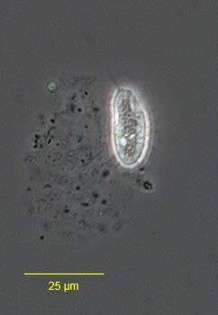
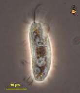
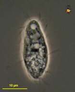
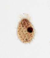
Kuklikophrya ougandae (DRAGESCO, 1972) FOISSNER, 1993. The organisms appear dark green in vivo due to large numbers of ingested cyanobacterial fragments.Collected from an organically enriched temporary freshwater pool near Boise, Idaho. May 2006.Phase contrast.