-
Herrera de Soria, Castille and Leon, Spain
-
Ribadelago, Castille and Leon, Spain
-
-
Herrera de Soria, Castille and Leon, Spain
-
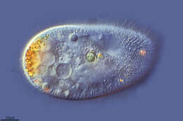
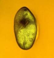
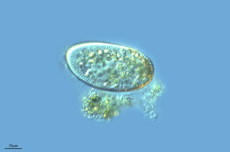
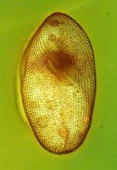
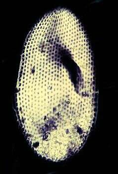
This cell has been fixed and stained with protargol which shows up cilia and other large microtubular structure. The image is of the ventral side, and shows the arrangem,ent of the cilia into kineties, the mpouth with more densely packed rows of cilia within the mouth, and the cytopyge or cell anus through which undigested residues of food will be discharged.
-
Ribadelago de Franco, Castilla y Len, Espaa
-
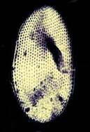
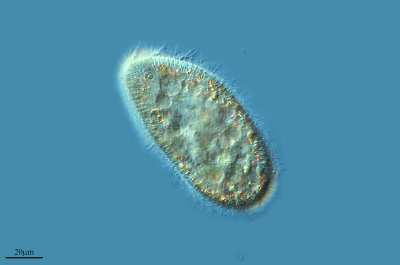
This image is of a cell that has been fixed (killed and reserved) and then stained with protargol which stains cilia and other microtubular and basic compounds a dark brown. The image shows the lines of cilia - the kineties, the mouth region and the cell anus or cytopyge, through which undigested residues of food are expelled.
-
A Veiga, Galicia, Spain
-
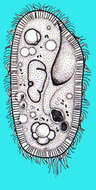
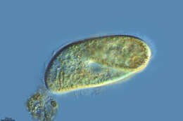
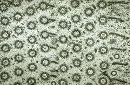
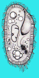
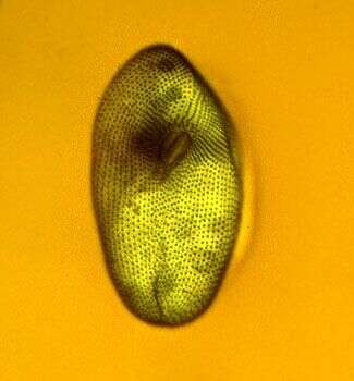
This cell has been placed in a suspension of a stain called nigrosin and then the preparation has been allowed to dry out. The stain dries around the cell showing the rows of depressions in the cell surface that correspond with where the cilia emerge. This species has two contractile vacuoles and they are unusual in the genus in not having radiating canals and because the pore is a long twisted structure. The pore of the posterior contractile vacuole can be seen as a twisted line in the posterior part of the cell.
-
A Veiga, Galicia, Spain
-
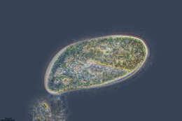
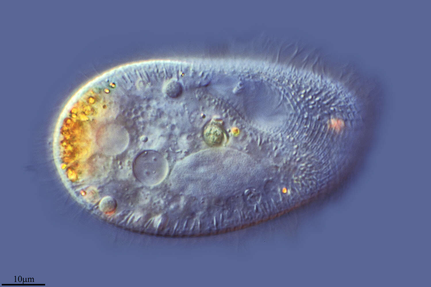
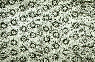
This is a transmission electron micrograph of a thin section through some of the cilia in the mouth. Paramecium cells collect food using densely packed cilia. There are three bands each four cilia wide. The image is of the two peniculi, the third band (the quadrulus) has more widely spaced cilia and is not visible.
-
Galende, Castille and Leon, Spain