-
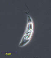
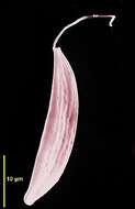
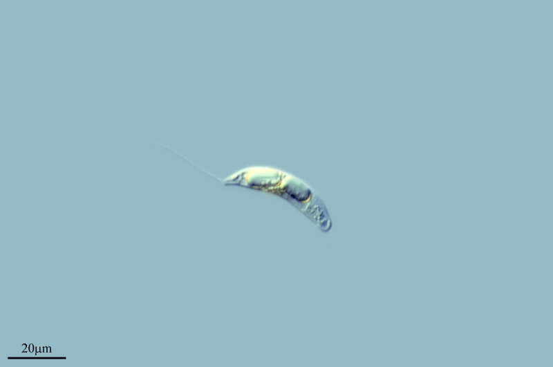
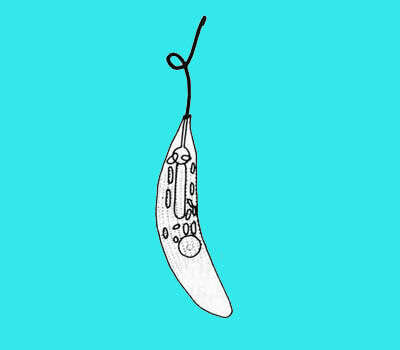
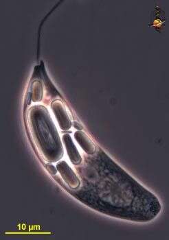
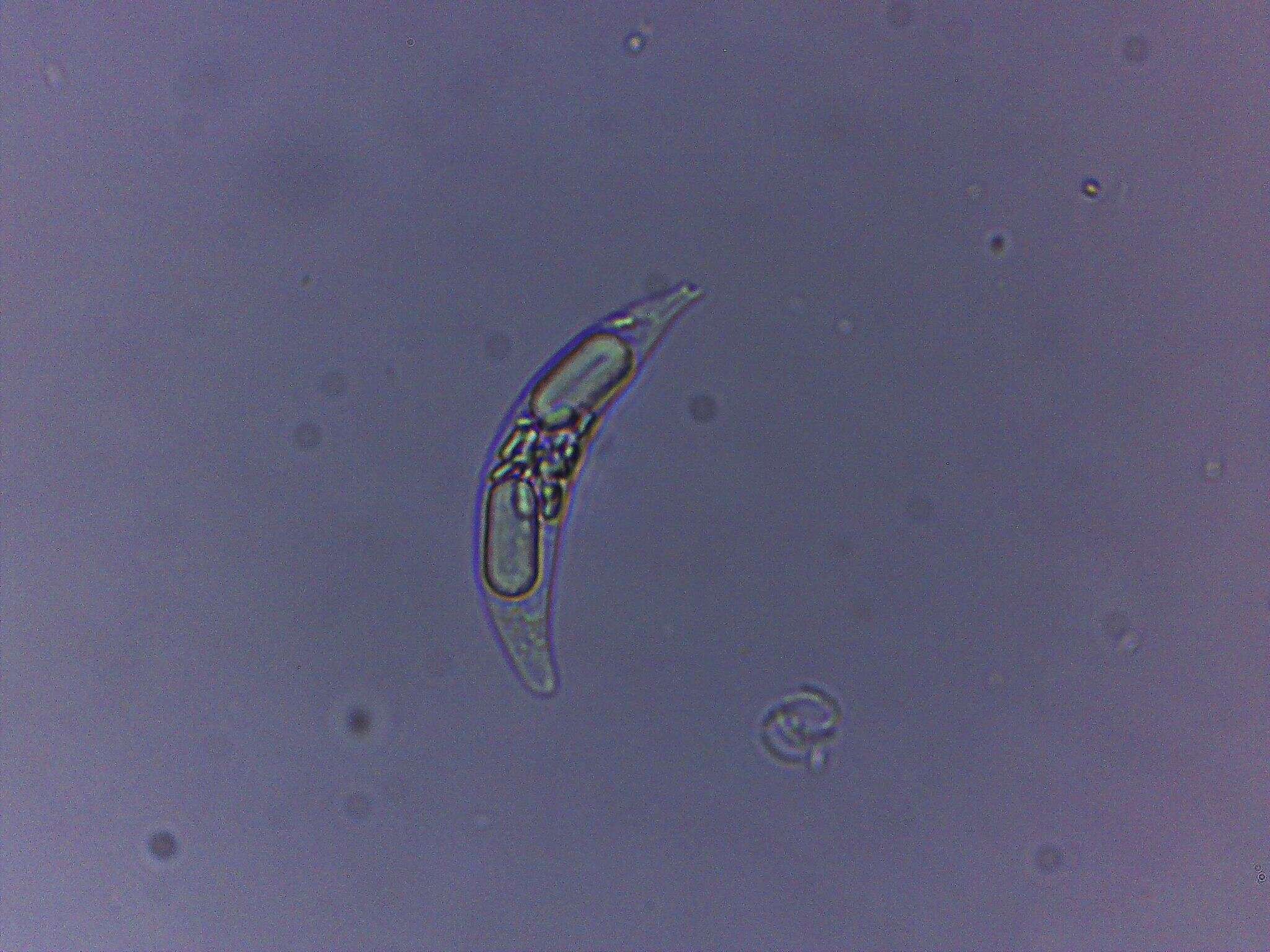
Portrait of the colorless euglenoid flagellate, Menoidium bibacillatum (Pringsheim, 1942). Strongly flattened. One side curved with the other more straight. One emergent flagellum. Stigma absent. Paramylon bodies are dimorphic with smaller round and larger elongate ring forms. Swims rotating on long axis. Highly refractile. From standing freshwater near Boise, Idaho. DIC.
-
Barrio Ballinas, Castille and Leon, Spain
-
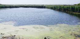
Ribadelago de Franco, Castille and Leon, Spain
-
Portrait of the colorles euglenoid flagellate,Menoidium bibacillatum (Pringsheim, 1942). Strongly flattened. One side curved with the other more straight. One emergent flagellum. Stigma absent. Paramylon bodies are dimorphic with smaller round and larger elongate ring forms. Swims rotating on long axis. Highly refractile. From standing freshwater near Boise, Idaho. Phase contrast.
-
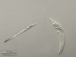
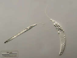
Scanning electron micrograph showing the anterior flagellum and the cell shape.
-
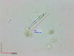
Portrait of Menoidium a colorless euglenoid flagellate. Strongly flattened. One side curved with the other more straight. One emergent flagellum. Stigma absent. Paramylon bodies are dimorphic with smaller round and larger elongate ring forms. Swims rotating on long axis. Highly refractile. From standing freshwater near Boise, Idaho. Brightfield.
-
Portrait of Menoidium a colorless euglenoid flagellate. Strongly flattened. One side curved with the other more straight. One emergent flagellum. Stigma absent. Paramylon bodies are dimorphic with smaller round and larger elongate ring forms. Swims rotating on long axis. From standing freshwater near Boise, Idaho. Phase contrast.
-
-
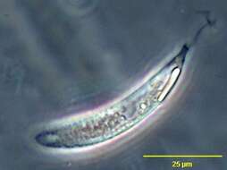
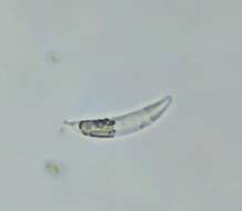
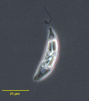
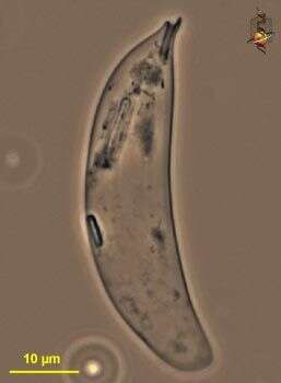
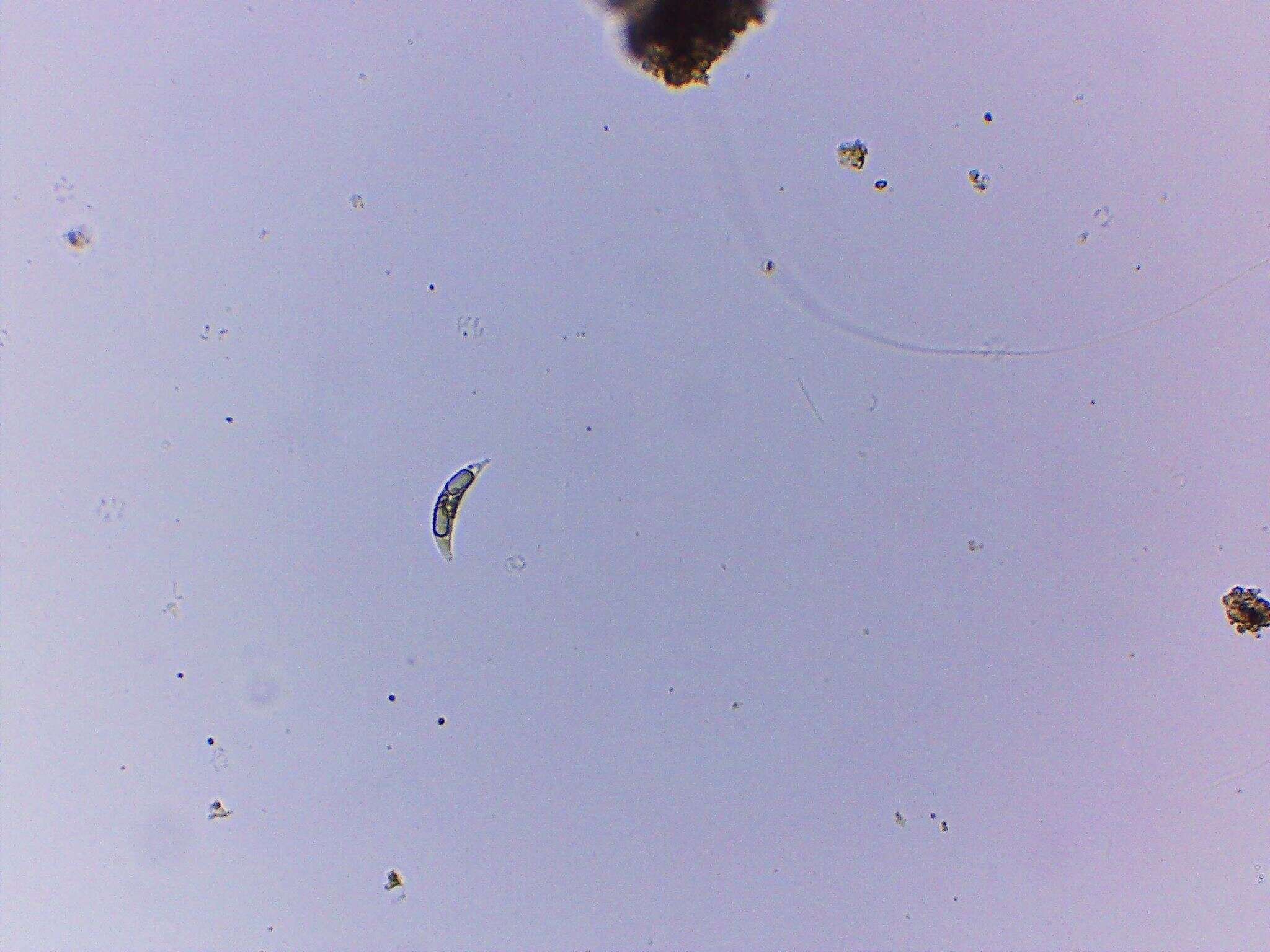
Menoidium pellucidum. Cell observed in freshwater habitats in the vicinity of Broome, Western Australia in September 2003. This image was taken using phase contrast optics. This work was supported by the Australian Biological Resources Study.
-
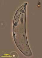
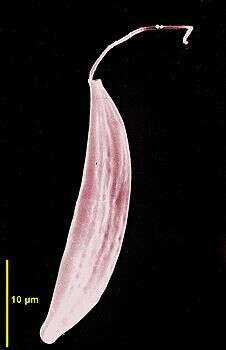
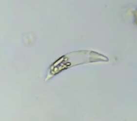
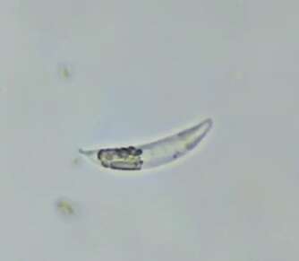
Menoidium pellucidum. Empty pellicle observed in freshwater habitats in the vicinity of Broome, Western Australia in September 2003. This work was supported by the Australian Biological Resources Study.
-
Menoidium pellucidum. Cell observed in freshwater habitats in the vicinity of Broome, Western Australia in September 2003. This image was taken using phase contrast optics. This work was supported by the Australian Biological Resources Study.
-
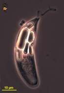
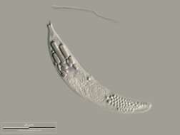
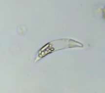
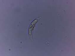
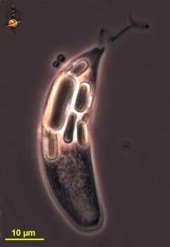
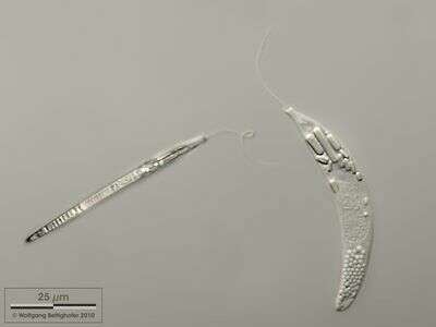
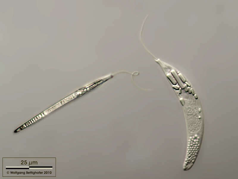
Top and lateral view of a cell. Sample from the pond Hegne Moor situated in the vicinity of Lake Constance (Bodensee, Southern Germany). Images were taken using Zeiss Universal with Olympus C7070 CCD camera.
-
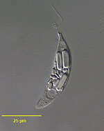
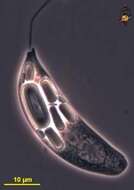
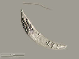
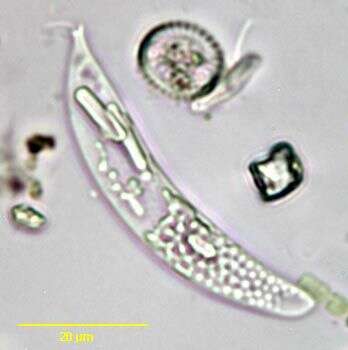
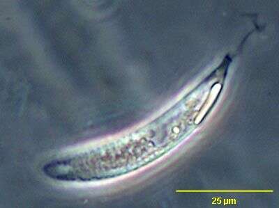
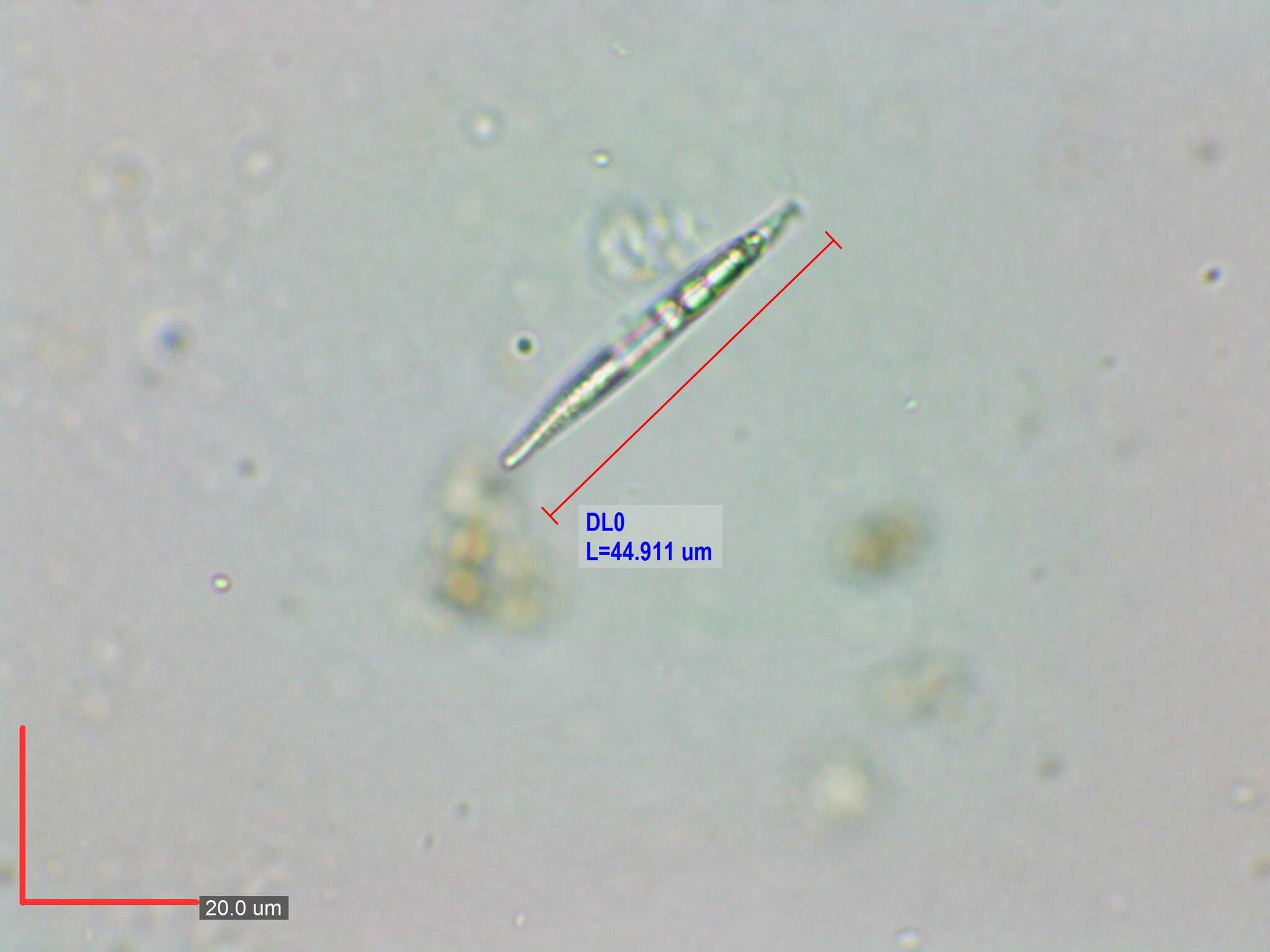
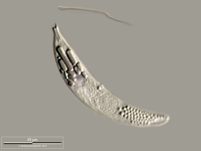
There are several paramylon bodies in the apical region, the nucleus is in the center of the cell. Scale bar indicates 25 µm. Sample from the pond Hegne Moor situated in the vicinity of Lake Constance (Bodensee, Southern Germany). Images were taken using Zeiss Universal with Olympus C7070 CCD camera.
-
-
-
-
-
-
-
-
-
Menoidium pellucidum Top and lateral view of a cell. Sample from the pond Hegne Moor situated in the vicinity of Lake Constance. Images were taken using Zeiss Universal with Olympus C7070 CCD camera.Image under Creative Commons License V 3.0 (CC BY-NC-SA). Place name: Bog Hegne Moor near Lake Constance (Germany) Latitude: 47.718106 Longitude: 9.093974 Drauf- und Seitenansicht einer Zelle. Zellen starr, nicht metabolisierend. Probe aus dem Simmelried nahe Konstanz. Mikrotechnik: Zeiss Universal, Kamera: Olympus C7070. Creative Commons License V 3.0 (CC BY-NC-SA). For permission to use of (high-resolution) images please contact postmaster@protisten.de.
-
Menoidium pellucidum There are several paramylon bodies in the apical region, the nucleus is in the center of the cell. Scale bar indicates 25 µm. Sample from ponds pond situated in the vicinity of Lake Constance. Images were taken using Zeiss Universal with Olympus C7070 CCD camera.Image under Creative Commons License V 3.0 (CC BY-NC-SA). Place name: Bog Hegne Moor near Lake Constance (Germany) Latitude: 47.718106 Longitude: 9.093974 Das Bild zeigt mehrere Paramylon-Körper (Speicherstoff) im apikalen Bereich (vorn, Nähe Mundöffnung), der Kern liegt in der Mitte der Zelle. Der Messbalken markiert eine Länge von 25 µm. Probe aus dem Simmelried nahe Konstanz. Mikrotechnik: Zeiss Universal, Kamera: Olympus C7070. Creative Commons License V 3.0 (CC BY-NC-SA). For permission to use of (high-resolution) images please contact postmaster@protisten.de.