-
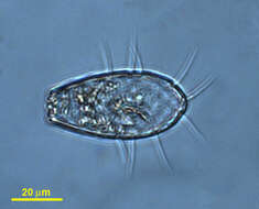
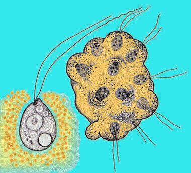
Assulina (ass-you-line-a) a testate amoeba with a subapical mouth located on the ventral surface of the slightly flattened lorica. Common in mosses. Phase contrast.
-
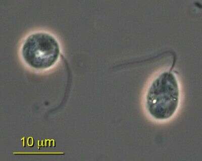
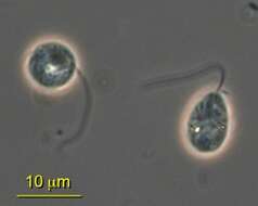
Quadricilia rotundata (Skuja, 1948) V+rs, 1992. Cells are 5-9 x 10-15 microns, globular cells 10-20 microns in diameter. Cell globular, or nearly so, with 4 (-8) unequal, smooth, acronematic flagella inserted anteriorly in a shallow depression. The flagella are 1-3 times the diameter of the cell body. Many thin, branched pseudopodia may be produced from any point of the cell surface. Cytoplasm sometimes highly vesiculate, nucleus central or in the cell anterior.
-
ATCC culture 50636.
-
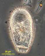
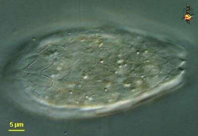
Euglypha (you-g-lie-fa) is a widespread and common testate amoeba. This one was found in a sample of moss, and this is a habitat in which testate amoebae are common. The aperture is to the top of the image. The test is made up of scale arranged a bit like fish scales, and their are marginal spine scales. This test no longer contains an amoeba. Phase contrast.
-
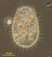
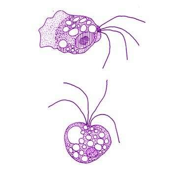
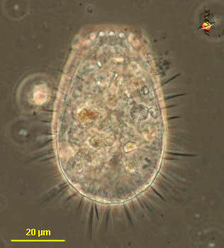
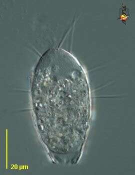
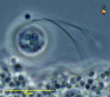
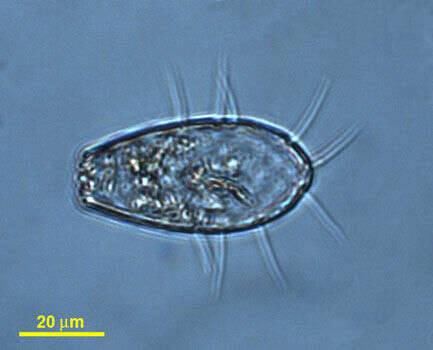
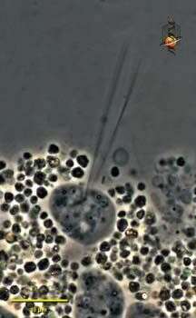
Abollifer prolabens Voers, 1992. The cell is about 8-12 x 10-20 microns It is ovoid-oval and dorso-ventrally flattened with a deep anterior or depression into which the flagellum inserts. The sides of this depression are swollen. The cell surface is rigid and granulated, the cell appears opaque and probably has a pellicle. The cell moves by gliding, but it may detach from the substrate and jerk through the water with an irregular sinusoidal flagellar beat, a second shorter trailing flagellum is sometimes present.
-
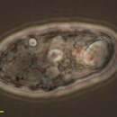
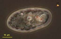
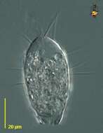
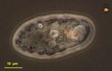
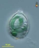
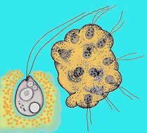
Paulinella (paul-in-ella) is a testate amoeba but this species is distinguished by the presence of (usually two) curved endosymbiotic blue green algae. The small aperture of the lorica is to the top of the image. Differential interference contrast.
-
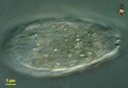
Euglypha (you-gly-fa) a shelled amoeba with filose pseudopodia (although this picture is only of the test) and with the test covered in small overlapping scales and a terminal aperture. The aperture through which the pseudopodia emerge is located to the bottom of the picture. Phase contrast.
-
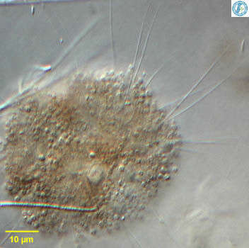
Spongomonas (spong-owe-moan-ass), is a solitary or colonial spongomonad flagellate, in which the cells are located within a more or less globular matrix formed from adhering small globules of mucilage. Many cells were dislodged while this sample was being prepared. Phase contrast.
-
Euglypha (you-gly-fa) a shelled amoeba with filose pseudopodia (although this picture is only of the test) and with the test covered in small overlapping scales and a terminal aperture. This species also with some spines. The aperture through which the pseudopodia emerge is located to the bottom of the picture. Phase contrast.
-
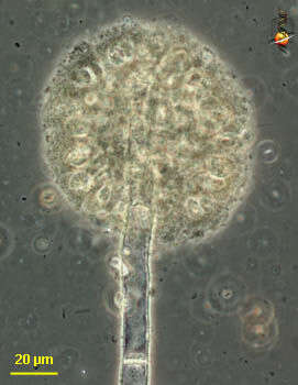
Spongomonas (spong-owe-moan-ass), is a solitary or colonial spongomonad flagellate, in which the cells are located within a more or less globular matrix formed from adhering small globules of mucilage. In this case the colony has formed at the end of some extraneous fibre. Phase contrast.
-
Euglypha (you-gly-fa) a shelled amoeba with filose pseudopodia (although this picture is only of the test) and with the test covered in small overlapping scales and a terminal aperture. The aperture through which the pseudopodia emerge is located to the bottom of the picture. Differential interference contrast.
-
Spongomonas (spong-owe-moan-ass), is a solitary or colonial spongomonad flagellate, in which the cells are located within a more or less globular matrix formed from adhering small globules of mucilage. This is an image of a cluster of cells picked from the surface of the pond. Phase contrast.
-
Euglypha (you-gly-fa) a shelled amoeba with filose pseudopodia (although this picture is only of the test) and with the test covered in small overlapping scales and a terminal aperture.Differential interference contrast.
-
Spongomonas (spong-owe-moan-ass), is a solitary or colonial spongomonad flagellate, in which the cells are located within a more or less globular matrix formed from adhering small globules of mucilage. This is an image of a thin cluster of cells. Phase contrast.
-
-
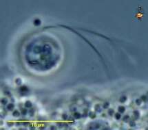
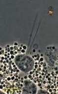
Spongomonas (spong-owe-moan-ass), is a solitary or colonial spongomonad flagellate, in which the cells are located within a more or less globular matrix formed from adhering small globules of mucilage. Detail of single cell showing the two flagella Phase contrast.
-
Cedar Swamp, Woods Hole, Massachusetts, USA. Photoed by Hwan Su Yoon.
-
-
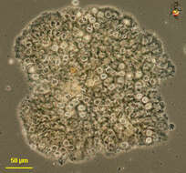
Samples from Sediment at Cedar swamps, Woods Hole, Massachusatts. Photographed by Hwan Su Yoon.
-
Spoongomonas, detail of several cells from within a globular colony. Each cell is more or less spherical and gives rise to two long flagella that are slightly different in length. The matrix of the colony is made up up of brown globular mucoid balls. From Lake Donghu, China. Differential interference contrast micrograph.
-
Samples from Sediment at Cedar swamps, Woods Hole, Massachusatts. Photographed by Hwan Su Yoon.
-
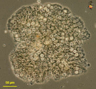
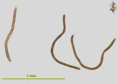
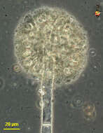
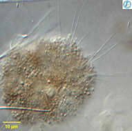
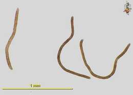
Spongomonas - a heterotrophic flagellate. The cells are spherical and give rise to two flagella that are slightly different in length. The cells form colonies, being embedded in a common mucous matrix that is made up of small globules of orange or brown mucus. This species forms sausage-shaped colonies that are up to a millimetre in length. Bright field illumination.
-
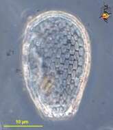
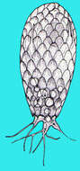
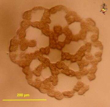
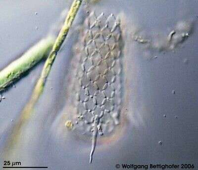
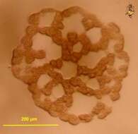
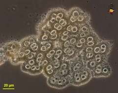
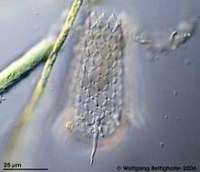
Members of testate amoebae group Euglyphidae form siliceous plates for constructing shell. Sample collection by Martin Kreutz from Simmelried near Konstanz(Baden-Wuerttemberg, Germany). This image was taken using Zeiss Universal with Olympus C7070 CCD camera.
-
Spongomonas - a heterotrophic flagellate. The cells are spherical and give rise to two flagella that are slightly different in length. The cells form colonies, being embedded in a common mucous matrix that is made up of small globules of orange or brown mucus. This species forms sausage-shaped colonies that are up to a millimetre in length. Phase contrast micrograph.