-
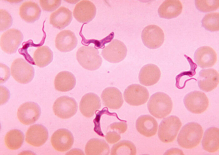
Trypanosoma forms in blood smear from patient with African trypanosomiasis
-
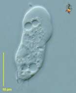
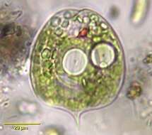
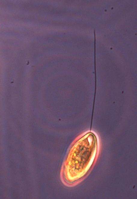
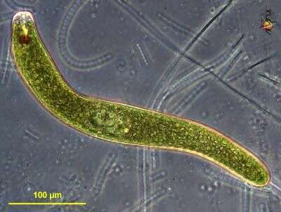
Phase contrast image of organism believed to be a euglenid flagellate (which is sarcasm for 'They're wrong').
-
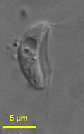
Trypanosoma cruzi amistigotes
-
Bodo saliens Larsen and Patterson, 1990. Bodo cells that are usually elongate elliptical and somewhat inflexible, and are 4 to 12 microns long (mostly 6 to 9 microns). Two flagella unequal in length emerge subapically from a shallow pocket. The anterior flagellum appears inactive, is as long as or sightly shorter than the cell and is held forwards with a single anterior curve held perpendicular to the substrate. The acronematic posterior flagellum is typically directed straight behind the cell and is about 2.2 to 3.5 times cell length. The cells swim in rapid darts in straight lines.
-
Vahlkampfia (vall-camp-fee-a) (= Schizopyrenus - skitz-owe-pyre-een-us ?) a heterolobose amoeba, pseudopodia are produced in sudden bulges, and are lobose. Large hyaline region. Also with posterior contractile vacuole, nucleus with nucleolus and various food vacuoles and inclusions. Also showing uroidal region (lower). Differential interference contrast.
-
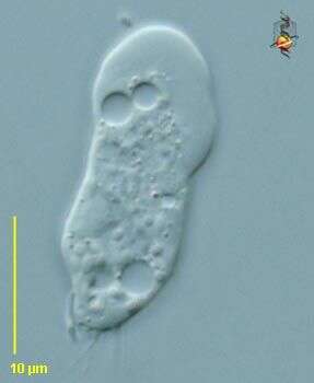
Stachyamoeba (stack-ee-a-me-ba) is a free-living amoeba, usually flattended, expanded anterior margin with broad hyaline zone. With filaments arising from the uroid. At one time treated as a gruberellid and probably related to the acrasids, it can be seen to have a flagellated stage with two flagella which indicates that it is a fairly conventional heteroloboseid. Phase contrast.
-
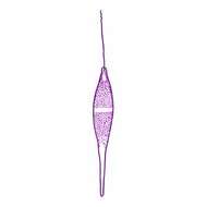
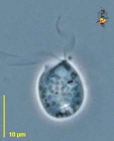
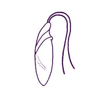
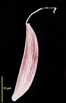
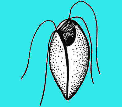
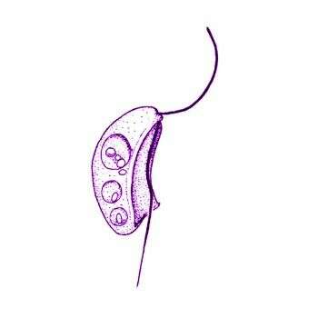
Pleurostomum flabellatum Ruinen, 1938. Cell 11-14 microns long, spindle-shaped and round in transverse section with two parallel flagella inserting apically. The flagella are 0.5-1.5 times body length. They are often, but not always, the same length. The flagella were usually observed stuck to the coverslip or slide. When free the flagella beat homodynamically. Subapically there is a wide and deep opening or groove that runs half to three quarters the way down the body in a half spiral and usually ends in close association with a food vacuole. The body was not observed to bend or flex except in one individual where the posterior third of the cell was stuck to the coverslip.
-
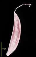
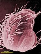
Scanning electron micrograph showing the anterior flagellum and the cell shape.
-
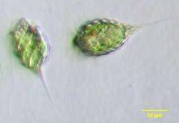
Phacus pleuronectes, euglenoid flagellate with a rigid, leaf-shaped pellicle with fine longitudinal striations and short curved spinous posterior. Many discoid chloroplasts. Usually with one large circular central paramylon body (although two are seen in this individual). Red stigma. From freshwater pond near Boise, Idaho. Brightfield
-
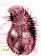
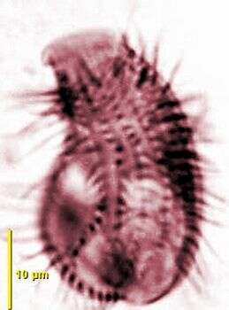
SCanning EM showing the dorsal face of the cell with ciliary lines and the anterior mouth bordered with lips.
-
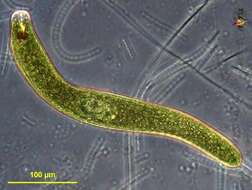
Distigma proteus Ehrenberg, 1838. Body highly metabolic, scarcely ever presenting the same contour, usually more or less elongate, with irregular constrictions and distensions, longer flagellum nearly equaling the body in length, the shorter one scarcely one quarter that length, endoplasm transparent, enclosing numerous dark coloured refringent corpuscles whose positions are constantly shifting from one extremely to the other in accordance with the peristaltic motions of the body, two minute, blackish, eye-like pigment-spots usually developed at the anterior extremity, tubular pharynx slender, greatly prolonged, contractile vacuole conspicuous, located close to the termination of the pharynx, nucleus ovate, subcentral. Length 44-106 microns
-
Euglena ehrenbergii. Cell observed in freshwater habitats in the vicinity of Broome, Western Australia in September 2003. This work was supported by the Australian Biological Resources Study.
-
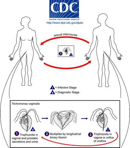
Centers for Disease Control/Division of Parasitic Diseases and Malaria
EOL staff
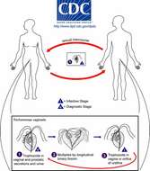
Life cycle of Trichomonas vaginalis, the cause of trichomoniasis in humansTrichomonas vaginalis resides in the female lower genital tract and the male urethra and prostate (1), where it replicates by binary fission (2). The parasite does not appear to have a cyst form, and does not survive well in the external environment. Trichomonas vaginalis is transmitted among humans, its only known host, primarily by sexual intercourse (3).From
Centers for Disease Control Parasites and Health website
-

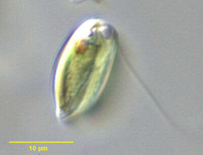
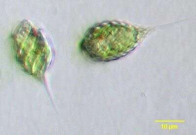
Carpediemonas (carp-ee-dee-a-moan-ass) bialata (Ruinen, 1938) Lee and Patterson, 2000. Cell outline is kidney-shaped. Cells are 6 to 14 microns long (mostly 9 to 12 microns), not rigid, and with a longitudinal ventral groove. A membrane moves down along the groove every 4 - 6 seconds. Two flagella emerge from the anterior part of the cell, the anterior flagellum bends backwards, is about the length of the cell and beats over the cell with a slow sweeping motion. The acronematic posterior flagellum beats asymmetrically and is about 1.5 times cell length. The posterior flagellum may vibrate actively in the groove when not beating. The cells consume bacteria up to 5 microns, and food materials are transferred by the moving membrane to the back of the cell. The cells may have many food vacuoles and attach to the substrate with the tip of the posterior flagellum. The cells move slowly by skidding or gliding with the anterior flagellum beating with a flicking motion. Commonly observed in late cultures.
-
Vahlkampfia (vall-camp-fee-a) (= Schizopyrenus - skitz-owe-pyre-een-us ?) a heterolobose amoeba, pseudopodia are produced in sudden bulges, and are lobose. Large hyaline regions. Also with posterior contractile vacuole, nucleus with nucleolus and various food vacuoles and inclusions. Usually with small uroids giving rise to filaments. Several cells. Phase contrast.
-
-
Cryptoglena pigra. Although Cryptoglena has been considered a euglenid flagellate the genus should probably be considered of uncertain affinity. The cells are solitary and have a single emergent flagellum slightly longer than the cell body. The cell body is rigid, dorsoventrally flattened and ovoid in outline with rounded anterior and bluntly pointed posterior. There is an obvious longitudinal furrow seen well in this image. Described as having one or two laminate parietal chloroplasts without pyrenoids. The posterior nucleus is not visible in this image. The prominent anterior stigma is seen well here. The genus has been described as monospecific by some authors and as containing five or six species by others. From a slow flowing freshwater stream near Boise, Idaho. Differential interference contrast illumination.
-
Phacus pyrum or P. rudicula, a small euglenoid flagellate having a pyriform rigid pellicle thrown into folds (P. pyrum is described as round in cross section while P. rudicula is said to be more flattened as is the cell shown here, but it is likely the two are different forms of the same species). With oblique ridges and tapering, pointed posterior. Red stigma. These individuals have shed their flagella which are usually about 1½ body length. From freshwater pond near Boise, Idaho. Oblique illumination.
-
Cell stained by protargol showing the kineties of cilia of one face of the cell and the anterior mouth.
-
Distigma proteus clavatum Playfair, 1921. Cells are 18-44 microns long and 8-12 microns wide. see Menoidium pellucidum var clavatum.
-
Euglena ehrenbergii. Cell observed in freshwater habitats in the vicinity of Broome, Western Australia in September 2003. This image was taken using differential interference contrast optics. This work was supported by
The Australian Biological Resources Study
-
Centers for Disease Control/Division of Parasitic Diseases and Malaria
EOL staff
Life cycle of Chilomastix mesnili The resistant cyst stage in the life cycle of Chilomastix is responsible for transmission. Both cysts and trophozoites can be found in the feces (diagnostic stages) (1). Infection occurs by the ingestion of cysts in contaminated water or food or by the fecal-oral route (via hands or fomites, i.e., inanimate objects such as towels that transmit infectious organisms to a host) (2). In the large (and possibly small) intestine, excystation releases trophozoites.From
Centers for Disease Control Parasites and Health website.
-
Carpediemonas bialata (Ruinen, 1938) Lee and Patterson, 2000. Cell outline is kidney-shaped. Cells are 6 to 14 microns long, not rigid, and with a longitudinal ventral groove. A membrane moves down along the groove every 4 - 6 seconds. Two flagella emerge from the anterior part of the cell, the anterior flagellum bends backwards, is about the length of the cell and beats over the cell with a slow sweeping motion. The acronematic posterior flagellum beats asymmetrically and is about 1.5 times cell length. The posterior flagellum may vibrate actively in the groove when not beating. The cells consume bacteria up to 5 microns long, and food materials are transferred by the moving membrane to the back of the cell. The cells may have many food vacuoles and attach to the substrate with the tip of the posterior flagellum. The cells move slowly by skidding or gliding with the anterior flagellum beating with a flicking motion.
-
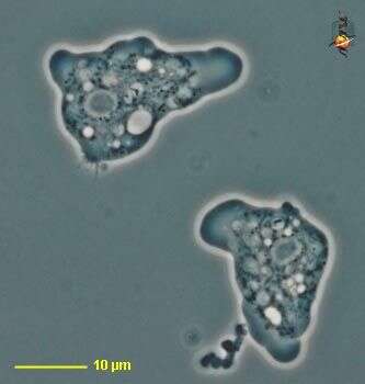
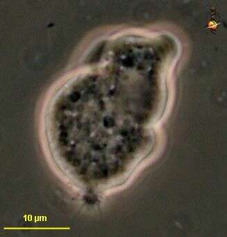
Heteramoeba (het-err-a-me-ba) a naked heterolobose amoeba, distinguished from other types of naked amoebae with lobose pseudopodia largely by ultrastructural features, but trophic heterolobose amoebae tend to form their pseudopodially suddenly rather than progressively. Phase contrast.