-
Ribadelago, Castille and Leon, Spain
-
Galende, Castile and Len, Spain
-
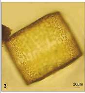
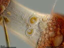
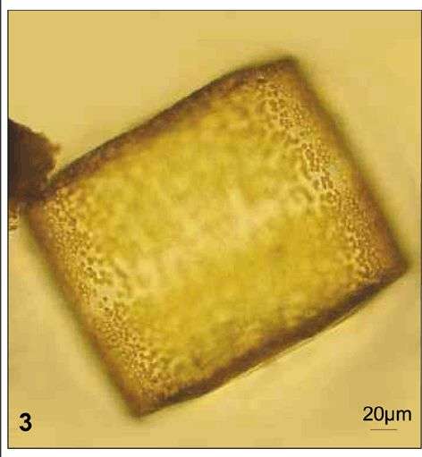
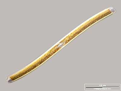
Fig 3: Coscinodiscus wailesii Light micrograph of a Lugol's preserved cell in girdle view
-
-
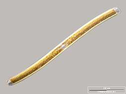
Cingular view. Scale bar indicates 100 µm. Sample from a wetland at the Pillersee (Tyrol, Austria). The image was built up using several photomicrographic frames with manual stacking technique. Images were taken using Zeiss Universal with Olympus C7070 CCD camera.Image under Creative Commons License V 3.0 (CC BY-NC-SA).
-
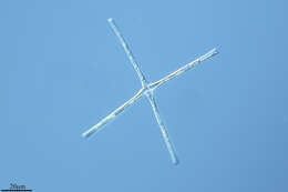
Corethron (core-eeth-ron) hystrix, centric diatom (stramenopile) with siliceous spines emerging from the border of the valves, many girdle bands (not visible here) make up the body of the cylinder. This image emphasizes the plastids. Differential interference microscopy.
data on this strain.
-
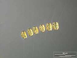
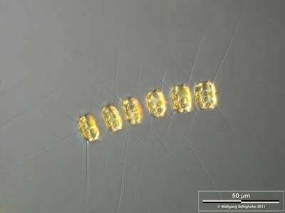
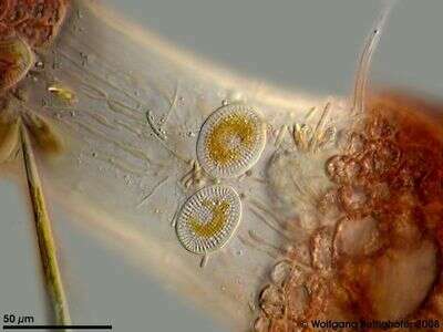
Scale bar indicates 50 µm. The image was built up using several photomicrographic frames with manual stacking technique. Sample from North Sea near Heligoland (spring diatom bloom). Images were taken using Zeiss Universal with Olympus C7070 CCD camera.
-
Fornæs, Djursland, Danmark
-
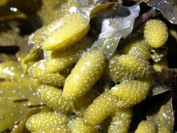
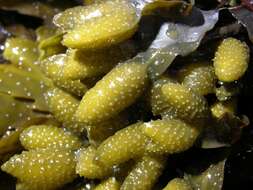
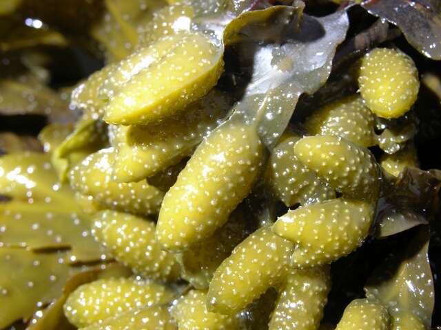
Spore Leaves of Alaria fistulosa. [There were about 220 on this plant.]
-
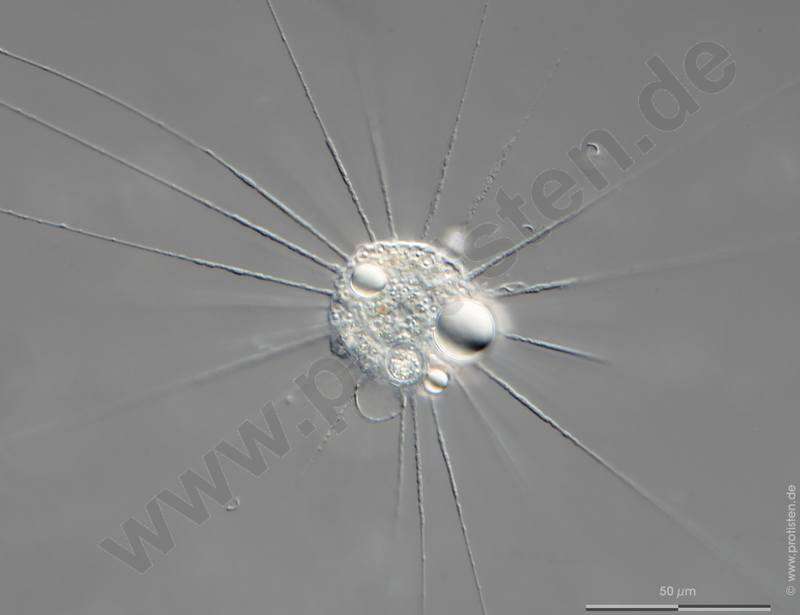
Actinophrys sol.Series (frame 2) of contracting vacuole. Scale bar indicates 50 m.Collected from on a pond near the Bodden bay Schwarzer Peter in the southern part of the isle Hiddensee (German Baltic Sea). The image was built up using several photomicrographic frames with manual stacking technique. Images were taken using Zeiss Axioplan with Olympus OM-D M5 MKII.For high-resolution images please ask postmaster@protisten.de.
-
Galende, Castille and Leon, Spain
-
San Martin De Castaneda, Castille and Leon, Spain
-
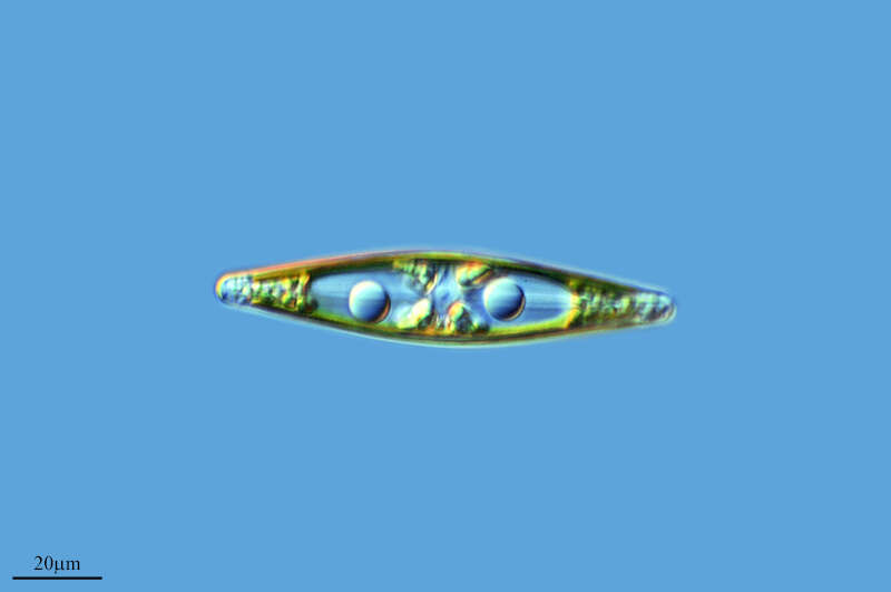
Fig 1: Schematic drawing of the cell in the valve view.
-
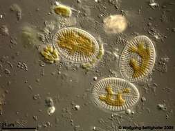
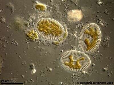
Cocconeis scutellum looking like footprints of the first man on the moon. This delicate Aufwuchs was grown on a microscope slide which was placed in a special slide holder hanging in the Bodden waters. Collected from Bodden, the brackish waters lying between the isles of Hiddensee and Ruegen (German Baltic Sea). This image was taken using Zeiss Universal with Olympus C7070 CCD camera.
-
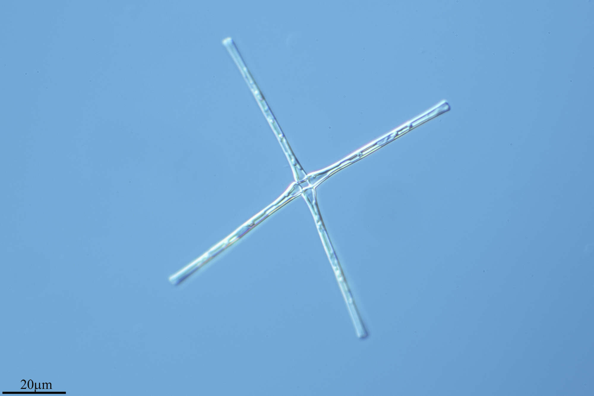
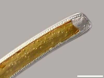
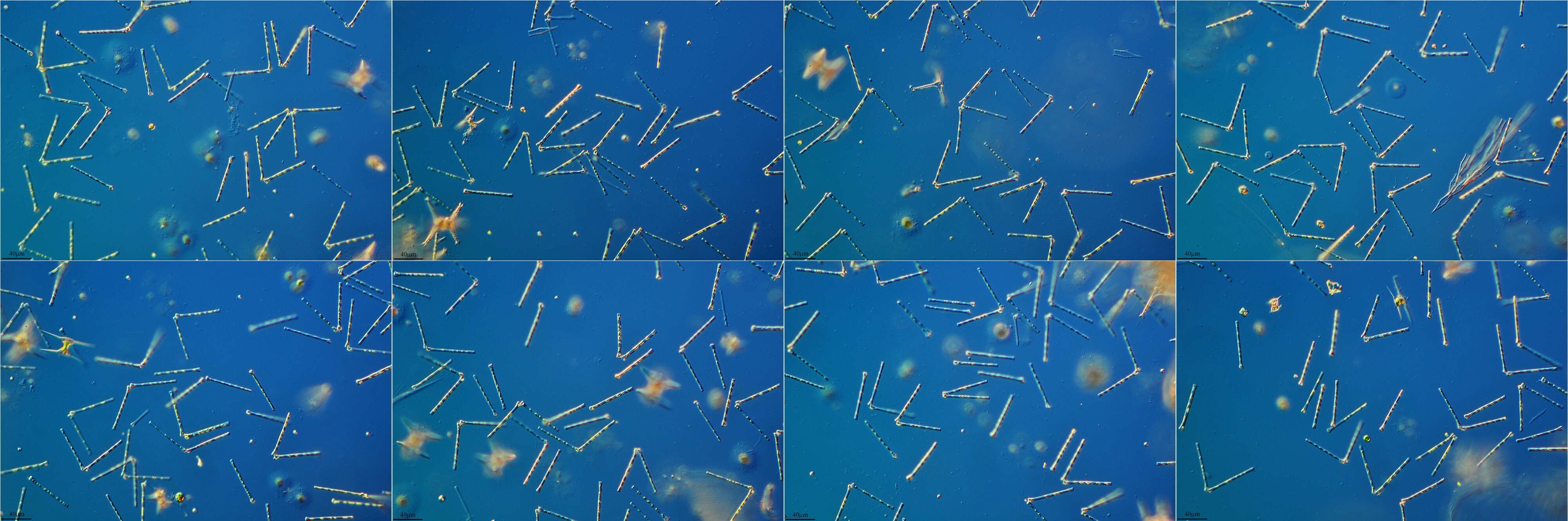
Detail: Apex in cingular view, displaying cingulum (central strae and the two canal raphes on the edges. Scale bar indicates 25 µm. Sample from a wetland at the Pillersee (Tyrol, Austria). The image was built up using several photomicrographic frames with manual stacking technique. Images were taken using Zeiss Universal with Olympus C7070 CCD camera.Image under Creative Commons License V 3.0 (CC BY-NC-SA).
-
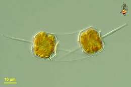
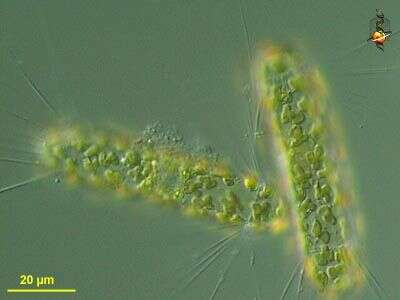
Ditylum (die-tie-lum) brightwellii. Marine centric diatom, cylindrical frustule from the ends of which is a long spine or labiate process. This contains two products of cell division, and each cell has numerous golden chloroplasts. Differential interference microscopy.
data on this strain.
-
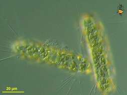
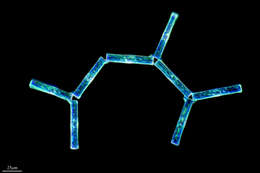
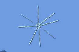
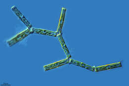
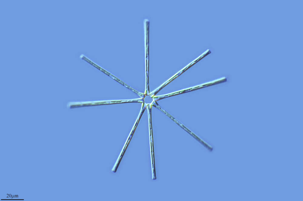
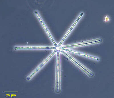
of the colonial diatom, Asterionella formosa (Hassall, 1850). The linear frustules have expanded ends. The frustules of the colony are connected by gelatinous cushions at the larger of their two ends in a radial array all in more or less the same plane The yellow chloroplasts are seen here. Blooms of this diatom may impart a disagreeable fishy taste to fresh water. Collected from freshwater pond near Boise, Idaho January 2003. Phase contrast illumination.
-
Fornæs, Djursland, Danmark
-
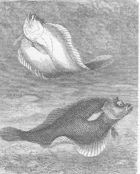
Dab (platessa Imanda).
-
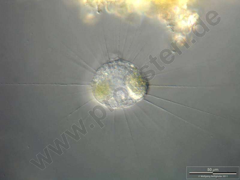
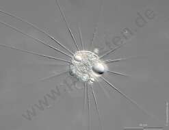
Actinophrys sol.Focus on food vacuole. Scale bar indicates 50 m.Sample from a pond situated in the vicinity of Lake Constance. The image was built up using several photomicrographic frames with manual stacking technique. Images were taken using Zeiss Universal with Olympus C7070 CCD camera.For high-resolution images please ask postmaster@protisten.de.
-
San Martin De Castaneda, Castille and Leon, Spain
-
San Martin De Castaneda, Castille and Leon, Spain
-
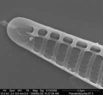
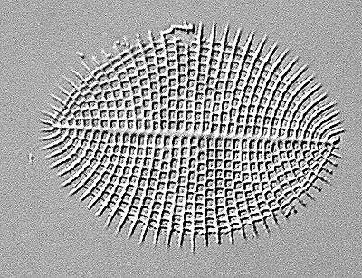
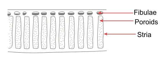
Fig 2: Electron micrograph image showing detail of the striae and fibulae
-
Cocconeis scutellum on a thallus segment of the red alga Ceramium. The two cell types of the alga's thallus are shown: one huge light axial cell with tubular rhodoplasts with the two terminal cortex structures built up by numerous little reddish rotund cells with their lenticular rhodoplasts.. Collected from Bodden, the brackish waters lying between the isles of Hiddensee and Ruegen (German Baltic Sea). This image was taken using Zeiss Universal with Olympus C7070 CCD camera.