-
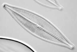
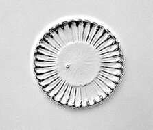
Craticula cuspidata is regarded as the senior synonym of Navicula cuspidata, under which name this asset was submitted. The change of name has been made by the micro*scope team.
-
-
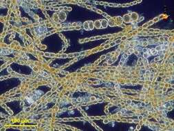
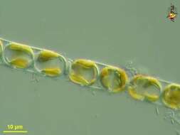
Melosira (mellow-sire-a) nummuloides, filament forming centric diatom, with multiple small plastids within the cell. Dark ground illumination. Leptosiropsis (leapt-owe-sire-op-sis) torulosa, green alga with organic wall that is produced in layers. Phase contrast microscopy.
data on this strain.
-
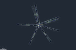
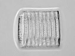
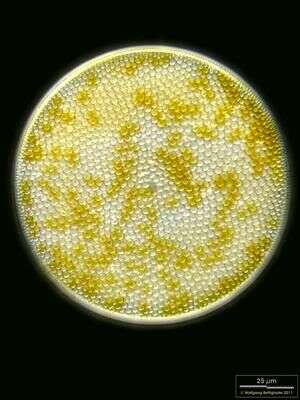
Valvar view. Scale bar indicates 25 µm. Sample from North Sea near Heligoland (spring diatom bloom). The image was built up using several photomicrographic frames with manual stacking technique. Images were taken using Zeiss Universal with Olympus C7070 CCD camera.
-
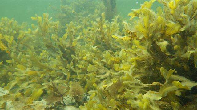
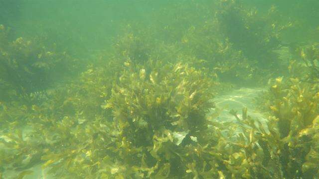
Fjellerup Strand
-
Santa Cruz de la Sierra, Extremadura, Espaa
-
San Martin De Castaneda, Castille and Leon, Spain
-
-
-
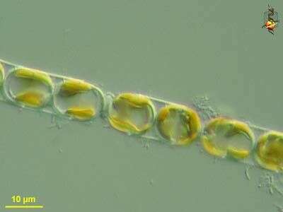
Melosira (mellow-sire-a) nummuloides, filament forming centric diatom, with multiple small plastids within the cell clearly shown in this micrograph. Differential interference microscopy.
data on this strain.
-
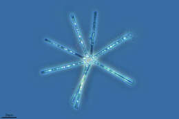
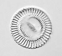
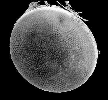
Scanning electron microscope image of valve. The organism is tentatively identified as C. radiatus. Sample taken from the water column off Martha's Vineyard, Massachusetts. Image by Charley O'Kelly and Shauna Murray.
-
Fjellerup Strand
-
Ribadelago, Castille and Leon, Spain
-
Ribadelago de Franco, Castille and Leon, Spain
-
-
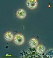
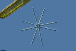
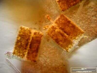
Scale bar indicates 100 µm. The image was built up using several photomicrographic frames with manual stacking technique. Sample from North Sea near Heligoland (Aufwuchs on red alga Ceramium spec.). Images were taken using Zeiss Universal with Olympus C7070 CCD camera.
-
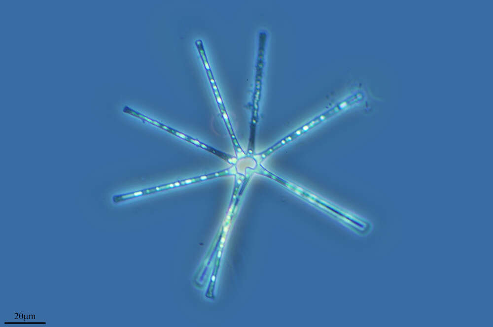
Ochromonas (ock-roe-moan-ass) sphaerocystis, iconic genus of the chrysophytes, body elongated or rounded, with two emergent flagella, golden plastids, sometimes with extrusible bodies under the cell surface. Phase contrast microscopy.
data on this strain.
-
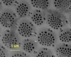
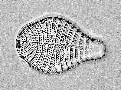
Scanning electron micrograph showing detail of the frustule of this diatom. The larger depressions are called areolae, and perforated region is called the cribrum, within which each perforation is referred to as a cribellum. The same term probably also refers to the perforations in the margins of the areolae. The species is probably C. radiatus. Sample from the water column off Martha's Vineyard. Images by Charley O'Kelly and Shauna Murray.
-
Fjellerup Strand
-
Galende, Castile and Len, Spain
-
Ribadelago de Franco, Castilla y Len, Espaa
-
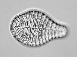
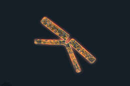
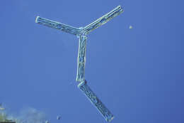
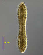
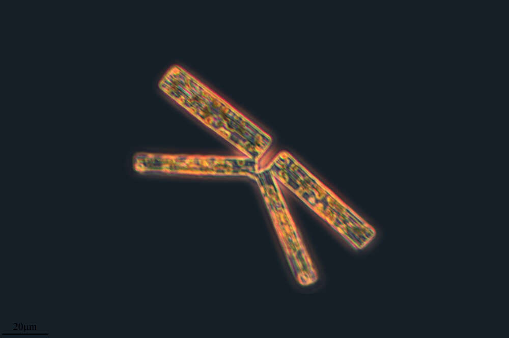
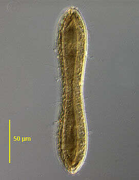
Valve view of the pennate diatom, Cymatopleura solea (Brébisson) W. Smith 1851. Valves are elliptical with a central waist and bluntly pointed apices. The valve surface is marked with several transverse undulations best seen in girdle view; striae and undulations are not interrupted in the median axis. Raphe are marginal. Very short costations appear as beading around the edge of the valve. There is a single lobulated plastid one half of which lies along the inner surface of the epivalve and the other along the inner surface of the hypovalve. Collected from the benthos of a freshwater pond near Boise, Idaho, January 2005. DIC.
-
Scale bar indicates 100 µm. The image was built up using several photomicrographic frames with manual stacking technique. Sample from North Sea near Heligoland (Aufwuchs on red alga Ceramium spec.). Images were taken using Zeiss Universal with Olympus C7070 CCD camera.
-
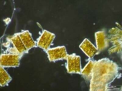
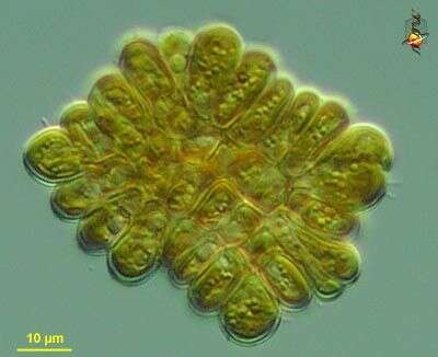
Phaeoplaca (fay-owe-plack-a) thallosa, a chrysophyte in which cells adhere to each other to form irregular rectangular sheets. Differential interference microscopy.
data on this strain.