-
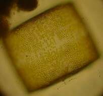
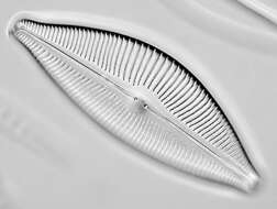
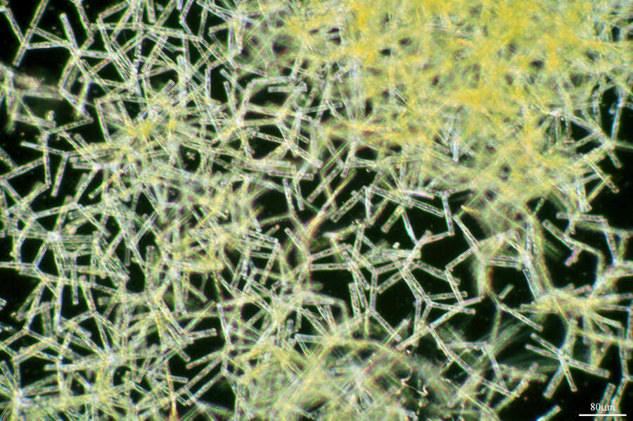
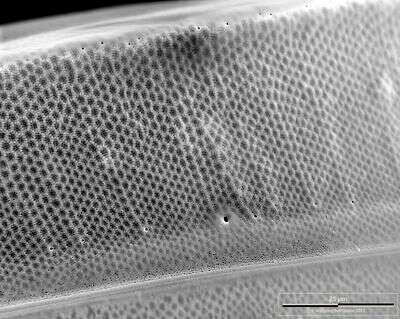
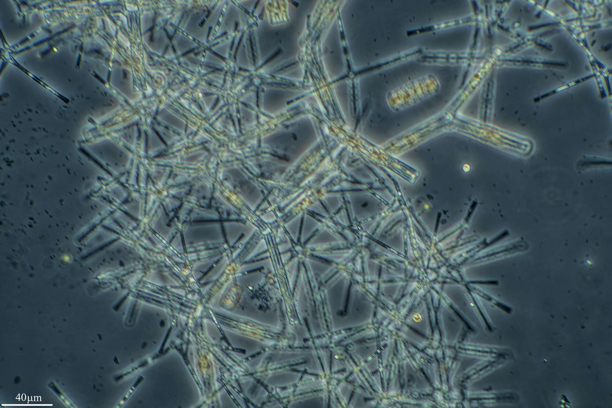
This species has a very fine aerolation. It can be distinguished from other species by the central hyaline area and the hyaline lines radiating from it between the areolae. It also has a distinctive shape in girdle view. The valve is very high (often higher than wide) and the valve margin appears to undulate slightly.
-
Lillebælt ved Nørreskoven på Als. Danmark
-
Galende, Castille and Leon, Spain
-
Ribadelago de Franco, Castilla y Len, Espaa
-
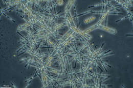
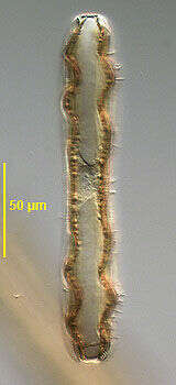
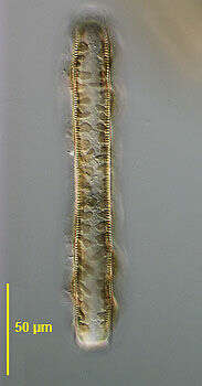
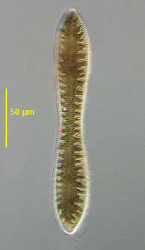
Optical section (girdle view) of the pennate diatom, Cymatopleura solea (Brébisson) W. Smith 1851. Valves are elliptical with a central waist and bluntly pointed apices. The valve surface is marked with several transverse undulations best seen in girdle view (seen well in this image). Striae and undulations are not interrupted in the median axis. Raphe are marginal. Very short costations appear as beading around the edge of the valve. The central nucleus is seen in this image. There is a single lobulated plastid one half of which lies along the inner surface of the epivalve and the other along the inner surface of the hypovalve. Collected from the benthos of a freshwater pond near Boise, Idaho, January 2005. DIC.
-
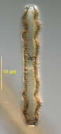
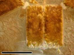
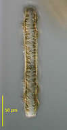
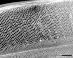
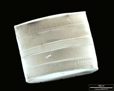
Pad of connecting mucilage is clearly visible. Scale bar indicates 100 µm. The image was built up using several photomicrographic frames with manual stacking technique. Sample from North Sea near Heligoland (Aufwuchs on red alga Ceramium spec.). Images were taken using Zeiss Universal with Olympus C7070 CCD camera.Image under Creative Commons License V 3.0 (CC BY-NC-SA).
-
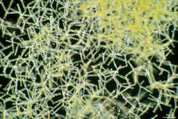
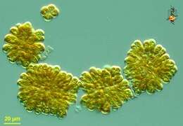
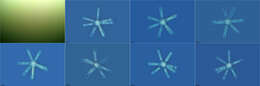
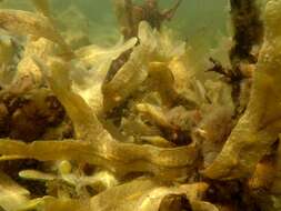
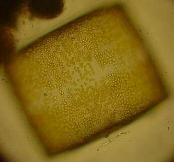
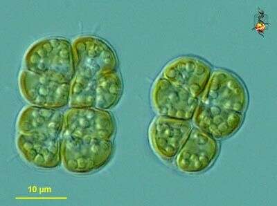
Phaeoplaca (fay-owe-plack-a) thallosa, a chrysophyte in which cells adhere to each other to form irregular rectangular sheets. Differential interference microscopy.
data on this strain.
-
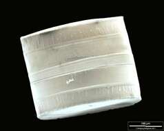
SEM of girdle view. The ligulae which fit in the open girdle bands are weakly visible. Scale bar indicates 100 µm. Sample from North Sea near Heligoland (spring diatom bloom). The image was built up using several photomicrographic frames with manual stacking technique. Use of SEM equipment courtesy of Lab Dr. Karl-Heinz Schäffner, Solingen, Germany.
-
Lillebælt ved Nørreskoven på Als. Danmark
-
Santa Cruz de la Sierra, Extremadura, Spain
-
Ribadelago de Franco, Castilla y Len, Espaa
-
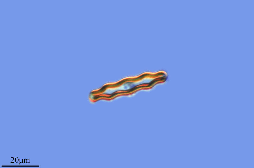
Optical section (girdle view) of the pennate diatom, Cymatopleura solea (Brébisson) W. Smith 1851. Valves are elliptical with a central waist and bluntly pointed apices. The valve surface is marked with several transverse undulations best seen in girdle view; striae and undulations are not interrupted in the median axis. Raphe are marginal. Very short costations appear as beading around the edge of the valve. There is a single lobulated plastid one half of which lies along the inner surface of the epivalve and the other along the inner surface of the hypovalve (seen well in this image). Collected from the benthos of a freshwater pond near Boise, Idaho, January 2005. DIC.
-
-
Phaeoplaca (fay-owe-plack-a) thallosa, a chrysophyte in which cells adhere to each other to form irregular rectangular sheets. Differential interference microscopy.
data on this strain.
-
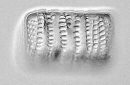
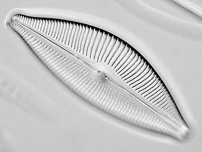
Closeup of the lateral side of the valve. Scale bar indicates 25 µm. Sample from North Sea near Heligoland (spring diatom bloom). The image was built up using several photomicrographic frames with manual stacking technique. Use of SEM equipment courtesy of Lab Dr. Karl-Heinz Schäffner, Solingen, Germany.
-
Lillebælt ved Nørreskoven på Als. Danmark
-
San Martin De Castaneda, Castille and Leon, Spain
-
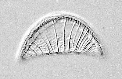
Optical section (valve view) of the pennate diatom, Cymatopleura solea (Brébisson) W. Smith 1851. Valves are elliptical with a central waist and bluntly pointed apices. The valve surface is marked with several transverse undulations best seen in girdle view; striae and undulations are not interrupted in the median axis. Raphe are marginal. Very short costations appear as beading around the edge of the valve. There is a single lobulated plastid one half of which lies along the inner surface of the epivalve and the other along the inner surface of the hypovalve (seen well in this image). Collected from the benthos of a freshwater pond near Boise, Idaho, January 2005. DIC.
-
-
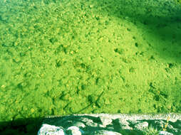
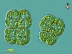
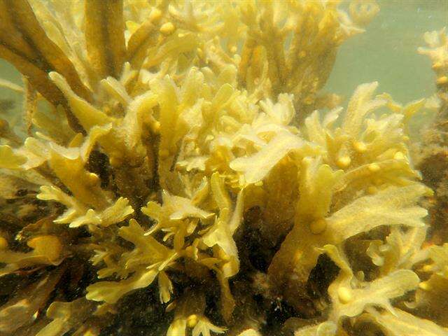
Phaeoschizochlamys (fay-owe-shit-sew-clam-ees), (pheothamniophyte), cells with golden plastids enclosed in mucus and forming aggregates. The mother cell wall is cast off in two halves (hemispheres) during cell division, and frequently these old walls are visible in the colony matrix. Differential interference microscopy.
data on this strain.
-
Valvar View. Scale bar indicates 100 µm. Sample from North Sea near Heligoland (spring diatom bloom). The image was built up using several photomicrographic frames with manual stacking technique. Use of SEM equipment courtesy of Lab Dr. Karl-Heinz Schäffner, Solingen, Germany.
-
Lillebælt ved Nørreskoven på Als. Danmark
-
Galende, Castile and Len, Spain
-