-
-
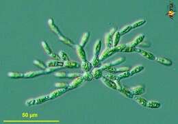
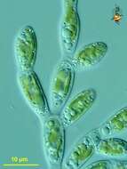
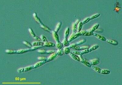
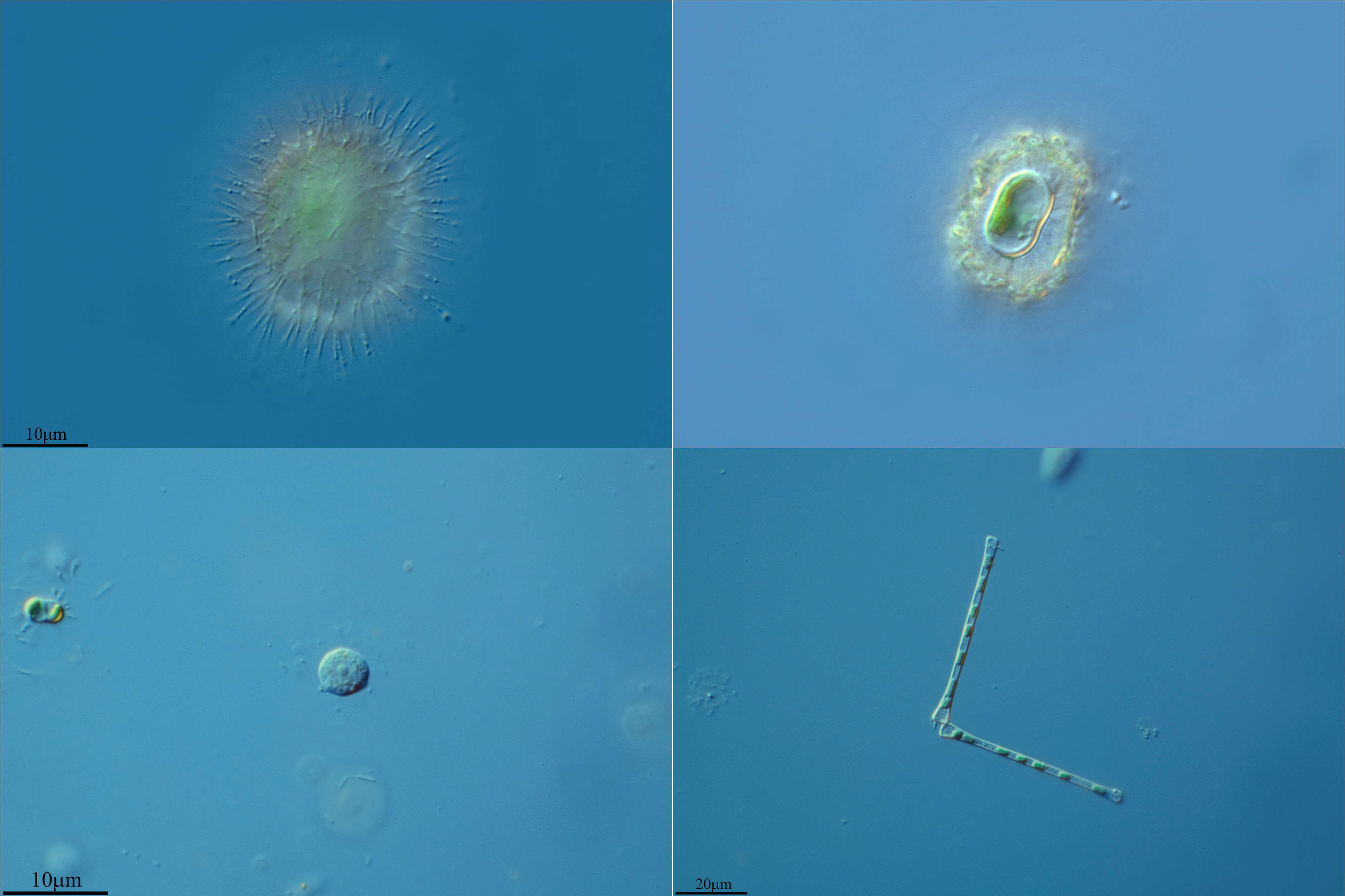
Phaeothamnion (fay-owe-tham-knee-um) branching filamentous pheothamniophyte alga. This species usually has a globular cell at the base of the filament, in this image, there are four globose cells, each giving rise to a branching filament. Differential interference microscopy.
data on this strain.
-
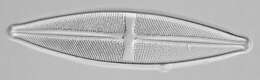
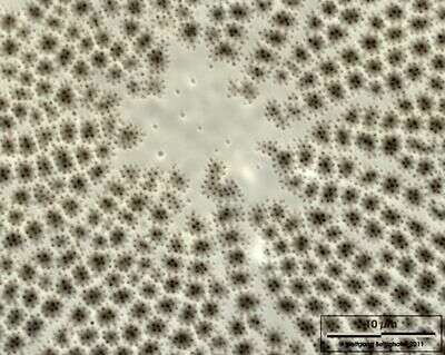
Closeup showing hyaline central area. Scale bar indicates 10 µm. Sample from North Sea near Heligoland (spring diatom bloom). Use of SEM equipment courtesy of Lab Dr. Karl-Heinz Schäffner, Solingen, Germany.
-
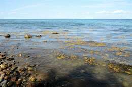
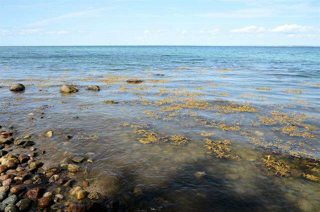
Lillebælt ved Nørreskoven på Als. Danmark
-
Galende, Castile and Len, Spain
-
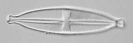
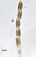
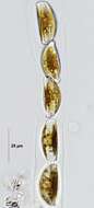
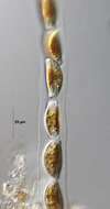
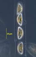
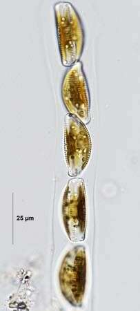
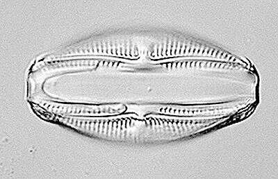
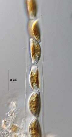
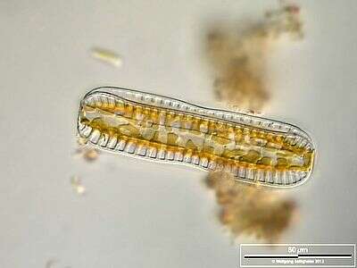
Cymbella tumida (Brébisson in Kützing) van Heurck 1882-1885. Frustules within gelatinous tube. Collected from detritus in a freshwater irrigation canal. Boise, Idaho. March 2009. Brightfield.
-
-
Phaeothamnion (fay-owe-tham-knee-um) branching filamentous pheothamniophyte alga, this micrograph showing details of plastids and the translucent vacuoles containing the storage carbohydrate (leucosin?). Differential interference microscopy.
data on this strain.
-
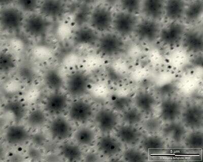
Closeup showing fine structure of valvar pores. Scale bar indicates 5 µm. Sample from North Sea near Heligoland (spring diatom bloom). Use of SEM equipment courtesy of Lab Dr. Karl-Heinz Schäffner, Solingen, Germany.
-
Galende, Castile and Len, Spain
-
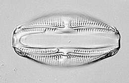
Cymbella tumida (Brébisson in Kützing) van Heurck 1882-1885. Cymbella tumida (Brébisson in Kützing) van Heurck 1882-1885. Frustules within gelatinous tube. Collected from detritus in a freshwater irrigation canal. Boise, Idaho. March 2009. Brightfield.
-
-
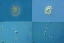
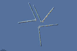
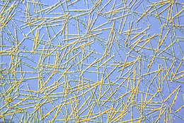
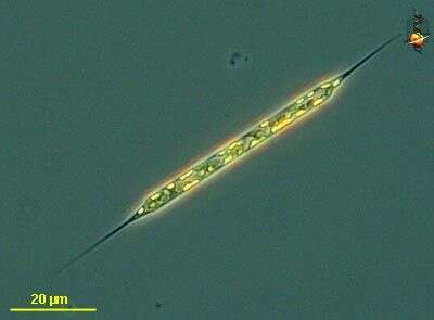
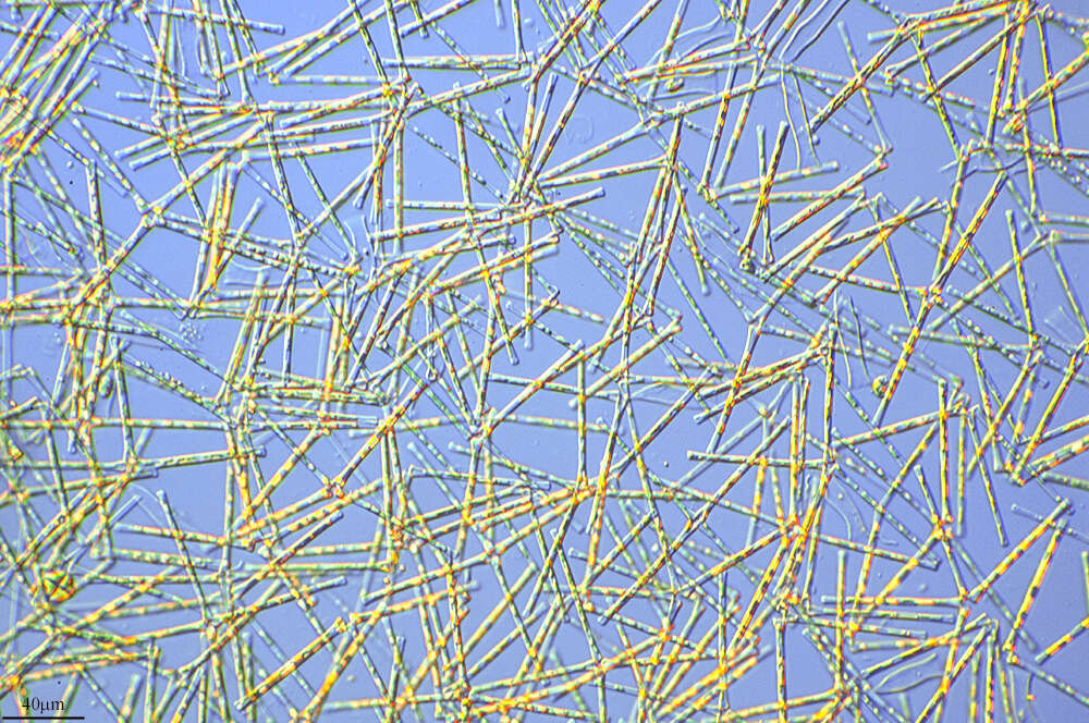
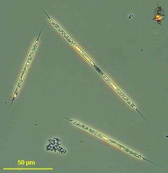
Rhizosolenia (rye-so-so-lean-ee-a) setigera, one of the common genera of marine phytoplantkonic diatoms, a centric diatom in which the valves, at the ends of the cells, are conical and give rise to spines. Much of the long cylindrical body is enclosed with hoop-shaped girdle bands. Phase contrast microscopy.
data on this strain.
-
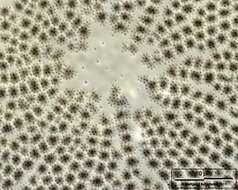
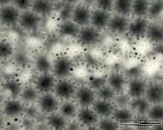
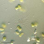
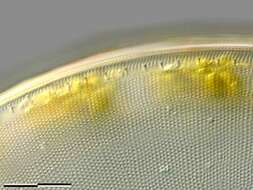
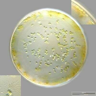
Valvar view. Insets showing marginal ring of labiate processes (upper right) and the hyaline central area (lower left). Scale bar indicates 100 µm. Sample from North Sea near Heligoland (spring diatom bloom). The image was built up using several photomicrographic frames with manual stacking and stitching technique. Images were taken using Zeiss Universal with Olympus C7070 CCD camera.
-
San Martn de Castaeda, Castilla y Len, Espaa
-
Cymbella tumida (Brébisson in Kützing) van Heurck 1882-1885. Frustules within gelatinous tube. Collected from detritus in a freshwater irrigation canal. Boise, Idaho. March 2009.DIC.
-
-
Rhizosolenia (rye-so-so-lean-ee-a) setigera, one of the common genera of marine phytoplantkonic diatoms, a centric diatom in which the valves, at the ends of the cells, are conical and give rise to spines. Much of the long cylindrical body is enclosed with hoop-shaped girdle bands. This image shows the plastids and the valve region of the cell. Differential interference microscopy.
data on this strain.
-
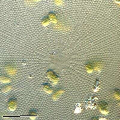
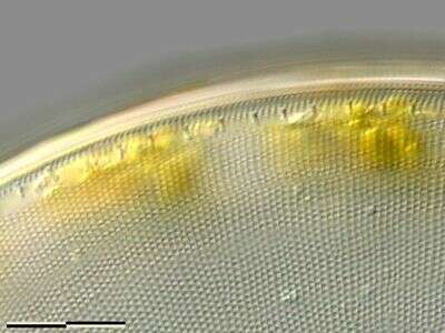
Closeup of the hyaline central area. Scale bar indicates 25 µm. Sample from North Sea near Heligoland (spring diatom bloom). The image was built up using several photomicrographic frames with manual stacking and stitching technique. Images were taken using Zeiss Universal with Olympus C7070 CCD camera.
-
San Martin De Castaneda, Castille and Leon, Spain
-
Cymbella tumida (Brébisson in Kützing) van Heurck 1882-1885. Frustules within gelatinous tube. Collected from detritus in a freshwater irrigation canal. Boise, Idaho. March 2009. Phase contrast.
-
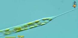
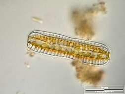
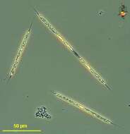
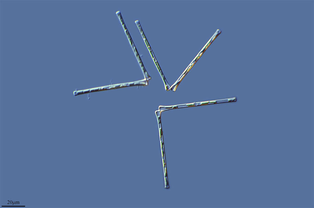
Cingular view displayin 3 of the 4 raphe keels. Scale bar indicates 50 µm. Sample from a wetland at the Pillersee (Tyrol, Austria). The image was built up using several photomicrographic frames with manual stacking technique. Images were taken using Zeiss Universal with Olympus C7070 CCD camera.Image under Creative Commons License V 3.0 (CC BY-NC-SA).
-
Rhizosolenia (rye-so-so-lean-ee-a) setigera, one of the common genera of marine phytoplantkonic diatoms, a centric diatom in which the valves, at the ends of the cells, are conical and give rise to spines. Much of the long cylindrical body is enclosed with hoop-shaped girdle bands. Phase contrast microscopy.
data on this strain.
-
Closeup of the marginal ring of labiate processes. Scale bar indicates 25 µm. Sample from North Sea near Heligoland (spring diatom bloom). The image was built up using several photomicrographic frames with manual stacking and stitching technique. Images were taken using Zeiss Universal with Olympus C7070 CCD camera.