-
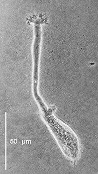
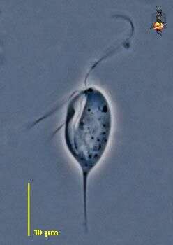
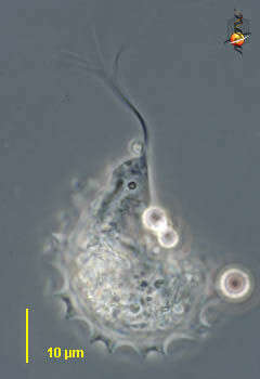
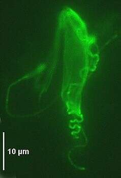
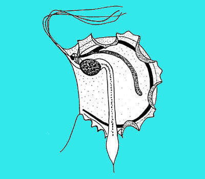
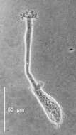
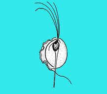
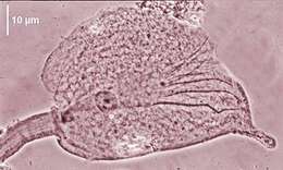
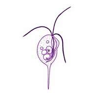
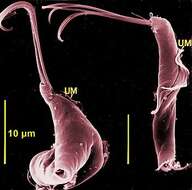
Oxymonas - oxymonad flagellates with a club-shaped cell, (5-165 µm), and an anterior extensile rostellum terminated by a holdfast that attaches to the cuticle of the host intestinal intima. The four relatively short flagella are inserted in two pairs at the base of the rostellum. The crystalline axostyle originates at the base of the rostellum and divides in thin branches traversing the cell to the posterior end where it is surrounded by a sheath. The nucleus is situated at the base of the rostellum. Oxymonas jouteli showing the long rostellum with the terminal finger-like holdfast (phase contrast).
-
-
-
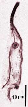
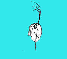
Oxymonas - oxymonad flagellates with a club-shaped cell, (5-165 µm), and an anterior extensile rostellum terminated by a holdfast that attaches to the cuticle of the host intestinal intima. The four relatively short flagella are inserted in two pairs at the base of the rostellum. The crystalline axostyle originates at the base of the rostellum and divides in thin branches traversing the cell to the posterior end where it is surrounded by a sheath. The nucleus is situated at the base of the rostellum. Oxymonas jouteli showing the anterior rostellum, the nucleus and the posterior axostyle with an arrow-shaped end (protargol).
-
-
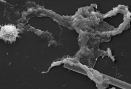
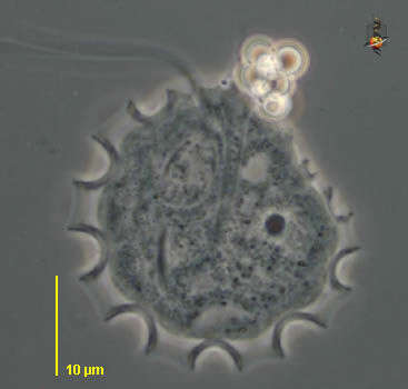
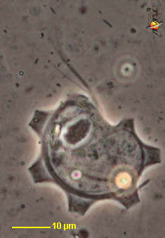
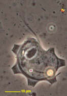
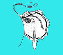
This scanning electron micrograph (SEM) of an untreated water specimen extracted from a wild stream mainly used to control flooding during inclement weather, revealed the presence of unidentified organisms, which included bacteria, protozoa, and algae. Occupying most of the field of view, an unidentified amorphous mucoidal biofilm was featured, which appeared to have enmeshed numbers of amoeboid organisms, while on the left was a strangely-beautiful microorganism displaying an outer surface studded with numerous projections, making it appear like a microscopic sea urchin. See PHIL 11715 for a colorized version of this image.Created: 2009
-
-
Oxymonas - oxymonad flagellates with a club-shaped cell, (5-165 µm), and an anterior extensile rostellum terminated by a holdfast that attaches to the cuticle of the host intestinal intima. The four relatively short flagella are inserted in two pairs at the base of the rostellum. The crystalline axostyle originates at the base of the rostellum and divides in thin branches traversing the cell to the posterior end where it is surrounded by a sheath. The nucleus is situated at the base of the rostellum. Oxymonas jouteli anterior rostellum, nucleus and subdivided posterior axostyle (protargol).
-
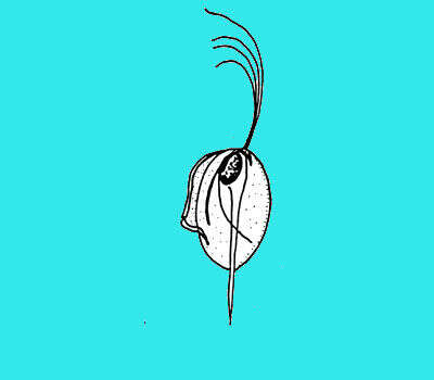
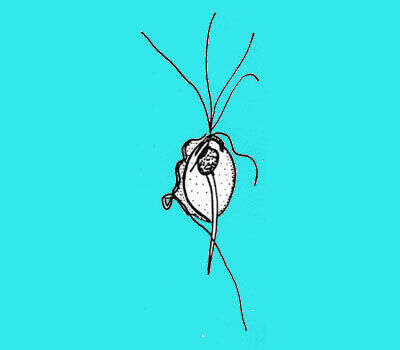
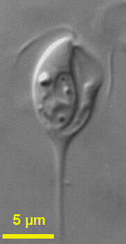
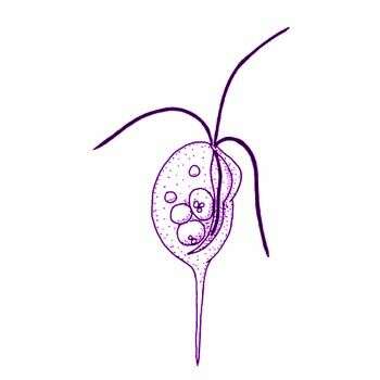
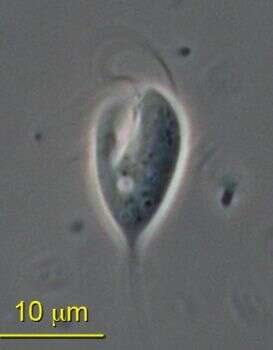
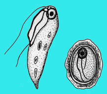
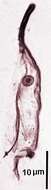
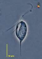
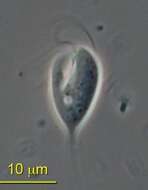
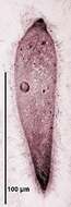
Chilomastix (kai-low-ma-sticks), a genus of retortamonad flagellates, mostly reported in association with animals, but we find this species (C. cuspidata) in anoxic sites. There are four flagella, we can only see three in this picture, inserting just subapically, and at the head of a large groove. One wall of the groove has a cusp. Posterior end drawn out as a long spike. Phase Contrast.
-
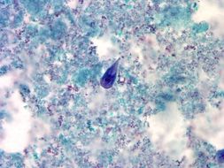
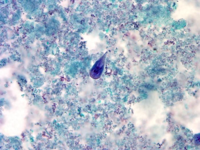
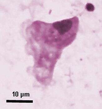
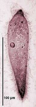
At a magnification of 1000X, this trichrome-stained photomicrograph revealed the morphologic characteristics of a blue-stained Giardia intestinalis protozoan trophozoite (center). In the small intestine, the protozoan cysts release trophozoites, with each cyst producing two trophozoites. Trophozoites multiply by longitudinal binary fission, remaining in the lumen of the proximal small bowel where they can be free, or attached to the mucosa by a ventral sucking disk. As the trophozoites mature, they are simultaneously migrating towards the colon, whereupon, they once again become thick-walled cysts, and are in this way, passed in the hosts stool into the environment. As cysts, these protozoan parasites can survive for many months until they are accidentally ingested by another unfortunate host.Created:
-
-
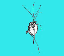
Pentatrichomonoides is a trichomonad flagellate (18-45 µm) with five anteriorly directed free flagella and a recurrent flagellum forming an undulating membrane. Anterior flagella arising from a gullet. Cell shape changeable, cigar-shaped, and truncated posteriorly or broadly triangular. Costa and undulating membrane often recurved posteriorly. No central axostyle, parabasal of variable shape. One species known at the time of writing - Pentatrichomonoides scroa from Mastotermes darwiniensis and Glyptotermes dudleyi, long form (Giemsa).
-
Chilomastix (kai-low-ma-sticks) cuspidata (Larsen and Patterson, 1990) Bernard et al., 1997. Cells are drop-shaped with a long posterior spike and about 20 - 32 microns long (including the spike) with a groove extending from the apex to the posterior end of the untapered part of the cell. The cells have 4 flagella inserting subapically and are directed anterior laterally, one is shorter than the cell and the other three are about the cell length. The short flagellum beats and lies within the ventral groove. The nucleus is situated subapically. Food vacuoles occur throughout the cell. The cells move slowly by swimming while rotating and may attach to the substrate by the tip of the spike.
-
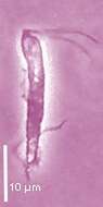
Trichomitopsis (trick-owe-mite-us) is a trichomonad flagellate. this genus has five flagella, four pointing forward and in this micrograph these adhere to each other along most of their lengths. There is also a recurrent flagellum which adheres to the surface of the cell and when it beats causes the margin of the cell to undulate. Flagellates ranging in size from 11-150 microns Costa stout, axostyle stout with a terminal segment often expanded into a pointed bulbous enlargement. The trichomonads often wrap around debris in the gut. They ingest particles of wood which gives some cells a very refractile appearance. From the termite Zootermopsis, supplied by Wards Natural Science Establishment, Rochester, New York, USA. Phase contrast.
-
Pentatrichomonoides is a trichomonad flagellate (18-45 µm) with five anteriorly directed free flagella and a recurrent flagellum forming an undulating membrane. Anterior flagella arising from a gullet. Cell shape changeable, cigar-shaped, and truncated posteriorly or broadly triangular. Costa and undulating membrane often recurved posteriorly. No central axostyle, parabasal of variable shape. One species known at the time of writing - Pentatrichomonoides scroa from Mastotermes darwiniensis and Glyptotermes dudleyi, triangular form (Giemsa).
-
Chilomastix cuspidata (Larsen and Patterson, 1990) Bernard et al., 1997. Cells are drop-shaped with a long posterior spike, they are about 20 - 32 microns long (including the spike) with a groove extending from the apex to the posterior end of the untapered part of the cell. The cells have 4 flagella inserting subapically and are directed anterior laterally, one is shorter than the cell and the other three are about the cell length. The short flagellum beats and lies within the ventral groove. The nucleus is situated subapically. Food vacuoles occur throughout the cell. The cells move slowly by swimming while rotating and may attach to the substrate by the tip of the spike.
-
Trichomitopsis (trick-owe-mite-us) is a trichomonad flagellate. this genus has five flagella, four pointing forward. There is also a recurrent flagellum which adheres to the surface of the cell and when it beats causes the margin of the cell to undulate - the feature that is emphasized in this image. The axostyle leading from the front to the rear is also evident as the stiff dark internal structure. From the termite Zootermopsis, supplied by Wards Natural Science Establishment, Rochester, New York, USA. Phase contrast.
-
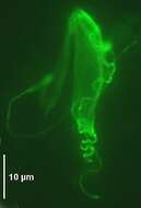
Pentatrichomonoides is a trichomonad flagellate (18-45 µm) with five anteriorly directed free flagella and a recurrent flagellum forming an undulating membrane. Anterior flagella arising from a gullet. Cell shape changeable, cigar-shaped, and truncated posteriorly or broadly triangular. Costa and undulating membrane often recurved posteriorly. No central axostyle, parabasal of variable shape. One species known at the time of writing - Pentatrichomonoides scroa from Mastotermes darwiniensis and Glyptotermes dudleyi. Pentatrichomonoides scroa from Mastotermes darwiniensis with undulating membrane and subjacent costa (immunofluorescence).
-
Chilomastix (kai-low-ma-sticks), a genus of retortamonad flagellates, mostly reported in association with animals, but we find this species (C. cuspidata) in anoxic sites. There are four flagella, we can only see three in this picture, inserting just subapically, and at the head of a large groove. One wall of the groove has a cusp. Posterior end drawn out as a long spike. ATCC 50927 was isolated from a salt marsh.
-
Trichomitopsis (trick-owe-mite-us) is a trichomonad flagellate. this genus has five flagella, four pointing forward. There is also a recurrent flagellum which adheres to the surface of the cell and when it beats causes the margin of the cell to undulate. Flagellates ranging in size from 11-150 microns Costa stout and very obvious here as a curving rod arising anterior to the nucleus. The trichomonads often wrap around debris in the gut. They ingest particles of wood which gives some cells a very refractile appearance. From the termite Zootermopsis, supplied by Wards Natural Science Establishment, Rochester, New York, USA. Phase contrast.
-
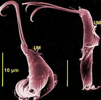
Scanning electron micrograph showing a slender form and a stumpy form with 5 anterior flagella and a recurrent one associated with an undulating membrane. Photograph from A. Breunig. More details in Brugerolle G., Breunig A., König H.(1994) Europ. J. Protistology 30, 372-378.
-
-
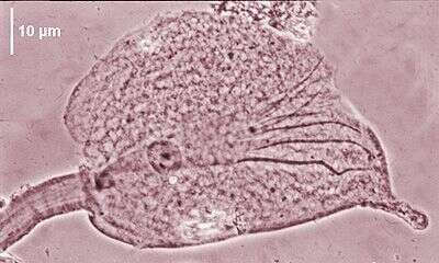
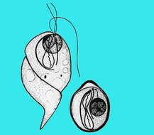
Pseudotrichomympha are hypermastigids of large size (300-500 µm) with a rostrum bearing long flagella separated from the post-rostral part which is covered with flagella arranged in longitudinal or oblique rows. Axostylar filament in the anterior half of the body, central nucleus. At the time of writing, with about 20 species occurring in several termites such as Coptotermes, Heterotermes. Pseudotrichonympha hertwigi from Coptotermes acinaciformis (phase contrast).
-
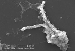
This scanning electron micrograph (SEM) of an untreated water specimen extracted from a wild stream mainly used to control flooding during inclement weather, revealed the presence of unidentified organisms, which included bacteria, protozoa, and algae. In this particular image, an unidentified amorphous mucoidal biofilm was featured, which appeared to have enmeshed numbers of amoeboid organisms.Created: 2009