-
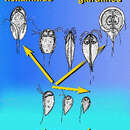
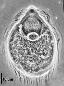
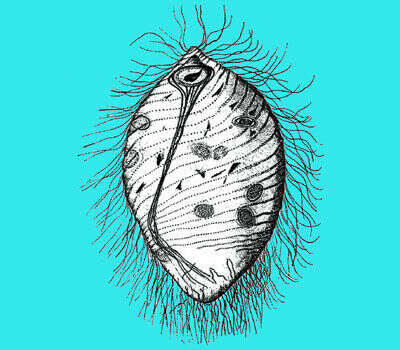
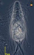
Trichonympha agilis from Reticulitermes lucifugus grassei, cell covered with flagella, anterior rostrum, parabasal fibres extending from the rostrum to the nucleus (phase contrast).
-
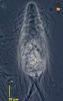
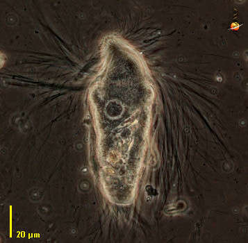
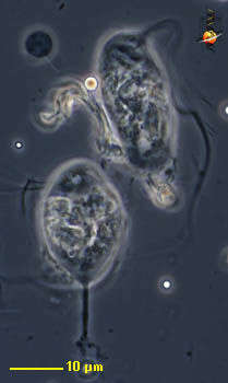
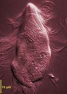
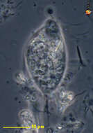
Spirotrichonympha (spire-o-trick-o-nympha-a) is a trichonymphid flagellate from the intestines of the termite Reticulotermes. These are hypermastigids in which the flagella insert in spiral arrays. Nucleus lies some distance behind the anterior end of the cell, but more or less determines the boundary between the front and the back of the cell. The back of the cell is capable of ingesting particles of wood. Phase contrast.
-
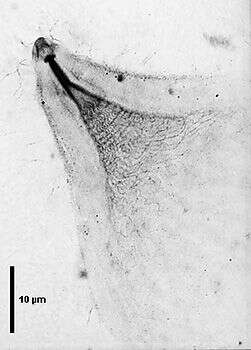
Trichonympha agilis, anterior part showing the parabasal cylinder posteriorly subdivided in many parabasal branches (silver staining).
-
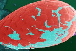
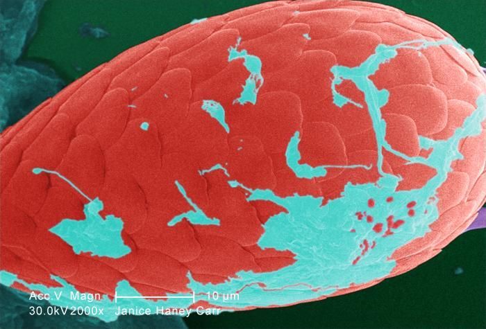
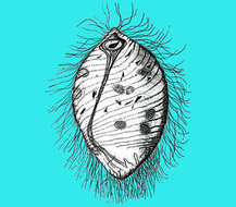
Under a moderate magnification of 2000X, this digitally-colorized scanning electron micrograph (SEM) of an untreated water specimen extracted from a wild stream mainly used to control flooding during inclement weather, revealed the presence of unidentified organisms, which included bacteria, protozoa, and algae. In this particular view, a single copepod-like microorganism was seen occupying the field of view, which seemed to be encased in an outer shell of armour-like plates, or scales. If you look closely, youll also notice the small grouping of bacteria, which had become enmeshed in a patch of biofilm on the dorsal surface of this creature's carapace.Created: 2009
-
Spirotrichonympha (spire-o-trick-o-nympha-a) is a trichonymphid flagellate from the intestines of the termite Reticulotermes. These are hypermastigids in which the flagella insert in spiral arrays. Nucleus lies some distance behind the anterior end of the cell, but more or less determines the boundary between the front and the back of the cell. The back of the cell is capable of ingesting particles of wood. Phase contrast.
-
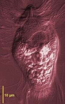
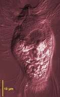
Differential interference microscopy showing the flagella of the rostrum separated from those of the cell body and the central nucleus.
-
-
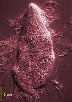
Differential interference microscopy showing the flagella of the rostrum separated from those of the cell body and the central nucleus.
-
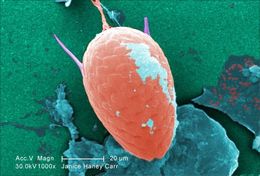
Under a moderate magnification of 1000X, this digitally-colorized scanning electron micrograph (SEM) of an untreated water specimen extracted from a wild stream mainly used to control flooding during inclement weather, revealed the presence of unidentified organisms, which included bacteria, protozoa, and algae. In this particular view, a single copepod-like microorganism was seen occupying the field of view, which seemed to be encased in an outer shell of armour-like plates, or scales. If you look closely, youll also notice the small grouping of bacteria, which had become enmeshed in a patch of biofilm.Created: 2009
-
-
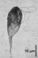
Tritrichomonads are small trichomonads (8-22 µm) with three anterior flagella and a recurrent flagellum forming a conspicuous undulating membrane with a posterior free portion. Costa stout or slender sustaining the undulating membrane; axostyle well developed; sausage-shaped parabasal. At the time of writing, there are about 20 species living in the intestinal tract of rodents, birds, reptiles and amphibians one species T. foetus is a parasite of the uro-genital tract of bovines. This image of Tritrichomonas augusta from the cloaca of amphibia and reptiles, three anterior flagella longer than those of Tritrichomonas muris, a recurrent flagellum forming an undulating membrane, posterior protruding axostyle (protargol and Giemsa staining).
-
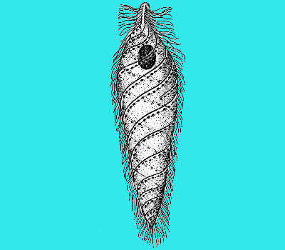
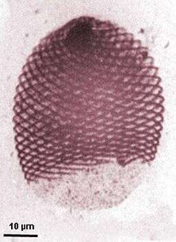
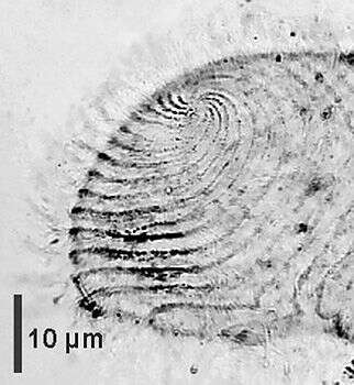
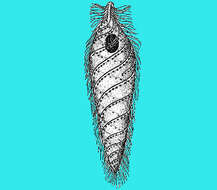
Large (80-180 µm) spirotrichonymphids with an anterior pole generally spiralled and bare, from which originate the helical flagellar rows that generally do not reach the posterior end. No rostrum or columella. Spot-shaped dictyosomes situated between the flagellar rows. Compound axostylar trunk that does not protrude at the posterior end. Occuring in termites, many species in Heterotermes, Coptotermes, Schedorhinotermes such as Holomastigotoides hemigymnum from Coptotermes sjoestedti. Whole cell with spiralled flagellar rows, posterior part free of flagella, anterior nucleus (protargol staining).
-
Tritrichomonads are small trichomonads (8-22 µm) with three anterior flagella and a recurrent flagellum forming a conspicuous undulating membrane with a posterior free portion. Costa stout or slender sustaining the undulating membrane; axostyle well developed; sausage-shaped parabasal. At the time of writing, there are about 20 species living in the intestinal tract of rodents, birds, reptiles and amphibians one species T. foetus is a parasite of the uro-genital tract of bovines. This image of Tritrichomonas augusta from the cloaca of amphibia and reptiles, three anterior flagella longer than those of Tritrichomonas muris, a recurrent flagellum forming an undulating membrane, posterior protruding axostyle (protargol and Giemsa staining).
-
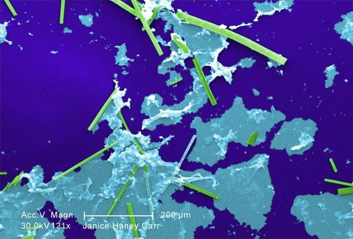
Under a relatively low magnification of 121X, this digitally-colorized scanning electron micrograph (SEM) of an untreated water specimen extracted from a wild stream mainly used to control flooding during inclement weather, revealed the presence of unidentified organisms, which included bacteria, protozoa, and algae. In this particular view, numbers of what appeared to be rod-shaped sections of various sizes were scattered throughout the field of view, which though unconfirmed, may have been vegetative in nature, and possibly algal organisms. There were also patches of biofilm present as well.Created: 2009
-
Large (80-180 µm) spirotrichonymphids with an anterior pole generally spiralled and bare, from which originate the helical flagellar rows that generally do not reach the posterior end. No rostrum or columella. Spot-shaped dictyosomes situated between the flagellar rows. Compound axostylar trunk that does not protrude at the posterior end. Occuring in termites, many species in Heterotermes, Coptotermes, Schedorhinotermes such as Holomastigotoides hemigymnum from Coptotermes sjoestedti. Anterior view showing the origin of the flagellar rows (silver staining).
-
Section of the "rail-type" undulating membrane by transmission EM
-
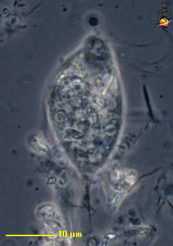
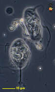
Foaina (foe-een-a) is one of the trichomonad flagellates - mostly endobiotic with four flagella arising from a point near the front of the cell. Three flagella project to the side or forwards, and one is directed to the rear. Usually with several cytoskeletal structures, an axial axostyle made of microtubules which encloses the nucleus anteriorly, as well as a costa which often lies under one of the flagella. The axostyle projects from the posterior end of the cell. From Cryptotermes. Phase contrast.
-
Undulating membrane of Tritrichomonas augusta from Amphibians and Reptiles by scanning EM
-
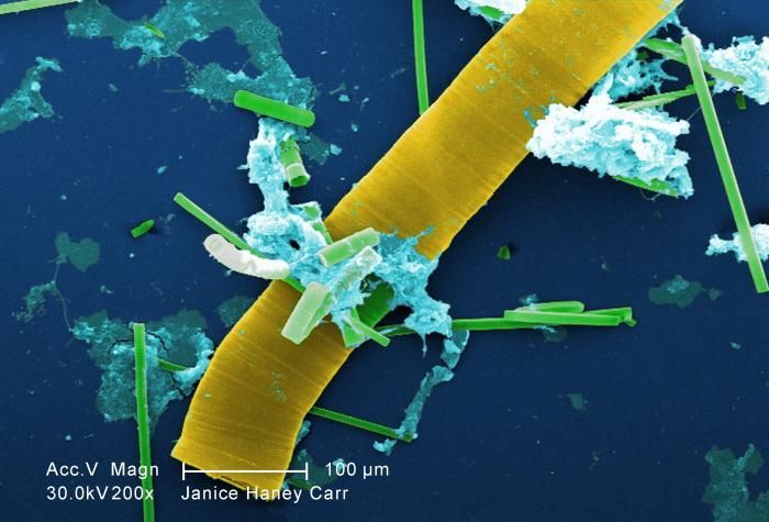
Under a relatively low magnification of 200X, this digitally-colorized scanning electron micrograph (SEM) of an untreated water specimen extracted from a wild stream mainly used to control flooding during inclement weather, revealed the presence of unidentified organisms, which included bacteria, protozoa, and algae. In this particular view, numbers of what appeared to be rod-shaped sections of various sizes were scattered throughout the field of view, which though unconfirmed, may have been vegetative in nature, and possibly algal organisms. There were also patches of biofilm present as well.Created: 2009
-
Foaina (foe-een-a) is one of the trichomonad flagellates - mostly endobiotic with four flagella arising from a point near the front of the cell. Three flagella project to the side or forwards - sometimes sticking together, and one is directed to the rear. Usually with several cytoskeletal structures, an axial axostyle made of microtubules which encloses the nucleus anteriorly, as well as a costa which often lies under one of the flagella. The axostyle projects from the posterior end of the cell and is adapted to form a holdfast which attaches the cell to a piece of debris. From the termite Cryptotermes. Phase contrast.
-
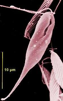
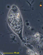
Chilomastix are retortamonad flagellates that have a pyriform and twisted cell body of about 20 µm in length with three anteriorly directed flagella and one short recurrent flagellum beating inside a ventral cytostomal pocket. The right fibril bordering the cytostomal pocket is thicker and forms a hook at its posterior end where food is phagocytosed. Cysts are pyriform and retain the internalised cytostomal fibers. They live in anoxic habitats but one species C. cuspidata is free-living. Among the 29 or so parasitic or endocommensal species described many live in the gut of vertebrates - such as C. mesnili in man and some in the gut of invertebrates such as C. aulastomi from the leech Aulastoma gulo. This species, Chilomastix caulleryi, is from the intestine of amphibia (haematoxylin staining).
-
Foaina (foe-een-a) is one of the trichomonad flagellates - mostly endobiotic with four flagella arising from a point near the front of the cell. Three flagella project to the side or forwards, and one is directed to the rear (lower left). Usually with several cytoskeletal structures, an axial axostyle made of microtubules which encloses the nucleus anteriorly, as well as a costa which often lies under one of the flagella. The axostyle projects from the posterior end of the cell and is adapted to form a holdfast which attaches the cell to a piece of debris. From the termite Cryptotermes. Phase contrast.
-
From toads, scanning EM showing the three anteriorly directed flagella, the ventral cytostomal aperture that contains the recurrent flagellum (not visible).
-
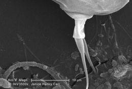
Under a moderate magnification of 3500X, this scanning electron micrograph (SEM) of an untreated water specimen extracted from a wild stream mainly used to control flooding during inclement weather, revealed the presence of unidentified organisms, which included bacteria, protozoa, and algae. In this particular view, the caudal end of a single copepod-like microorganism was seen occupying the field of view. Also, if you look closely towards the lower right corner, youll also notice the small grouping of bacteria, which had become enmeshed in a patch of biofilm.Created: 2009