-
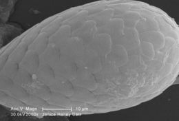
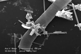
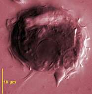
Under a moderate magnification of 2000X, this scanning electron micrograph (SEM) of an untreated water specimen extracted from a wild stream mainly used to control flooding during inclement weather, revealed the presence of unidentified organisms, which included bacteria, protozoa, and algae. In this particular view, a single copepod-like microorganism was seen occupying the field of view, which seemed to be encased in an outer shell of armour-like plates, or scales. See PHIL 11788 for a colorized view of this image.Created: 2009
-
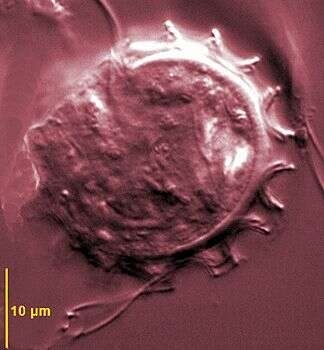
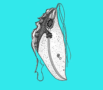
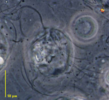
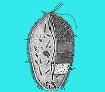
Caduceia (cad-you-see-a) a devescovinid flagellate from the termite Cryptotermes. With three free anterior flagella and a thick cord-like or ribbon-like recurrent flagellum (not visible here). The recurrent flagellum adheres to the cell body along a variable length which is underlain by a cresta. There is no real undulating membrane. The body may be covered with short spirochaetes and short rod-like bacteria adhering in some areas. Axostyle fine, extending through body to project from the back of the cell. There is a microtubular axostyle extending the length of the body, but anteriorly it wraps around the nucleus. The front end of the cell can move freely, and for this reason Tamm called these organisms Rubberneckia, a term which has since become a verb to refer to motorway-accident-voyeurism. There is some sign of the dictyosomes around the nucleus, although are better illustrated in other micrographs. Phase contrast.
-
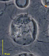
Undulating membrane, DIC image
-
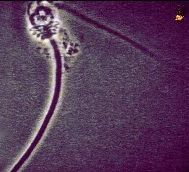
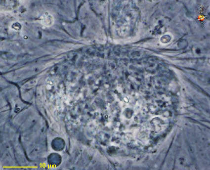
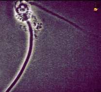
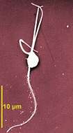
Caduceia (cad-you-see-a) a devescovinid flagellate from the termite Cryptotermes with three free anterior flagella and a thick cord-like or ribbon-like recurrent flagellum. This is the nucleus, flagellum, dictyosome and axostyle complex, sometimes called the karyomastigont, which has been extracted from cells. Phase contrast.
-
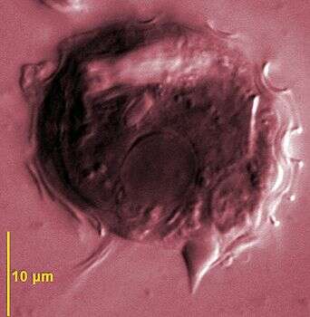
DIC image showing bulbous posterior axostyle
-
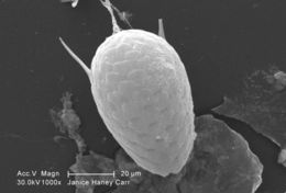
Under a moderate magnification of 1000X, this scanning electron micrograph (SEM) of an untreated water specimen extracted from a wild stream mainly used to control flooding during inclement weather, revealed the presence of unidentified organisms, which included bacteria, protozoa, and algae. In this particular view, a single copepod-like microorganism was seen occupying the field of view, which seemed to be encased in an outer shell of armour-like plates, or scales. If you look closely, youll also notice the small grouping of bacteria, which had become enmeshed in a patch of biofilm. See PHIL 11787 for a colorized version of this image.Created: 2009
-
-
Scanning electron micrograph showing recurrent flagellum associated with the conspicuous undulating membrane, posterior axostyle and cytoplsam filled with wood particles.
-
-
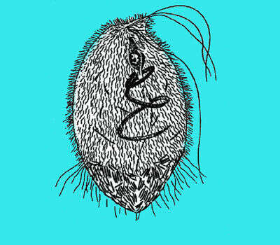
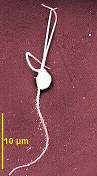
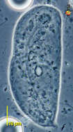
Tricercomitus are very small parabasalid flagellates (3-6 µm) with 3 anterior flagella and a recurrent one very long and adhering for most of its length along the body. Tricercomitus divergens is from Kalotermes flavicollis. This scanning EM shows the three anterior flagella and the long adhering recurrent flagellum bearing down-like hairs.
-
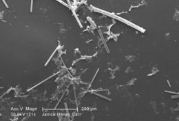
Under a relatively low magnification of 121X, this scanning electron micrograph (SEM) of an untreated water specimen extracted from a wild stream mainly used to control flooding during inclement weather, revealed the presence of unidentified organisms, which included bacteria, protozoa, and algae. In this particular view, numbers of what appeared to be rod-shaped sections of various sizes were scattered throughout the field of view, which though unconfirmed, may have been vegetative in nature, and possibly algal organisms. There were also patches of biofilm present as well. See PHIL 11786 for a colorized version of this image.Created: 2009
-
-
-
Under a relatively low magnification of 200X, this scanning electron micrograph (SEM) of an untreated water specimen extracted from a wild stream mainly used to control flooding during inclement weather, revealed the presence of unidentified organisms, which included bacteria, protozoa, and algae. In this particular view, numbers of what appeared to be rod-shaped sections of various sizes were scattered throughout the field of view, which though unconfirmed, may have been vegetative in nature, and possibly algal organisms. There were also patches of biofilm present as well. See PHIL 11785 for a colorized version of this image.Created: 2009
-
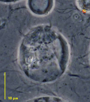
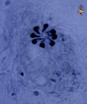
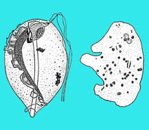
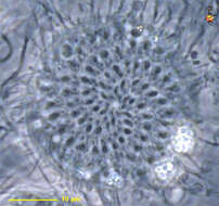
Coronympha (corr-o-nymph-a), a calonymphid flagellate which is a multinucleated trichomonad flagellate. All members of this group have multiple nuclei each with its own set of flagella. This genus has 8 or 16 karyomastigonts arranged in a single anterior circle. Each karyomastigont comprises three anterior flagella, a thick recurrent flagellum with an adherent proximal portion underlain by a cresta, an axostyle-pelta complex, and a drop-shaped parabasal close to the nucleus. The axostylar trunks are independent and only meet posteriorly, sometimes forming a caudal projection (see thumbnail). Phase contrast.
-
Coronympha (corr-o-nymph-a), a calonymphid flagellate which is a multinucleated trichomonad flagellate. All members of this group have multiple nuclei each with its own set of flagella. This genus has 8 or 16 karyomastigonts arranged in a single anterior circle. Each karyomastigont comprises three anterior flagella, a thick recurrent flagellum with an adherent proximal portion underlain by a cresta, an axostyle-pelta complex, and a drop-shaped parabasal close to the nucleus. The axostylar trunks are independent and only meet posteriorly, sometimes forming a caudal projection (see thumbnail). Four sets of flagella can be seen in this image, and a set seems to include 4 flagella (lower right). Phase contrast.
-
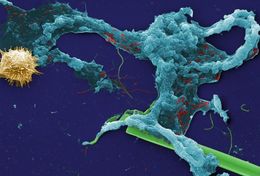
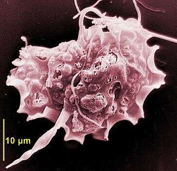
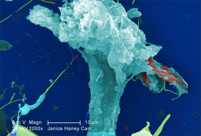
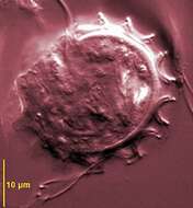
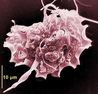
This digitally-colorized scanning electron micrograph (SEM) of an untreated water specimen extracted from a wild stream mainly used to control flooding during inclement weather, revealed the presence of unidentified organisms, which included bacteria, protozoa, and algae. Occupying most of the field of view, an unidentified amorphous mucoidal biofilm was featured, which appeared to have enmeshed numbers of amoeboid organisms, while on the right was a strangely-beautiful microorganism displaying an outer surface studded with numerous projections, making it appear like a microscopic sea urchin.Created: 2009
-
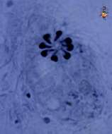
Coronympha (corr-o-nymph-a), a calonymphid flagellate which is a multinucleated trichomonad flagellate. All members of this group have multiple nuclei each with its own set of flagella. This genus has 8 or 16 karyomastigonts arranged in a single anterior circle. Each karyomastigont comprises three anterior flagella, a thick recurrent flagellum with an adherent proximal portion underlain by a cresta, an axostyle-pelta complex, and a drop-shaped parabasal close to the nucleus. The karyomastigonts have been stained in this fixed preparation
-
-
At a magnification of 2000X, this digitally-colorized scanning electron micrograph (SEM) of an untreated water specimen extracted from a wild stream, which is mainly used to control flooding during inclement weather, revealed the presence of unidentified organisms, which included bacteria, protozoa, and algae. In this particular image, an expanding amorphous organic biofilm was featured.Created: 2009
-
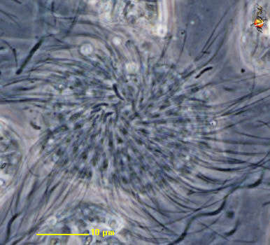
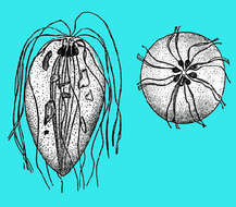
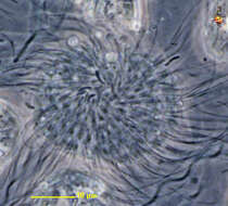
Metacoronympha (met-a-caw-row-nymph-a) one of the multinucleated trichomonad flagellates, always with many flagella. Flagella in small groups of 4, three extending outwards and one recurrent. The flagella are linked to nuclei which are arranged in a spiral around the anterior end of the cell. Axostyles are independent, but grouped at the posterior end. Parabasal bodies dot-shaped and located against the nuclei. Probably M. senta, from the termite Incisitermes (in America. Phase contrast.
-
Metacoronympha (met-a-caw-row-nymph-a) one of the multinucleated trichomonad flagellates, always with many flagella. Flagella in small groups of 4, three extending outwards and one recurrent. The flagella, as evident here, are linked to nuclei which are arranged in a spiral around the anterior end of the cell. as is evident from this image. Axostyles are independent, but grouped at the posterior end. Parabasal bodies dot-shaped and located against the nuclei. Probably M. senta, from the termite Incisitermes (in America. Phase contrast.
-
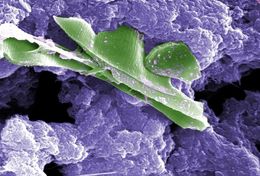
This digitally-colorized scanning electron micrograph (SEM) of an untreated water specimen extracted from a wild stream mainly used to control flooding during inclement weather, revealed the presence of unidentified organisms, which included bacteria, protozoa, and algae. In this particular image, unidentified sheets of algae were wrapped in a mass of what appeared to be a mucoid amorphous biofilm.Created: 2009
-
Metacoronympha (met-a-caw-row-nymph-a) one of the multinucleated trichomonad flagellates, always with many flagella. Flagella in small groups of 4, three extending outwards and one recurrent. The flagella are linked to nuclei which are arranged in a spiral around the anterior end of the cell as is evident from this image. Probably M. senta, from the termite Incisitermes. Phase contrast.