-
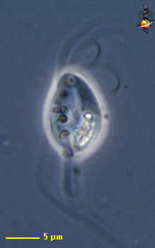
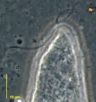
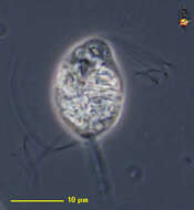
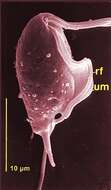
Foaina (foe-een-a) is one of the trichomonad flagellates - mostly endobiotic with four flagella arising from a point near the front of the cell. Three anterior free flagella and one recurrent one adhering to the cell body only on its proximal part. Usually with several cytoskeletal structures, an axial axostyle made of microtubules which encloses the nucleus anteriorly, as well as a costa which often lies under one of the flagella. The axostyle projects from the posterior end of the cell and may be adapted to form a holdfast which attaches the cell to a piece of debris. Parabasal body (dictyosomes) rod-, disc- or V-shaped. Consumes particles of wood. his image shows the three anterior flagella clearly. From the termite Cryptotermes. Phase contrast.
-
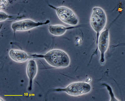
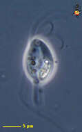
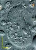
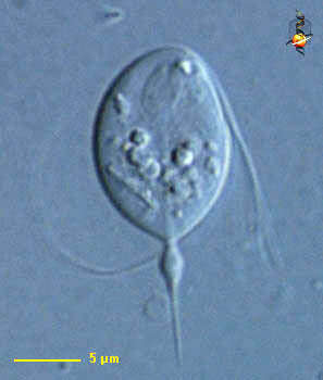
Hexamastix are small (10-30 µm) trichomonad flagellates with five grouped anteriorly directed flagella and a recurrent non-adhering flagellum. Axostyle well developed and parabasal V-shaped. Among the 11 species described, several live in Invertebrates such as H. claviger in termites (Kirby, 1930) and in vertebrates such as H. caviae in rodents (Nie, 1950). Hexamastix claviger from Kalotermes flavicollis (phase contrast).
-
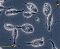
Foaina (foe-een-a) is one of the trichomonad flagellates - mostly endobiotic with four flagella arising from a point near the front of the cell. Three anterior free flagella and one recurrent one adhering to the cell body only on its proximal part. Usually with several cytoskeletal structures, an axial axostyle which usually projects from the posterior end of the cell and may be adapted to form a holdfast. Group of cells from the termite Cryptotermes. Phase contrast.
-
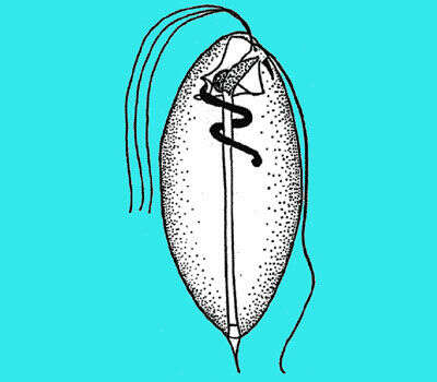
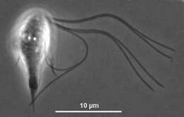
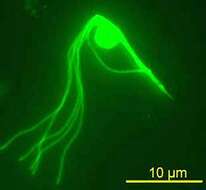
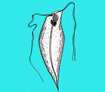
Hexamastix termitis showing the five anterior flagella, the recurrent flagellum, the nucleus and the axostyle.
-
Under a moderate magnification of 3500X, this scanning electron micrograph (SEM) of an untreated water specimen extracted from a wild stream mainly used to control flooding during inclement weather, revealed the presence of unidentified organisms, which included bacteria, protozoa, and algae. In this particular view, the caudal end of a single copepod-like microorganism was seen occupying the field of view. Also, if you look closely towards the upper right corner, youll also notice the small grouping of bacteria, which had become enmeshed in a patch of biofilm.Created: 2009
-
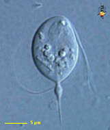
Foaina (foe-een-a) is one of the trichomonad flagellates - mostly endobiotic with four flagella arising from a point near the front of the cell. Three anterior free flagella and a recurrent one adhering to the cell body only on its proximal part. Three flagella are tightly grouped together, separating only at their distal ends. Usually with several cytoskeletal structures, an axial axostyle made of microtubules which encloses the nucleus anteriorly, as well as a costa which often lies under one of the flagella. The axostyle projects from the posterior end of the cell and may be adapted to form a holdfast which attaches the cell to a piece of debris. Parabasal body (dictyosomes) rod-, disc- or V-shaped. Electron microscopy has shown the trichomonad characters and particularly the presence of an infrakinetosomal body similar to that of Tritrichomonas. About 20 species living in the intestinal tract of vertebrates. other species live in the gut of invertebrates, especially arthropods such as termites and roaches, coleoptera, tipulid larvae and myriapods. Nucleus evident near front of cell, axostyle extending along the axis of the cell and out of the posterior end. From the termite Cryptotermes. Differential interference contrast.
-
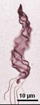
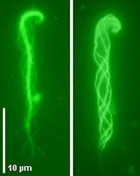
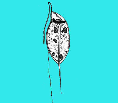
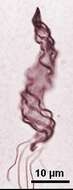
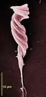
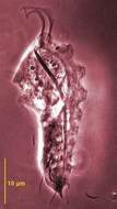
Oxymonad flagellates with a spirally twisted and contractile cell body of 20-100 µm in length and 3 -10 µm in breath. Four flagella arise at the anterior end and adhere to the cell body in gutters but have a posterior free portion. The nucleus is anterior or median and the cell is traversed by a contractile axostyle generally not protruding posteriorly. There is no developed anterior holdfast in contrast to Pyrsonympha. It phagocytoses wood particles, about six species, occuring in several termites such as Reticulitermes and Hodotermopsis species. Dinenympha exilis from Hodotermopsis sjoestedti, Giemsa.
-
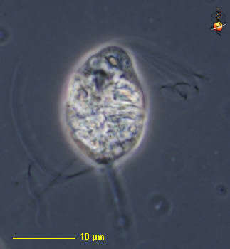
Foaina (foe-een-a) is one of the trichomonad flagellates - mostly endobiotic with four flagella arising from a point near the front of the cell. Three anterior free flagella and one recurrent one adhering to the cell body only on its proximal part. Usually with several cytoskeletal structures, an axial axostyle made of microtubules which encloses the nucleus anteriorly, as well as a costa which often lies under one of the flagella. The axostyle projects from the posterior end of the cell and may be adapted to form a holdfast which attaches the cell to a piece of debris. Parabasal body (dictyosomes) rod-, disc- or V-shaped. Consumes particles of wood. From the termite Kalotermes. Phase contrast.
-
Oxymonad flagellates with a spirally twisted and contractile cell body of 20-100 µm in length and 3 -10 µm in breath. Four flagella arise at the anterior end and adhere to the cell body in gutters but have a posterior free portion. The nucleus is anterior or median and the cell is traversed by a contractile axostyle generally not protruding posteriorly. There is no developed anterior holdfast in contrast to Pyrsonympha. It phagocytoses wood particles, about six species, occuring in several termites such as Reticulitermes and Hodotermopsis species. Dinenympha exilis from Hodotermopsis sjoestedti showing the central axostyle (left) and the four flagella adhering to the cell body (right) (immunofluorescence micrograph).
-
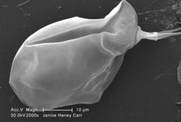
Under a moderate magnification of 2000X, this scanning electron micrograph (SEM) of an untreated water specimen extracted from a wild stream mainly used to control flooding during inclement weather, revealed the presence of unidentified organisms, which included bacteria, protozoa, and algae. In this particular view, a single copepod-like microorganism was seen occupying the field of view. Also, if you look closely towards the upper right corner, youll also notice the small grouping of bacteria, which had become enmeshed in a patch of biofilm. See PHIL 11791 for a colorized version of this image.Created: 2009
-
-
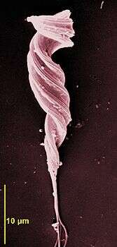
Scanning EM showing the adhering flagella to the twisted cell body with a posterior protruding axostyle
-
-
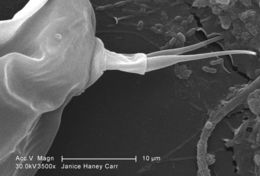
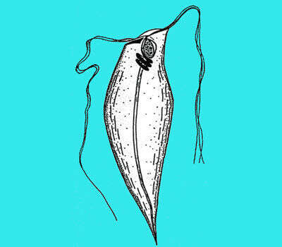
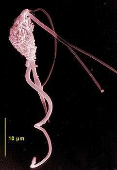
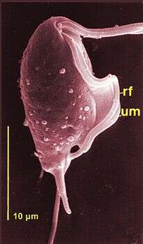
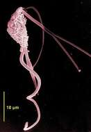
Neotermes joutelli, scanning EM showing the three anterior flagella and the long cord-like recurrent flagellum.
-
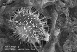
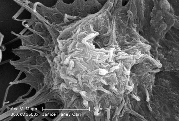
Under a moderately-high magnification of 2500X, this scanning electron micrograph (SEM) of an untreated water specimen extracted from a wild stream mainly used to control flooding during inclement weather, revealed the presence of unidentified organisms, which included bacteria, protozoa, and algae. In this particular view, a microorganism is featured, the exterior of which is covered by numerous projections imparting an appearance of a sea urchin. This microscopic pin cushion was teathered to its surroundings by a biofilm within which many bacteria, and amoeboid protozoa could be seen enmeshed as well. See PHIL 11781 for a greater magnification of this organisms exterior. See PHIL 11789 for a colorized version of this image.Created: 2009
-
-
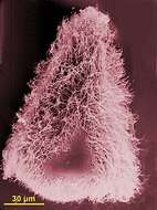
Koruga bonita is the type species and is a symbiont of Mastotermes darwiniensis, it has the same characters as Deltotrichonympha and is difficult to distinguish from it. The cell is triangular with an anterior dome and covered with flagella except the posterior end. There is a central axostyle and several other axostylar fibres.
-
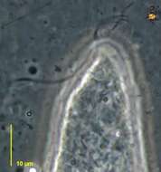
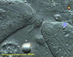
Caduceia (cad-you-see-a) a devescovinid flagellate from the termite Cryptotermes. There is a microtubular axostyle extending the length of the body, but anteriorly it wraps around the nucleus. The globules around the nucleus are elements of the Golgi apparatus (dictyosomes). Finely granular region around the axostyle just posterior to the nucleus is the bacterial cup with large numbers of endosymbiotic bacteria. There is a small indentation just below the apex and to the upper left. This is the point of insertion of the flagella. Food vacuoles contain bacteria or small pieces of wood. Differential interference contrast.
-
Section of the lamellar-like undulating membrane by transmission electron microscopy
-
Under a relatively-high magnification of 6500X, this scanning electron micrograph (SEM) of an untreated water specimen extracted from a wild stream mainly used to control flooding during inclement weather, revealed the presence of unidentified organisms, which included bacteria, protozoa, and algae. In this particular view, a single amoeboid-like vegetative organism appeared to have blossomed from the underlying biofilm mass occupying the right side of the image.Created: 2009
-
Caduceia (cad-you-see-a) a devescovinid flagellate from the termite Cryptotermes with three free anterior flagella and a thick cord-like or ribbon-like recurrent flagellum. Three of the flagella can be seen extending to the right, and one lies adjacent to the left side of the body. Phase contrast.
-
Scanning EM showing the anterior flagella, the recurrent flagellum attached to the lamellar undulating membrane, the posteriorly pointing axostyle.
-
Caduceia (cad-you-see-a) a devescovinid flagellate from the termite Cryptotermes. The anterior regions of two cells show the axostyles, the nucleus, bacterial cups - the finely granular regions posterior to the nucleus. The irregular region around the left hand nucleus is formed by the dictyosomes. Differential interference contrast.
-
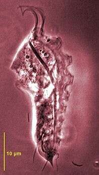
Trichomitpsis termopsidis from Hodotermopsis sjoestedti - all cells have two flagellar apparatuses comprising two undulating membranes and two costa and axostyles. Phase contrast microscopy.