-
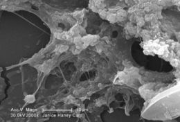
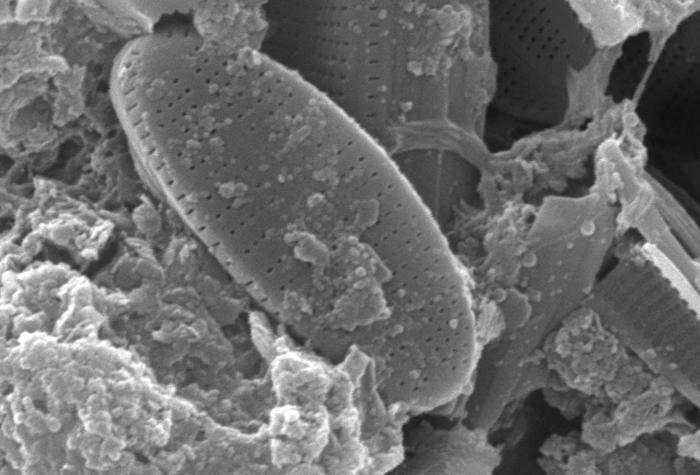
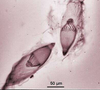
This scanning electron micrograph (SEM) of an untreated water specimen extracted from a wild stream mainly used to control flooding during inclement weather, revealed the presence of unidentified organisms, which included bacteria, protozoa, and algae. In this particular image, a number of unidentified oblong elliptical-shaped diatoms were featured.Created: 2009
-
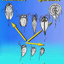
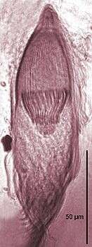
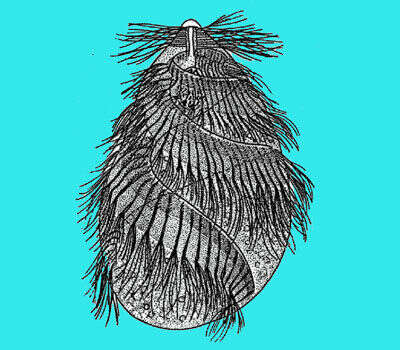
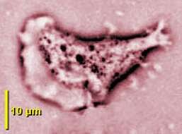
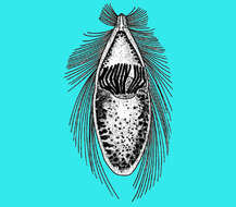
Trichonympha (trick-owe-nymph-a) is one of the hypermastigid flagellates - flagellates with large numbers of flagella. They and the trichomonads made up the group called the parabasalids - almost all of which are endobiotic or parasitic. There is an anterior symmetrical rostrum, and the numerous flagella arise from this region and from the region of the body immediately behind. There are also spirochaetes attached to the back of the body. The nucleus is a large structure lying some distance behind this. Cytoskeletal fibres with associated dictyosomes form bands running from the points of flagellar insertion through the anterior part of the body. Ingesta (food vacuoles) are found in the posterior part of the body. From the wood-eating roach, Cryptocercus. Phase contrast.
-
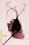
Cell showing the four anterior flagella, the short undulating membrane, the nucleus, the parabasal apparatus with the long parabasal fibre and the axostyle (protargol staining).
-
Trichonympha (trick-owe-nymph-a) is one of the hypermastigid flagellates - flagellates with large numbers of flagella. They and the trichomonads made up the group called the parabasalids - almost all of which are endobiotic or parasitic. There is an anterior symmetrical rostrum, and the numerous flagella arise from this region and from the region of the body immediately behind. The nucleus is a large structure lying some distance behind this. Cytoskeletal fibres with associated dictyosomes form bands running from the points of flagellar insertion through the anterior part of the body. Ingesta (food vacuoles) are found in the posterior part of the body. From the wood-eating roach, Cryptocercus. Phase contrast.
-
Cell stained by protargol, showing the four anterior flagella, the short undulating membrane and the subjacent costa.
-
Trichonympha (trick-owe-nymph-a) is one of the hypermastigid flagellates - flagellates with large numbers of flagella. They and the trichomonads made up the group called the parabasalids - almost all of which are endobiotic or parasitic. There is an anterior symmetrical rostrum, and the numerous flagella arise from this region and from the region of the body immediately behind. There are also spirochaetes attached to the back of the body. The nucleus is a large structure lying some distance behind this. Cytoskeletal fibres with associated dictyosomes form bands running from the points of flagellar insertion through the anterior part of the body. Ingesta (food vacuoles) are found in the posterior part of the body. From the wood-eating roach, Cryptocercus. Phase contrast.
-
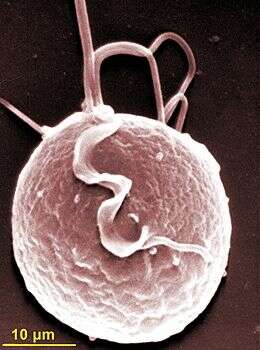
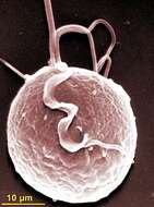
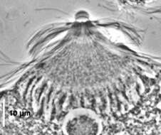
Scanning electron micrograph showing anterior flagella and the recurrent flagellum adhering to the short lamellar undulating membrane.
-
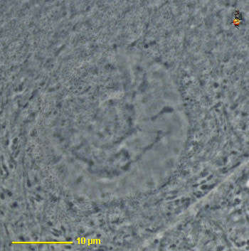
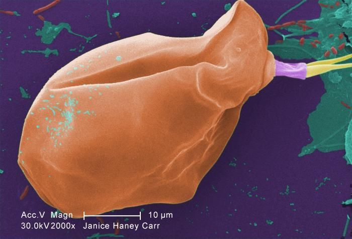
At a magnification of 2000X, this scanning electron micrograph (SEM) of an untreated water specimen extracted from a wild stream, which is mainly used to control flooding during inclement weather, revealed the presence of unidentified organisms, which included bacteria, protozoa, and algae.Created: 2009
-
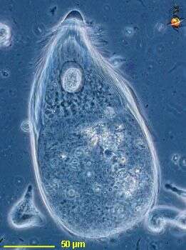
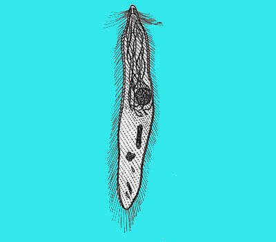
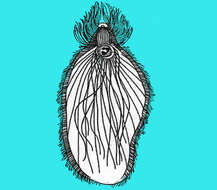
Trichonympha (trick-owe-nymph-a) is one of the hypermastigid flagellates - flagellates with large numbers of flagella. The nucleus and cytoplasm - with numerous hydrogenosomes. From the termite Zootermopsis, supplied by Ward s Natural Science Establishment, Rochester, New York, USA. Phase contrast.
-
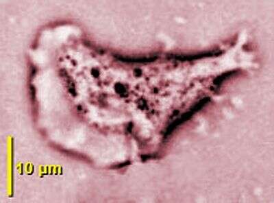
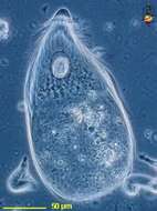
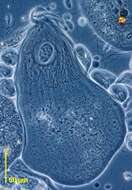
Adherent amoeboid cell by phase contrast microscopy.
-
-
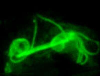
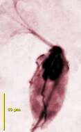
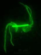
Division stages by immunofluorescence showing the two sets of flagella separated by a thick paradesmose.
-
This scanning electron micrograph (SEM) of an untreated water specimen extracted from a wild stream, which is mainly used to control flooding during inclement weather, revealed the presence of unidentified organisms, which included bacteria, protozoa, and algae. For a digitally-colorized version of this image see PHIL 11695.Created: 2009
-
-
Division, by immunofluorescence, showing the two sets of flagella separated by a thick paradesmose.
-
-
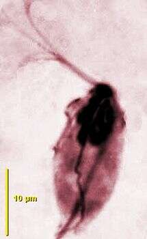
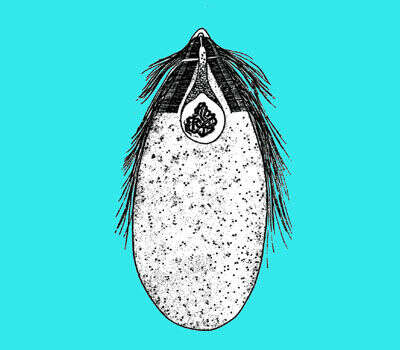
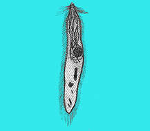
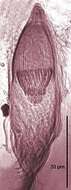
Trichonympha agilis from Reticulitemes lucifugus grassei, anterior rostrum, central nucleus surrounded by parabasal apparatus branches (protargol staining).
-
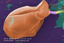
Under a moderate magnification of 2000X, this digitally-colorized scanning electron micrograph (SEM) of an untreated water specimen extracted from a wild stream mainly used to control flooding during inclement weather, revealed the presence of unidentified organisms, which included bacteria, protozoa, and algae. In this particular view, a single copepod-like microorganism was seen occupying the field of view. Also, if you look closely towards the upper right corner, youll also notice the small grouping of bacteria, which had become enmeshed in a patch of biofilm.Created: 2009
-
-
Trichonympha agilis from Reticulitemes lucifugus grassei, anterior rostrum, central nucleus surrounded by parabasal apparatus branches (protargol staining).
-
-
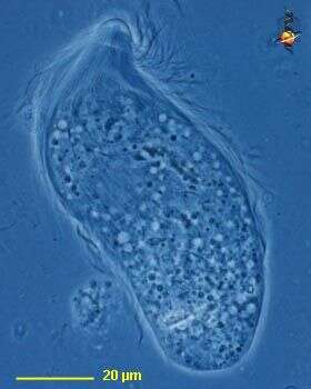
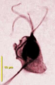
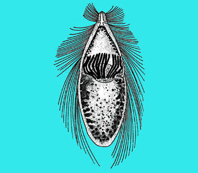
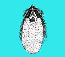
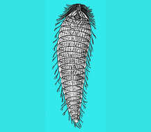
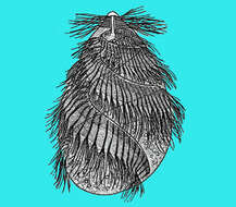
Trichonympha agilis from Reticulitermes lucifugus grassei, cell covered with flagella, anterior rostrum, parabasal fibres extending from the rostrum to the nucleus (phase contrast).
-
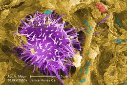
Under a moderately-high magnification of 2500X, this digitally-colorized scanning electron micrograph (SEM) of an untreated water specimen extracted from a wild stream mainly used to control flooding during inclement weather, revealed the presence of unidentified organisms, which included bacteria, protozoa, and algae. In this particular view, a microorganism is featured, the exterior of which is covered by numerous projections imparting an appearance of a sea urchin. This microscopic pin cushion was teathered to its surroundings by a biofilm within which many bacteria, and amoeboid protozoa could be seen enmeshed as well. See PHIL 11781 for a greater magnification of this organisms exterior.Created: 2009
-