When first discovered in 1977, these microscopic single-celled organisms were classified as Bacteria. However, later genetic and biochemical analyses showed that they were more closely related to the Eukaryotes (which include humans). The researchers who discovered this, Woese and his colleagues, proposed the addition of the “domain” as a taxonomic level above that of kingdom, with all life classified within one of three domains, now called Bacteria, Eukaryotes, and Archaea. As of 2010 more than 250 species of Archaea had been described, most fitting into one of two groups, Euryarchaeota and Crenarchaeota. Many of these live in extreme environments – places where we once believed no life could exist.
(Woese et al., 1990; Kimball, 2010; UCMP, accessed 2011)
Apparently nearly anywhere and eveywhere on Earth.
Because Archaea can thrive in extreme environments, they occur in places we didn’t think were habitats – very low (4 °C) and very high (120 °C) temperatures of polar seas, hydrothermal vents and hot sulfur springs; high and very low pH; hypersaline water, and anaerobic environments like human colons and termite guts. They also live in moderate habitats in the oceans, soils, freshwater (Kimball, 2010).
Archaea are generally less than one micron long, may or may not have flagella, and cell-shapes include spherical, box-like, triangular, and slender filamentous forms (UCMP, accessed 2011).
PHYLA / DIVISIONS
Euryarchaeota: methanogens, halophiles, thermophiles
Crenarchaeota: hyperthermophiles, cool-water marine plankton, nitrifying soil/sediment microbes
Nanoarchaeota: thermophiles
Korarchaeota (proposed): thermophiles
Thaumarchaeota (proposed): chemolithoautotrophic ammonia-oxidizers
(Hohn et al., 2002; Huber et al., 2002; Brochier-Armanet et al., 2008; Kimball, 2010; Spang et al., 2010; Auchtung et al., 2011)
Archaea is ’n domein van eensellige mikroörganismes. Dié mikrobes is prokariote, wat beteken hulle het geen selkern of enige ander organelle in hul selle wat deur ’n membraan omsluit word nie.
Archaea is aanvanklik as bakterieë geklassifiseer en het die naam Archaebacteria (in die koninkryk Monera) gekry, maar dié klassifikasie is verouderd.[1] Hul selle het unieke eienskappe wat hulle skei van die ander twee domeins van lewe: Bacteria en Eukaryota. Archaea word verder verdeel in vier filums.
Archaea is eers beskou as ekstremofiele wat in uiters ongunstige toestande soos warmwaterbronne en soutmere voorkom, maar hulle is sedertdien ontdek in ’n groot verskeidenheid habitats, onder meer in die menslike kolon en naeltjie.[2]
Hulle is veral talryk in die oseane, en die archaea in plankton kan een van die grootste groepe organismes op die planeet wees. Archaea is ’n belangrike deel van lewe op Aarde en kan ’n rol speel in beide die koolstof- en stikstofsiklus.
Archaea is ’n domein van eensellige mikroörganismes. Dié mikrobes is prokariote, wat beteken hulle het geen selkern of enige ander organelle in hul selle wat deur ’n membraan omsluit word nie.
Archaea is aanvanklik as bakterieë geklassifiseer en het die naam Archaebacteria (in die koninkryk Monera) gekry, maar dié klassifikasie is verouderd. Hul selle het unieke eienskappe wat hulle skei van die ander twee domeins van lewe: Bacteria en Eukaryota. Archaea word verder verdeel in vier filums.
Archaea is eers beskou as ekstremofiele wat in uiters ongunstige toestande soos warmwaterbronne en soutmere voorkom, maar hulle is sedertdien ontdek in ’n groot verskeidenheid habitats, onder meer in die menslike kolon en naeltjie.
Hulle is veral talryk in die oseane, en die archaea in plankton kan een van die grootste groepe organismes op die planeet wees. Archaea is ’n belangrike deel van lewe op Aarde en kan ’n rol speel in beide die koolstof- en stikstofsiklus.
Les archaea (o arquées) son microorganismos unicelulares. Al igual que les bacteries, les archaea carecen de nucleu y son por tantu procariontes. D'otra miente, les diferencies a nivel molecular ente arquées y bacteries son tan fundamentales que se-yos clasifica en grupos estremaos. De fechu, estes diferencies son mayores de les qu'hai, por exemplu, ente una planta y un animal. Anguaño considérase que les archaea tan filoxenéticamente más prósimes a los eucariontes qu'a les bacterias.
Les archaea foren escubiertes orixinariamente n'ambientes llímite, pero dende entós se-yos tien atopao en too tipu d'hábitats y podríen contribuyir fasta'l 20% de la biomasa.[2] Son particularmente comunes nos océanos y puen ser un de los más abundantes grupos d'organismos nel planeta.[3] Dalgunes especies son hipertermófiles y puen sobrevivir y prosperar a temperatures selemente superiores a los 100 °C y se-yos atopa en xéiseres, fontes hidrotermales y pozos de petroleu.[4] Otres estremófiles s'atopen n'agua hiper-salina, ácida o alcalina. Ensin embargu, otres son mesófiles o psicrófiles y prosperen n'ambientes tales como marismes, agües residuales, agua de mar y nel suelu. A les metanóxenes se-yos pue atopar nel tractu dixestivu d'animales tales como rumiantes, termites y seres humanos. Nun se conocen patóxenos pa los seres humanos,[5][6] anque se tien propuesto dalguna rellación ente los metanóxenos y los trastornos periodontales humanos.[7]
Archaea constitúi un de los dominios nos que se dividen los seres vivos. Tiempu atrás clasificábense como pertenecientes al reinu Monera na taxonomía tradicional de los cinco reinos. En 1990 propúnxose consideralos un dominiu separtáu, según el sistema de tres dominios de Carl Woese.[8][9]
Les archaea (o arquées) son microorganismos unicelulares. Al igual que les bacteries, les archaea carecen de nucleu y son por tantu procariontes. D'otra miente, les diferencies a nivel molecular ente arquées y bacteries son tan fundamentales que se-yos clasifica en grupos estremaos. De fechu, estes diferencies son mayores de les qu'hai, por exemplu, ente una planta y un animal. Anguaño considérase que les archaea tan filoxenéticamente más prósimes a los eucariontes qu'a les bacterias.
Les archaea foren escubiertes orixinariamente n'ambientes llímite, pero dende entós se-yos tien atopao en too tipu d'hábitats y podríen contribuyir fasta'l 20% de la biomasa. Son particularmente comunes nos océanos y puen ser un de los más abundantes grupos d'organismos nel planeta. Dalgunes especies son hipertermófiles y puen sobrevivir y prosperar a temperatures selemente superiores a los 100 °C y se-yos atopa en xéiseres, fontes hidrotermales y pozos de petroleu. Otres estremófiles s'atopen n'agua hiper-salina, ácida o alcalina. Ensin embargu, otres son mesófiles o psicrófiles y prosperen n'ambientes tales como marismes, agües residuales, agua de mar y nel suelu. A les metanóxenes se-yos pue atopar nel tractu dixestivu d'animales tales como rumiantes, termites y seres humanos. Nun se conocen patóxenos pa los seres humanos, anque se tien propuesto dalguna rellación ente los metanóxenos y los trastornos periodontales humanos.
Archaea constitúi un de los dominios nos que se dividen los seres vivos. Tiempu atrás clasificábense como pertenecientes al reinu Monera na taxonomía tradicional de los cinco reinos. En 1990 propúnxose consideralos un dominiu separtáu, según el sistema de tres dominios de Carl Woese.
Arxelər və ya arxebakteriyalar - təkhüceyrəli mikroorqanizmlər qrupu. Həm aləm, həm də aləmüstü hesab edilirlər. Prokaryotik və eukaryotik orqanizmlərin xüsusiyyətlərini daşıyırlar. Ümumiyyətlə, eksterm canlılar olaraq bilinirlər. Əsasən metasintezedici orqanizmlərdir. 1990-cı ildə kəşf olunmuşlar və bakteriyalara aid edilmişlər.
Arxelərin əsasən dörd növü ayırd edilir: metanogen, qalofil, termofil və psixofil arxelər.
Həyat fəaliyyəti nəticəsində metan CH4 əmələ gətirən arxelərdir.
Arxelər və ya arxebakteriyalar - təkhüceyrəli mikroorqanizmlər qrupu. Həm aləm, həm də aləmüstü hesab edilirlər. Prokaryotik və eukaryotik orqanizmlərin xüsusiyyətlərini daşıyırlar. Ümumiyyətlə, eksterm canlılar olaraq bilinirlər. Əsasən metasintezedici orqanizmlərdir. 1990-cı ildə kəşf olunmuşlar və bakteriyalara aid edilmişlər.
Els arqueobacteris o arqueus[1] (Archaea) són un grup de microorganismes unicel·lulars; el terme arxibacteri n'és una denominació desestimada. Els arqueobacteris, com els eubacteris, són procariotes que manquen de nucli cel·lular o qualsevol altre orgànul dins les cèl·lules. En el passat, se'ls considerà un grup inusual de bacteris, però com que tenen una història evolutiva independent i presenten moltes diferències en la seva bioquímica respecte a la resta de formes de vida, actualment se'ls classifica com un domini distint en el sistema de tres dominis. En aquest sistema, presentat per Carl Woese, les tres branques evolutives principals són els arqueobacteris, els bacteris i els eucariotes. Els arqueobacteris estan subdividits en quatre filums, dels quals dos, els crenarqueots i els euriarqueots, són estudiats més intensivament. La classificació dels arqueobacteris roman dificultosa, car la immensa majoria d'aquests organismes no han sigut mai estudiats al laboratori i només se'ls ha detectat per mitjà de l'anàlisi dels seus àcids nucleics en mostres del medi ambient.
En general, els arqueobacteris i bacteris són bastant semblants en forma i en mida, tot i que alguns arqueobacteris tenen formes molt inusuals, com ara les cèl·lules planes i quadrades d'Haloquadra walsbyi. Malgrat aquesta semblança visual amb els bacteris, els arqueobateris tenen gens i diverses rutes metabòliques que són més properes a les dels eucariotes; notablement, els enzims implicats en la transcripció i la traducció. Altres aspectes de la bioquímica dels arqueobacteris són únics, com ara els èters lípids de les seves membranes cel·lulars. Els arqueobacteris exploten una varietat de recursos molt més grans que els eucariotes; des de compostos orgànics familiars com ara els sucres, fins a l'ús d'amoníac, ions de metalls o fins i tot hidrogen com nutrients. Els arqueobacteris tolerants de la sal (els halobacteris) utilitzen la llum solar com font d'energia, i altres espècies d'arqueobacteris fixen carboni; tanmateix, a diferència de les plantes i els cianobacteris, no es coneix cap espècie d'arqueobacteri que sigui capaç d'ambdues coses. Els arqueobacteris es reprodueixen asexualment i es divideixen per fissió binària, fragmentació o gemmació; a diferència dels bacteris i els eucariotes, no es coneix cap espècie d'arqueobacteri que formi espores.
Originalment, els arqueobacteris eren vists com a extremòfils que vivien en ambients severs, com ara aigües termals i llacs salats, però posteriorment se'ls ha observat en una gran varietat d'hàbitats, com ara sòls, oceans i aiguamolls. Els arqueobacteris són especialment nombrosos als oceans, i els que es troben al plàncton podrien ser un dels grups d'organismes més abundants del planeta. Aquests procariotes són actualment considerats una part important de la vida a la Terra i podrien tenir un paper important tant en el cicle del carboni com en el cicle del nitrogen. No es coneixen exemples clars de patògens o paràsits arquobacterians, però solen ser mutualistes o comensals. En són exemples els arqueobacteris metanògens que viuen a l'intestí dels humans i els remugants, on estan presents en grans quantitats i contribueixen a digerir l'aliment. Els arqueobacteris tenen la seva importància en la tecnologia; hi ha metanògens que són utilitzats per produir biogàs i com a part del procés de depuració, i els enzims d'arqueobacteris extremòfils capaços de resistir a temperatures elevades i solvents orgànics són explotats en la biotecnologia.
L'any 2012 es va demostrar per primera vegada, que els arqueobacteris són consumits per organismes vius, en concret per part de cucs poliquets dins la família Dorvilleidae. L'estudi es va publicar al Society for Microbial Ecology Journal, de la revista Nature.[2]
A principis del segle XX, els procariotes eren considerats un únic grup d'organismes i classificats segons la seva bioquímica, morfologia i metabolisme. Per exemple, els microbiòlegs intentaven classificar els microorganismes segons l'estructura de la seva paret cel·lular, la seva forma, i les substàncies que consumeixen.[3] Tanmateix, el 1965 es proposà un nou sistema,[4] utilitzant les seqüències genètiques d'aquests organismes per esbrinar quins procariotes estan realment relacionats entre ells. Aquest mètode, conegut com a filogènia, és el mètode principal utilitzat actualment.
Els arqueobacteris foren classificats per primer cop com un grup separat de procariotes el 1977 per Carl Woese i George E. Fox en arbres filogenètics basats en les seqüències de gens d'ARN ribosòmic (ARNr).[5] Aquests dos grups foren originalment denominats arqueobacteris i eubaceris i tractats com regnes o subregnes, que Woese i Fox denominaren "regnes originals". Woese argumentà que aquest gup de procariotes és un tipus de vida fonamentalment distint. Per emfatitzar aquesta diferència, aquests dos dominis foren posteriorment anomenats Archaea i Eubacteria.[6] El nom científic Archaea prové del grec antic ἀρχαῖα, que vol dir "els antics".[7] El terme "arqueobacteri" prové de la combinació d'aquesta arrel i del terme grec baktērion, que significa "petit bastó".
Originalment, només es classificaren els metanògens en aquest nou domini, i els arqueobacteris eren considerats extremòfils que només viuen en hàbitats com ara aigües termals i llacs salats. A finals del segle XX, els microbiòlegs s'adonaren que els arqueobacteris són un grup d'organismes gran i divers àmpliament distribuïts a la natura, i que són comuns en hàbitats molt menys extrems, com ara sòls i oceans.[8] Aquesta nova presa de consciència de la importància i l'omnipresència vingué de l'ús de la reacció en cadena de la polimerasa per detectar procariotes en mostres d'aigua o sòl a partir únicament dels seus àcids nucleics. Això permet detectar i identificar organismes que no poden ser cultivats al laboratori, que sol ser difícil.[9][10]
La classificació dels arqueobacteris, i dels procariotes en general, és un camp en constant moviment i controvertit. Els sistemes actuals de classificació intenten organitzar els arqueobacteris en grups d'organismes que comparteixen trets estructurals i avantpassats comuns.[11] Aquestes classificacions es basen significativament en l'ús de seqüències de gens d'ARN ribosòmic per revelar les relacions entre els organismes (filogenètica molecular).[12] La majoria d'espècies d'arqueobacteris cultivables i ben investigades són membres de dos fílums principals, els euriarqueots i crenarqueots. S'han proposat temptativament altres grups. Per exemple, a la peculiar espècie Nanoarchaeum equitans, que fou descoberta el 2003, se li ha atribuït el seu propi fílum, el dels nanoarqueots.[13] També s'ha proposat el nou fílum dels korarqueots, que conté un nombre reduït d'inusuals espècies termòfiles que comparteixen trets dels dos fílums principals, però que són més properes als crenarqueots.[14][15] Altres espècies d'arqueobacteris recentment descobertes només tenen una relació distant amb qualsevol d'aquests grups, com ara els nanoorganismes arquobacterians acidòfils de Richmond Mine (ARMAN), que foren descoberts el 2006.[16]
La classificació dels arqueobacteris en espècies també és controvertida. En biologia, una espècie és un grup d'organismes relacionats. Una definició popular d'espècie entre els animals és un conjunt d'organismes que poden reproduir-se entre ells i que estan reproductivament aïllats d'altres grups d'organismes (és a dir, no poden reproduir-se amb altres espècies).[17] Tanmateix, els esforços per classificar els procariotes tals com els arqueobacteris en espècies es compliquen pel fet que són asexuals i que presenten un alt nivell de transferència horitzontal de gens entre llinatges. Aquest tema és controvertit; per exemple, algunes dades suggereixen que en arqueobacteris com ara Ferroplasma, es poden agrupar cèl·lules individuals en poblacions de genoma molt similar i que rarament transfereixen gens a grups més divergents de cèl·lules.[18] Alguns argumenten que aquests grups de cèl·lules són anàlegs a espècies. D'altra banda, estudis d'Halorubrum descobriren un intercanvi genètic significatiu entre aquestes poblacions.[19] Aquests resultats han portat a pensar que classificar aquests grups d'organismes com espècies tindria poc sentit pràctic.[20]
El coneixement actual sobre la diversitat dels arqueobacteris és fragmentari, i no es pot estimar amb cap mena de precisió el nombre total d'espècies d'arqueobacteris.[12] Fins i tot les estimacions del nombre total de fílums arquobacterians varien entre 18 i 23, dels quals només vuit tenen representants que s'han cultivat i estudiat directament. Molts d'aquests grups hipotètics són coneguts únicament a partir d'una sola seqüència d'ARNr, cosa que indica que la diversitat d'aquests organismes roman completament desconeguda.[21] El problema de com estudiar i classificar microbis no cultivats també es dóna en els bacteris.[22]
Els arqueobacteris són una forma de vida molt antiga. S'han datat fòssils probables d'aquestes cèl·lules de fa gairebé 3.500 milions d'anys,[23] i s'han detectat restes de lípids que podrien ser arquobacterians o eucariotes en esquists de fa 2.700 milions d'anys.[24] Com que la majoria de procariotes no tenen una morfologia característica, la forma dels fòssils no serveix per identificar-los com arqueobacteris. En canvi, fòssils químics, en forma dels lípids únics dels arqueobacteris, ofereixen més informació car aquests compostos no apareixen en altres grups d'organismes.[25] Aquests lípids han sigut detectats en roques que daten del Precambrià. Les restes més antigues conegudes d'aquests lípids d'isoprè daten del cinturó d'Isua, a l'oest de Groenlàndia, que inclouen sediments formats fa 3.800 milions d'anys que són els més antics del món.[26] L'origen dels arqueobacteris sembla extremament remot en el temps, i el llinatge arqueobacterià podria ser el més antic de la Terra.[27]
Woese argumentà que els bacteris, arqueobacteris i eucariotes representen cadascun un llinatge evolutiu distint que divergí fa molts milions d'anys d'una colònia ancestral d'organismes.[29][30] Tanmateix, alguns biòlegs han argumentat que els arqueobacteris i eucariotes sorgiren d'un grup de bacteris.[31] Thomas Cavalier-Smith proposà el clade dels neomurs per representar aquesta teoria. Neomura vol dir "parets noves" i es refereix a la teoria que els arqueobacteris i els eucariotes haurien derivat de bacteris que (entre altres adaptacions) havien substituït les parets de peptidoglicà per altres glicoproteïnes. És possible que l'últim avantpassat comú dels bacteris i els arqueobacteris fos un termòfil, cosa que presenta la possibilitat que les temperatures baixes siguin "ambients extrems" pels arqueobacteris, i que els organismes que viuen en ambients més freds apareguessin més tard en la història de la vida a la Terra.[32] Com que els arqueobacteris i els bacteris no estan relacionats més estretament els uns amb els altres que amb els eucariotes, s'ha argumentat que el terme "procariota" no té un sentit evolutiu autèntic i que caldria descartar-lo completament.[33]
La relació entre arqueobacteris i eucariotes roman un problema important. A part de les semblances en l'estructura i les funcions cel·lulars que es discuteixen més avall, molts arbres genètics agrupen els dos clades. Algunes de les primeres anàlisis fins i tot suggerien que la relació entre els eucariotes i el fílum arqueobacterià dels euriarqueots és més propera que la relació entre els euriarqueots i els crenarqueots.[34] Tanmateix, actualment es considera més versemblant que l'avantpassat dels eucariotes divergís bastant aviat dels arqueobacteris.[35][36] El descobriment de gens similars als dels arqueobacteris en alguns grups de bacteris, com ara Thermotoga maritima, fa que aquestes relacions siguin difícils de determinar, car ha tingut lloc una transferència horitzontal de gens.[37] Alguns han suggerit que els eucariotes sorgiren de la fusió d'un arqueobacteri i un bacteri, que esdevingueren el nucli i el citoplasma; això explica diverses semblances genètiques però presenta dificultats a l'hora d'explicar l'estructura cel·lular.[38]
La mida dels arqueobacteris varia entre 0,1 micròmetres (μm) i més de 15 μm de diàmetre, i existeixen en diverses formes, habitualment esferes, bastons, espirals o plaques.[39] Altres morfologies dels crenarqueots inclouen cèl·lules lobulades de forma irregular en Sulfolubus, filaments prims semblants a agulles de menys d'un micròmetre de diàmetre en Thermofilum, i barres gairebé perfectament rectangulars a Thermoproteus i Pyrobaculum.[40] Fins i tot existeix una espècie d'arqueobacteri plana i quadrada anomenada Haloquadra walsbyi, que viu en basses hipersalines.[41] Aquestes formes inusuals són suportades probablement tant per les seves parets cel·lulars com per un citoesquelet procariota. En els arqueobacteris existeixen proteïnes relacionades amb els components del citoesquelet d'altres organismes,[42] i es formen filament dins de les seves cèl·lules,[43] però a diferència d'altres organismes, aquestes estructures cel·lulars no estan ben compreses en els arqueobacteris.[44]
Algunes espècies d'arqueobacteris formen agregats o filaments de cèl·lules de fins a 200 μm de llargada,[39] i aquests organismes poden ser membres prominents de les comunitats microbials que produeixen biofilms.[45] N'és un exemple extrem Thermococcus coalescens, car agregats d'aquestes cèl·lules es fusionen en cultiu, formant cèl·lules úniques gegants.[46] Una forma especialment elaborada de colònia pluricel·lular és la dels arqueobacteris del gènee Pyrodictium. Les seves cèl·lules produeixen agrupacions de llargs i prims tubs buits que reben el nom de cànules, que sobresurten de la superfície de les cèl·lules i les connecten en una densa colònia semblant a un arbust.[47] Es desconeix la funció d'aquestes cànules, però podrien permetre a les cèl·lules comunicar-se o intercanviar nutrients amb les seves veïnes.[48] També es poden formar colònies per associació entre espècies diferents. Per exemple, en una comunitat que fou descoberta el 2001 en un aiguamoll alemany, hi havia colònies blanques i rodones d'una nova espècie d'arqueobacteri del fílum dels euriarqueots escampades al llarg de filaments prims que poden mesurar fins a 15 cm de llarg; aquests filaments es componen d'una espècie particular de bacteris.[49]
Els arqueobacteris són semblants als bacteris en la seva estructura cel·lular general, però la composició i organització d'algunes d'aquestes estructures diferencien els arqueobacteris. Com els bacteris, els arqueobacteris manquen de membranes interiors, de manera que les seves cèl·lules no contenen orgànuls.[33] També s'assemblen als bacteris en què la seva membrana cel·lular està habitualment delimitada per una paret cel·lular i en què neden per mitjà d'un o més flagels.[50] En la seva estructura general, els arqueobacteris s'assemblen especialment als bacteris grampositius, car la majoria tenen una única membrana plasmàtica i paret cel·lular, i manquen d'espai periplàsmatic; l'excepció d'aquesta regla general és l'arqueobacteri Ignicoccus, que té un espai periplasmàtic particularment gran que conté vesícules limitades per membranes, i que queda tancat per una membrana exterior.[51]
Les membranes arqueobacterianes es componen de molècules que difereixen molt de les que es troben en altres formes de vida, cosa que és una prova que els arqueobacteris només tenen una relació distant amb els bacteris i eucariotes.[52] En tots els organismes, les membranes cel·lulars es componen de molècules conegudes com a fosfolípids. Aquestes molècules tenen una part polar que es dissol en l'aigua (el "cap" de fosfats), i una part "greixosa" no polar que no es dissol en l'aigua (la "cua" de lípids). Aquestes parts diferents queden connectades per un grup glicerol. Dins l'aigua, els fosfolípids s'aglomeren, amb els caps de fosfats polars vers l'aigua i les cues lipídiques no polars lluny d'ella. Això fa que s'estructurin en capes. L'estructura principal de la membrana cel·lular és una doble capa d'aquests fosfolípids, que rep el nom de bicapa lipídica.
Els fosfolípids de les membranes arqueobacterianes són inusuals en quatre coses. Primerament, els bacteris tenen membranes compostes principalment de lípids èster amb glicerol, mentre que els arqueobacteris les tenen compostes de lípids èter amb glicerol.[53] La diferència entre aquests dos tipus de fosfolípids és el tipus d'enllaç que uneix els lípids al grup glicerol; aquests dos tipus d'enllaç apareixen en groc a la imatge de la dreta. En els lípids èster hi ha un enllaç èster, mentre que en la resta de lípids hi ha un enllaç èter. Els enllaços èter tenen una resistència química superior a la dels enllaços èster, cosa que podria contribuir a la capacitat d'alguns arqueobacteris de sobreviure a temperatures extremes o en ambients molt àcids o alcalins.[54] Els eubacteris i eucariotes també contenen alguns lípids èter, però a diferència dels arqueobacteris, aquests lípids no formen una part important de les seves membranes.
Segonament, els lípids arquobacterians són únics perquè l'estereoquímica del grup glicerol és la inversa que la que s'observa en altres organismes. El grup glicerol pot existir en dues formes que són la imatge inversa l'una de l'altra, i que es poden denominar formes dretana i esquerrana; en llenguatge químic se les anomena enantiòmers. De la mateixa manera que una mà dreta no entra fàcilment dins un guant per la mà esquerra, una molècula de glicerol dretana generalment no pot ser utilitzada o creada per enzims adaptats per la forma esquerrana. Això suggereix que els arqueobacteris utilitzen enzims completament diferents per sintetitzar els seus fosfolípids dels que utilitzen els bacteris i eucariotes; com que aquests enzims es desenvoluparen molt al principi de la història de la vida, això suggereix al seu torn que els arqueobacteris se separaren molt aviat dels altres dos dominis.[52]
Tercerament, les cues lipídiques dels fosfolípids dels arqueobacteris tenen una composició química diferent de les d'altres organismes. Els lípids arquobacterians es basen en la cadena lateral isoprenoide i són llargues cadenes amb múltiples branques laterals i, a vegades, fins i tot anells de ciclopropà o ciclohexà.[55] Això contrasta amb els àcids grassos que hi ha a les membranes d'altres organismes, que tenen cadenes rectes sense branques ni anells. Tot i que els isoprenoides tenen un paper important en la bioquímica de molts organismes, només els arqueobacteris els utilitzen per produir fosfolípids. Aquestes cadenes amb branques podrien ajudar a evitar que les membranes arqueobacterianes tinguin fuites a altes temperatures.[56]
Finalment, en alguns arqueobacteris la bicapa lipídica és substituïda per una única monocapa. De fet, els arqueobacteris fusionen les cues de dues molècules fosfolipídiques independents en una única molècula amb dos caps polars; aquesta fusió podria fer la seva membrana més rígida i més apta per resistir ambients severs.[57] Per exemple, tots els lípids de Ferroplasma són d'aquest tipus, cosa que es creu que ajuda aquest organisme a sobreviure en els medis extraordinàriament àcids en què habita.[58]
La majoria d'arqueobacteris tenen una paret cel·lular; les excepcions són Thermoplasma i Ferroplasma.[59] En la majoria d'arqueobacteris, la paret es compon de proteïnes de capa de superfície, que formen una capa S.[60] Una capa S és una agrupació rígida de molècules proteíniques que cobreixen l'exterior de la cèl·lula com una cota de malla.[61] Aquesta capa ofereix una protecció química i física, i pot servir de barrera, impedint que entrin en contacte macromolècules amb la membrana cel·lular.[62] A diferència dels bacteris, la majoria d'arqueobacteris no tenen peptidoglicà a la paret cel·lular.[63] L'excepció és el pseudopeptidoglicà, que es troba en els metanobacterials, però aquest polímer és diferent del peptidoglicà bacterià, ja que manca de D-aminoàcids i àcid N-acetilmuràmic.[62]
Els arqueobacteris també tenen flagels, que funcionen d'una manera semblant als flagels bacterians – són llargs palets moguts per motors rotatoris situats a la base dels flagels. Aquests motors són impulsats pel gradient de protons de la membrana. Tanmateix, els flagels arquobacterians són notablement diferents en la seva composició i el seu desenvolupament.[50] Cada tipus de flagel evolucionà d'un avantpassat diferent; el flagel bacterià evolucionà d'un sistema de secreció de tipus III, mentre que els flagels arquobacterians semblen haver evolucionat dels pili bacterians de tipus IV.[64] A diferència del flagel bacterià, que és un palet buit i que està format per subunitats que es mouen per la cavitat central i després s'afegeixen a la punta del flagel, els flagels arquobacterians se sintetitzen per mitjà de l'addició de subunitats a la seva base.[65]
Els arqueobacteris presenten una gran varietat de reaccions químiques en el seu metabolisme, i utilitzen moltes fonts d'energia diferents. Aquestes formes de metabolisme es classifiquen en grups nuricionals, segons la font de l'energia i del carboni. Alguns arqueobacteris obtenen l'energia de composts inorgànics com ara el sofre o l'amoníac (són litòtrofs). Aquests arqueobacteris inclouen nitrificants, metanògens i oxidants anaeròbics de metà.[66] En aquestes reaccions, un compost passa electrons a l'altre (en una reacció redox), alliberant energia que és utilitzada per alimentar les activitats de les cèl·lules. Un compost actua com donador d'electrons i l'altre com acceptor d'electrons. Una característica comuna de totes aquestes reaccions és que l'energia alliberada és utilitzada per generar trifosfat d'adenosina (ATP) per mitjà de la quimiosmosi, que és el mateix procés bàsic que té lloc als mitocondris de les cèl·lules animals.[67]
Altres grups d'arqueobacteris utilitzen la llum solar com font d'energia (són fotòtrofs). Tanmateix, cap d'aquests organismes no presenta una fotosíntesi generadora d'oxigen.[67] Moltes de les rutes metabòliques bàsiques són compartides per totes les formes de vida; per exemple, els arqueobacteris utilitzen una forma modificada de la glicòlisi (la via Entner-Doudoroff), i un cicle de Krebs complet o parcial.[68] Aquestes semblances amb la resta d'organismes probablement reflecteixen tant l'evolució primerenca d'aquestes parts del metabolisme en la història de la vida, com el seu alt nivell d'eficiència.[69]
Tipus nutricionals del metabolisme arqueobacterià Tipus nutricional Font d'energia Font de carboni Exemples Fotòtrofs Llum solar Compostos orgànics Halobacteria Litòtrofs Compostos inorgànics Compostos inorgànics o fixació del carboni Ferroglobus, Methanobacteria o Pyrolobus Organòtrofs Compostos orgànics Compostos orgànics o fixació del carboni Pyrococcus, Sulfolobus o MethanosarcinalesAlguns euriarqueots són metanògens i produeixen gas metà en ambients anaeròbics com ara aiguamolls. Aquest tipus de metabolisme evolucionà aviat, i fins i tot és possible que el primer organisme de vida lliure fos un metanogen.[70] Una reacció típica d'aquests organismes implica l'ús de diòxid de carboni com receptor d'electrons per oxiditzar hidrogen. La metanogènesi implica una varietat de coenzims que són únics a aquests arqueobacteris, com ara el coenzim M o el metanofuran.[71] Altres compostos orgànics com ara alcohols, àcid acètic o àcid fòrmic són utilitzats com acceptors d'electrons pels metanògens. Aquestes reaccions són habituals en els arqueobacteris intestinals. L'àcid acètic també és descompost en metà i diòxid de carboni directament, pels arqueobacteris acetòtrofs. Aquests acetòtrofs són arqueobacteris de l'ordre dels metanosarcinals, i són una part important de les comunitats de microorganismes productores de biogàs.[72]
Altres arqueobacteris utilitzen el CO2 de l'atmosfera com font de carboni, en un procés anomenat fixació del carboni (són autòtrofs). En els arqueobacteris, aquest procés implica o bé una forma molt modificada del cicle de Calvin,[74] o una ruta metabòlica recentment descoberta coneguda com a cicle del 3-hidroxipropionat/4-hidroxibutirat.[75] Els crenarqueots també utilitzen el cicle de Krebs invers i els euriarqueots també utilitzen la via reductora acetil-CoA.[76] En aquests organismes, la fixació del carboni és alimentada per fonts inorgàniques d'energia, en lloc de ser alimentada per la llum solar com és el cas en les plantes i cianobacteris. No es coneixen arqueobacteris que puguin dur a terme la fotosíntesi, que és quan la llum és utilitzada pels fotoautòtrofs com font d'energia a més de font d'aliment per la fixació del diòxid de carboni.[77] Les fonts d'energia utilitzades pels arqueobacteris per fixar el carboni són extremament diverses, i van de l'oxidació de l'amoníac (per part dels nitrosopumilals en un metabolisme anammox)[78][79] fins a l'oxidació d'àcid sulfhídric o sofre elemental per part d'espècie de Sulfolobus, utilitzant oxigen o ions metàl·lics com acceptors d'electrons.[67]
Els arqueobacteris fotòtrofs utilitzen llum per produir energia química en forma d'ATP. En els halobacteris hi ha bombes d'ions que s'activen per la llum, com ara la bacteriorodopsina i l'halorodopsina, que generen gradients d'ions per mitjà del bombament d'ions vers l'exterior de la cèl·lula a través de la membrana plasmàtica. L'energia emmagatzemada en aquests gradients electroquímics és posteriorment convertida en ATP per l'ATP sintasa.[39] Aquest procés és una forma de fotofosforilació. L'estructura i el funcionament d'aquestes bombes activades per la llum han sigut estudiats en gran detall, cosa que ha revelat que la seva capacitat de moure ions a través de les membranes depèn en uns canvis produïts per la llum en l'estructura d'un cofactor de retinol sepel·lit al centre de la proteïna.[80]
Els arqueobacteris solen tenir un únic nucleoide, mal anomenat cromosoma circular,[81] la mida del qual pot arribar a 5.751.492 parells de bases en Methanosarcina acetivorans,[82] que té el genoma arqueobacterià més gran seqüenciat fins a present. El genoma de Nanoarchaeum equitans és el genoma arqueobacterià més petit conegut amb només 490.885 parells de bases, i es calcula que només conté 537 gens codificadors de proteïnes.[83] En els arqueobacteris també s'observen petits fragments independents d'ADN, anomenats plasmidis. Els plasmidis poden moure's d'una cèl·lula a l'altra pel contacte físic, en un procés que pot ser similar a la conjugació bacteriana.[84][85]
Els arqueobacteris poden ser infectats per virus d'ADN bicatenari, que no estan relacionats a cap altra forma de virus i que tenen una varietat de formes inusuals; alguns semblen ampolles, barres ganxudes o llàgrimes.[87] Aquests virus han sigut estudiats amb més detall en els arqueobacteris termòfils, especialment en els ordres dels sulfolobals i els termoproteals.[88] Les defenses contra aquests virus poden implicar l'ús d'ARN interferent de seqüències d'ADN repetitiu dels genomes arquobacterians relacionades amb els gens dels virus.[89][90]
Els arqueobacteris són genèticament distints dels bacteris i eucariotes, i fins a un 15% de les proteïnes que codifica un genoma arqueobacterià són únics als arqueobacteris, tot i que la majoria d'aquests gens únics manquen de funció coneguda.[91] De la resta de gens únics als arqueobacteris que tenen una funció identificada, la majoria treballen en la metanogènesi. Els gens compartits pels arqueobacteris, bacteris i eucariotes formen un nucli comú de funcions cel·lulars, implicat principalment en la transcripció, la traducció, i el metabolisme dels nucleòtids.[92] Altres característiques distintives dels genomes arquobacterians són l'organització de gens amb funcions relacionades – com ara enzims que catalitzen fases de la mateixa ruta metabòlica – en nous operons, així com grans diferències en els seus gens d'ARNt i les seves aminoacil ARNt sintetases.[92]
La transcripció i la traducció en els arqueobacteris són més similars als processos dels eucariotes que els dels bacteris, i l'ARN polimerasa i els ribosomes arquobacterians són molt propers als seus homòlegs eucariotes.[81] Tot i que els arqueobacteris només tenen un tipus d'ARN polimerasa, la seva estructura i funció en la transcripció sembla propera a la de l'ARN polimerasa II dels eucariotes, amb assemblatges proteics similars (els factors de transcripció generals) que dirigeixen la unió de l'ARN polimerasa al promotor d'un gen.[93] Tanmateix, altres factors de transcripció arquobacterians són més propers als que tenen els bacteris.[94] La modificiació posttranscripcional és més simple que en els eucariotes, car la majoria de gens arquobacterians manquen d'introns, tot i que hi ha molts introns en elseus gens de l'ARN de transferència i ARN ribosòmic,[95] i pot haver-hi introns en alguns dels seus gens codificadors de proteïnes.[96][97]
Els arqueobacteris es reprodueixen asexualment per fissió binària o múltiple, fragmentació, o gemmació; no experimenten meiosi, de manera que si una espècie d'arqueobacteri existeix en més d'una forma, totes elles tindran el mateix material genètic.[39] La divisió cel·lular és controlada en els arqueobacteris en un cicle cel·lular; un cop el cromosoma de la cèl·lula s'ha replicat i els dos cromosomes fills han quedat separats, la cèl·lula es divideix.[98] Els detalls del cicle cel·lular arqueobacterià només han sigut investigats en el gènere Sulfolobus, però aquest té característiques que són similars tant als sistemes bacterians com als eucariotes. En aquest arqueobacteri, els cromosomes es repliquen a partir de múltiples punts d'inici (orígens de replicació) utilitzant ADN polimerases que s'assemblen als enzims eucariotes equivalents.[99] Tanmateix, les proteïnes que controlen la divisió cel·lular, com ara la proteïna FtsZ, que forma un anell contraent al voltant de la cèl·lula, i els components del septe que es forma a través del centre de la cèl·lula, són similars als seus homòlegs bacterians.[98]
Tant els bacteris com els eucariotes produeixen espores, però aquest fet no ha sigut observat en cap arqueobacteri conegut.[100] Algunes espècies d'haloarqueus experimenten dimorfisme fenotípic i creixen com diversos tipus diferents de cèl·lula, incloent-hi estructures amb parets gruixudes que són resistents als xocs osmòtics i que permeten als arqueobacteris sobreviure en aigües amb concentracions baixes de sal, però aquestes no són estructures reproductives, sinó que podrien contribuir amb la dispersió a nous hàbitats.[101]
Els arqueobacteris existeixen en una gran varietat d'hàbitats, són una part important dels ecosistemes globals,[8] i podrien representar fins a un 20% del total de biomassa de la Terra.[102] Nombrosos arqueobacteris són extremòfils, i aquest tipus d'hàbitat fou vist històricament com el seu nínxol ecològic.[66] De fet, alguns arqueobacteris sobreviuen a altes temperatures, com la soca 121, sovint per sobre de 100 °C, com les que hi ha als guèisers, xemeneies mineralitzants, i pous de petroli. Altres viuen en hàbitats molt freds, i d'altres en aigües altament salines àcides o alcalines. Tanmateix, altres arqueobacteris són mesòfils que viuen en condicions molt més suaus; aiguamolls, clavegueres, els oceans i el sòl.[8]
Els arqueobacteris extremòfils són membres de quatre grups fisiològics principals. Són els halòfils, termòfils, alcalífils i acidòfils.[103] Aquests grups no són incloents ni tenen una relació amb el fílum al qual pertanyi un determinat arqueobacteri, ni són mútuament exclusius, car alguns arqueobacteris pertanyen a diversos d'aquests grups. Tanmateix, són útils com punt de partida per la classificació d'aquests organismes.
Els halòfils, incloent-hi el gènere Halobacterium, viuen en ambients extremament salins, com ara llacs salats, i comencen a superar els seus homòlegs bacterians a salinitats superiors al 20-25%.[66] Els termòfils prosperen a temperatures per sobre de 45 °C, en llocs com ara aigües termals; els arqueobacteris hipertermòfils són els que prosperen a temperatures superiors a 80 °C.[104] La Soca 116 arqueobacteriana de Methanopyrus kandleri creix a 122 °C, que és la temperatura més alta registrada a la qual prospera un organisme.[105] Altres arqueobacteris existeixen en condicions molt àcides o alcalines.[103] Per exemple, un dels acidòfils arquobacterians més extrems és Picrophilus torridus, que creix a un pH 0, cosa que equival a prosperar en àcid sulfúric amb una concentració molar d'1,2.[106]
Aquesta resistència a ambients extrems ha convertit els arqueobacteris en el centre d'especulació sobre les possibles propietats de la vida extraterrestre.[107] Aquesta s'ha centrat en la possibilitat que existeixi vida microbial a Mart,[108] i fins i tot s'ha arribat a suggerir que microbis viables podrien viatjar entre planetes en meteorits.[109]
Recentment, diversos estudis han demostrat que els arqueobacteris no existeixen únicament en medis mesòfils i termòfils, sinó que també estan presents, a vegades en altes quantitats, a temperatures baixes. Per exemple, els arqueobacteris són comuns en ambients oceànics freds com ara els mars polars.[110] Encara són més significants les grans quantitats d'aqueobacteris que viuen arreu del món en la comunitat planctònica (com a part del picoplàncton).[111] Tot i que aquests arqueobacteris poden ser presents en quantitats extremament grans (fins a un 40% de la biomassa microbial), gairebé cap d'aquestes espècies no ha sigut aïllada i estudiada en un cultiu pur.[112] Per consegüent, la comprensió actual del paper dels arqueobacteris en l'ecologia dels oceans és rudimentària, de manera que la seva influència completa sobre els cicles biogeoquímics globals roman en gran part sense explorar.[113] Tanmateix, un estudi recent ha demostrat que un grup de crenarqueots marins són capaços de dur a terme la nitrificació, suggerint que aquests organismes podrien ser importants en el cicle del nitrogen oceànic.[114] També es troben grans quantitats d'arqueobacteris als sediments que cobreixen el fons marí, i aquests organismes formen la majoria de cèl·lules vivents a profunditats de més d'un metre dins aquest sediment.[115][116]
Els arqueobacteris formen part dels sistemes de la Terra que reciclen elements com ara carboni, nitrogen i sofre dels diversos hàbitats de cada ecosistema. Tot i que aquestes activitats són vitals pel funcionament normal dels ecosistemes, els arqueobacteris també poden contribuir als canvis que els humans causen a l'ambient, i fins i tot provocar contaminació.
Els arqueobacteris duen a terme múltiples passos del cicle del nitrogen; això inclou tant reaccions dissimilatòries que eliminen nitrogen dels ecosistemes (com ara la respiració basada en nitrats i la desnitrificació), com processos assimilatoris que introdueixen nitrogen, com ara l'assimilació de nitrogen i la fixació de nitrogen.[117] El paper dels arqueobacteris en les reaccions d'oxidació d'amoníac fou descobert recentment; és particularment important als oceans.[78][118] Sembla que els arqueobacteris també tenen una especial importància en l'oxidació d'amoníac al sòl; produeixen nitrits, que són posteriorment oxidats en nitrats per altres microbis, i després absorbits per plantes i altres organismes.[119]
En el cicle del sofre, els arqueobacteris que creixen oxidant compostos de sofre són importants car alliberen aquest element de les roques, fent-lo disponible per altres organismes. Tanmateix, els arqueobacteris que fan això, com ara Sulfolobus, poden causar danys ambientals, car produeixen àcid sulfúric com producte residual, i el creixement d'aquests organismes en mines abandonades pot causar un drenatge miner àcid.[120]
En el cicle del carboni, els arqueobacteris metanògens són rellevants com productors de metà. La capacitat d'aquests arqueobacteris d'eliminar hidrogen és important en la degradació de matèria orgànica per part de les poblacions de microorganismes que actuen com desintegradors en ecosistemes anaeròbics, com ara sediment, aiguamolls o instal·lacions de tractament de residus.[121] Tanmateix, el metà és un dels gasos d'efecte hivernacle més abundants de l'atmosfera terrestre, representant el 18% del total global.[122] És un gas d'efecte hivernacle 25 vegades més potent que el diòxid de carboni.[123] Els metanògens són la font principal de metà atmosfèric, i són els responsables de gran part de les emissions anuals de metà del món.[124] Per tant, aquests arqueobacteris contribueixen a les emissions globals de gasos d'efecte hivernacle i a l'escalfament global.
Les interaccions ben definides entre arqueobacteris i altres organismes són o bé mutualistes o bé comensalistes. El 2007, encara no es coneixia cap exemple clar de patogen o paràsit arqueobacterià.[125][126] Tanmateix, s'ha suggerit una relació entre la presència d'algunes espècies de metanògens i les infeccions a la boca,[127][128] i Nanoarchaeum equitans podria ser parasitari, car només sobreviu i es reprodueix dins les cèl·lules del crenarqueot Ignicoccus hospitalis,[129] i sembla no oferir cap benefici al seu hoste.[130]
Un exemple ben comprès de mutualisme és la interacció entre els protozous i els arqueobacteris metanògens del sistema digestiu d'animals que digereixen cel·lulosa, com ara remugants i tèrmits.[131] En aquests ambients anaeròbics, els protozous descomponen la cel·lulosa de material vegetal per obtenir-ne energia. Aquest procés allibera hidrogen com producte residual, però els nivells alts d'hidrogen redueixen l'energia generada per aquesta reacció. Quan els metanògens converteixen l'hidrogen en metà, els protozous se'n beneficien car podran obtenir més energia de la descomposició de la cel·lulosa..[132]
Aquestes associacions entre metanògens i protozous van un pas més enllà en diverses espècies de protozous anaeròbics, com ara Plagiopyla frontata; en aquest cas, els arqueobacteris viuen dins el protozou i consumeixen l'hidrogen produït als seus hidrogenosomes.[133][134] S'estan descobrint associacions similars amb organismes més grans, amb el descobriment que l'arqueobacteri marí Cenarchaeum symbiosum viu a dins de (és un endosimbiont de) l'esponja Axinella mexicana.[135]
Els arqueobacteris també poden ser comensals, beneficiant-se d'una associació sense ajudar o danyar l'altre organisme. Per exemple, el metanogen Methanobrevibacter smithii és de llarg l'arqueobacteri més comú de la flora humana, representant aproximadament un 10% de tots els procariotes de l'intestí humà.[136] Com en el cas dels tèrmits, és possible que aquests metanògens siguin en realitat mutualistes en els humans, interaccionant amb altres microbis de l'intestí per facilitat la digestió dels aliments.[137] També hi ha comunitats arqueobacterianes associades amb una varietat d'altres organismes, com ara a la superfície de coralls,[138] i a la part del sòl que envolta les arrels de les plantes (la rizosfera).[139][140]
Els arqueobacteris extremòfils, especialment els que resisteixen la calor o acideses i alcalinitats extremes, són una font d'enzims que funcionen en aquestes condicions severes.[141][142] Aquests enzims tenen una gran varietat d'usos. Per exemple, ADN polimerases termoestables, com ara la Pfu ADN polimerasa de Pyrococcus furiosus, han revolucionat la biologia molecular permetent que es faci servir la reacció en cadena de la polimerasa com tècnica senzilla i ràpida per clonar ADN. En la indústria, amilases, galactosidases i pul·lulanases d'altres espècies de Pyrococcus que funcionen per sobre de 100 °C permeten tractar aliments a altres temperatures, com en la producció de llet i xerigot pobres en lactosa.[143] Els enzims d'aquests arqueobacteris termòfils també tendeixen a ser molt estables en dissolvents orgànics, permetent-ne la utilització en processos respectuosos amb el medi ambient en química verda que sintetitzen compostos orgànics.[142] L'estabilitat dels enzims termòfils també fa que siguin més fàcils d'utilitzar en la biologia estructural, de manera que els enzims homòlegs d'enzims bacterians o eucariotes d'arqueobacteris extremòfils siguin utilitzats en estudis estructurals.[144]
En contrast amb la varietat d'usos dels enzims arquobacterians, els usos dels organismes en si en la biotecnologia són més limitats. Tanmateix, els arqueobacteris metanògens són una part vital del tractament de residus, car formen part de la comunitat de microorganismes que duen a terme la digestió anaeròbica i produeixen biogàs.[145] En la mineralúrgia, els arqueobacteris acidòfils són prometedors per l'extracció de metalls dels minerals, incloent-hi or, cobalt i coure.[146]
S'ha descobert una nova classe d'antibiòtics potencialment útils en els arqueobacteris. S'han descrit algunes d'aquestes arqueocines, però es creu que n'existeixen centenars més, especialment en Haloarchaea i Sulfolobus.[147] Aquests compostos són importants car tenen una estructura diferent de la dels antibiòtics bacterians, de manera que poden tenir un mode d'acció diferent. A més, podrien permetre la creació de nous marcadors seleccionables per utilitzar-los en la biologia molecular arqueobacteriana. El descobriment de noves arqueocines depèn de la recol·lecció i cultiu reeixits de noves espècies d'arqueobacteris del medi ambient.[148]
Els arqueobacteris o arqueus (Archaea) són un grup de microorganismes unicel·lulars; el terme arxibacteri n'és una denominació desestimada. Els arqueobacteris, com els eubacteris, són procariotes que manquen de nucli cel·lular o qualsevol altre orgànul dins les cèl·lules. En el passat, se'ls considerà un grup inusual de bacteris, però com que tenen una història evolutiva independent i presenten moltes diferències en la seva bioquímica respecte a la resta de formes de vida, actualment se'ls classifica com un domini distint en el sistema de tres dominis. En aquest sistema, presentat per Carl Woese, les tres branques evolutives principals són els arqueobacteris, els bacteris i els eucariotes. Els arqueobacteris estan subdividits en quatre filums, dels quals dos, els crenarqueots i els euriarqueots, són estudiats més intensivament. La classificació dels arqueobacteris roman dificultosa, car la immensa majoria d'aquests organismes no han sigut mai estudiats al laboratori i només se'ls ha detectat per mitjà de l'anàlisi dels seus àcids nucleics en mostres del medi ambient.
En general, els arqueobacteris i bacteris són bastant semblants en forma i en mida, tot i que alguns arqueobacteris tenen formes molt inusuals, com ara les cèl·lules planes i quadrades d'Haloquadra walsbyi. Malgrat aquesta semblança visual amb els bacteris, els arqueobateris tenen gens i diverses rutes metabòliques que són més properes a les dels eucariotes; notablement, els enzims implicats en la transcripció i la traducció. Altres aspectes de la bioquímica dels arqueobacteris són únics, com ara els èters lípids de les seves membranes cel·lulars. Els arqueobacteris exploten una varietat de recursos molt més grans que els eucariotes; des de compostos orgànics familiars com ara els sucres, fins a l'ús d'amoníac, ions de metalls o fins i tot hidrogen com nutrients. Els arqueobacteris tolerants de la sal (els halobacteris) utilitzen la llum solar com font d'energia, i altres espècies d'arqueobacteris fixen carboni; tanmateix, a diferència de les plantes i els cianobacteris, no es coneix cap espècie d'arqueobacteri que sigui capaç d'ambdues coses. Els arqueobacteris es reprodueixen asexualment i es divideixen per fissió binària, fragmentació o gemmació; a diferència dels bacteris i els eucariotes, no es coneix cap espècie d'arqueobacteri que formi espores.
Originalment, els arqueobacteris eren vists com a extremòfils que vivien en ambients severs, com ara aigües termals i llacs salats, però posteriorment se'ls ha observat en una gran varietat d'hàbitats, com ara sòls, oceans i aiguamolls. Els arqueobacteris són especialment nombrosos als oceans, i els que es troben al plàncton podrien ser un dels grups d'organismes més abundants del planeta. Aquests procariotes són actualment considerats una part important de la vida a la Terra i podrien tenir un paper important tant en el cicle del carboni com en el cicle del nitrogen. No es coneixen exemples clars de patògens o paràsits arquobacterians, però solen ser mutualistes o comensals. En són exemples els arqueobacteris metanògens que viuen a l'intestí dels humans i els remugants, on estan presents en grans quantitats i contribueixen a digerir l'aliment. Els arqueobacteris tenen la seva importància en la tecnologia; hi ha metanògens que són utilitzats per produir biogàs i com a part del procés de depuració, i els enzims d'arqueobacteris extremòfils capaços de resistir a temperatures elevades i solvents orgànics són explotats en la biotecnologia.
L'any 2012 es va demostrar per primera vegada, que els arqueobacteris són consumits per organismes vius, en concret per part de cucs poliquets dins la família Dorvilleidae. L'estudi es va publicar al Society for Microbial Ecology Journal, de la revista Nature.
Grŵp o organebau ungellog, microsgopig, procaryotig yw'r Archaea. Fe'u dosbarthwyd fel bacteria tan yn ddiweddar ond fe'u dosberthir mewn parth eu hunain bellach. Maent yn debyg i facteria o ran golwg ond mae gwahaniaethau yn eu DNA ac RNA, yn eu prosesau genetig fel trawsgrifiad ac yn strwythur eu cellfuriau a'u cellbilenni. Mae llawer o'r Archaea'n byw mewn amgylcheddau eithafol megis tarddellau poeth neu mewn dŵr asidig neu halwynog.
Archea (Archaea, z řec. ἀρχαῖα, archaia — starobylý[1]), jednotné číslo archeon či v latinizované podobě archeum[pozn. 2], dříve též archebakterie (Archaebacteria), je rozsáhlá skupina (doména) prokaryotických jednobuněčných organismů, jejíž nezávislost na ostatních doménách života (bakterie a eukaryota) byla zjištěna teprve roku 1977. Od r. 2015 existují silné indicie pro fakt, že uvnitř archeí, v nadkmeni Asgard, má svůj původ i prapředek všech eukaryot (mitochondrii však získal endosymbiózou od bakterií).
Velikost buněk se u různých zástupců značně liší, obvykle se pohybuje od 0,1 do 15 mikrometrů. Od bakterií a eukaryot je odlišuje stavba jejich cytoplazmatické membrány, buněčné stěny, genom a některé metabolické pochody. Rozmnožují se binárním dělením.
Na Zemi se archea objevila již před 3,5 miliardami let. Ačkoliv se mnohá archea vyskytují v pestrém spektru různých prostředí, dnes jsou známa především jako extrémofilní organismy. Vyhledávají extrémní stanoviště s vysokou teplotou, extrémním pH či vysokým obsahem solí. Hrají významnou roli v koloběhu prvků, zejména uhlíku, dusíku a síry. Některá archea či jejich enzymy našly užití i v technologiích a průmyslu.
Na začátku dvacátého století byla všechna prokaryota (dnes archea a bakterie) považována na základě biochemických, morfologických a fyziologických znaků za stejnorodou skupinu organismů. Vědce k tomu vedlo například složení jejich buněčné stěny, tvar buňky a druh přijímané potravy.[5] Ke zmíněné úrovni poznání archeí přispívala i skutečnost, že je archea těžké kultivovat: vyžadují specifickou teplotu, obsah solí a množství kyslíku.[6] V roce 1965 byl navržen nový typ klasifikace,[7] který spočíval ve zjišťování genových sekvencí nukleových kyselin daného organismu. Na základě genetického materiálu lze totiž poznat, která prokaryota jsou si příbuzná více a která méně. Tento tzv. fylogenetický typ systematiky se užívá dodnes.
Carl Woese a George E. Fox v roce 1977 na základě sekvenace genů kódujících rRNA (ribozomální RNA) vytvořili termín archebakterie pro popis organismů, které jsou fylogeneticky nezávislé na pravých eubakteriích (Eubacteria).[8] Dnes se pro archebakterie spíše používá taxon Archaea a pro tehdejší eubakterie se razí termín bakterie, čímž se zdůrazňuje vzájemná nezávislost těchto skupin organismů. Tři skupiny života, tedy archea, bakterie a eukaryota, se často označují jako domény.[1]
Zpočátku se do nové domény řadila pouze methanogenní prokaryota, protože se předpokládalo, že archea žijí jen v extrémních podmínkách (tzv. extrémofilové), jako jsou např. horká vřídla či slaná jezera. Na konci dvacátého století se však zjistilo, že se archea běžně vyskytují v půdě jako součást edafonu či v mořské vodě jako součást planktonu.[9] Archea zde byla objevena na základě sběru vzorků nukleových kyselin z prostředí. Ty byly namnoženy pomocí polymerázové řetězové reakce a identifikovány; většinu archeí totiž nelze kultivovat v laboratoři.[10][11]
Klasifikace archeí je i dnes neustále v pohybu. Současné systémy se snaží seskupovat organismy na základě jejich skutečné příbuznosti.[13] Velkou roli v systematice hrají sekvence genů pro ribozomální RNA, díky nimž lze příbuzenské vztahy odhalit.[14] Většina z těch nejznámějších archeí se dnes řadí do dvou kmenů, Euryarchaeota a Crenarchaeota. Přesto existují a jsou nadále objevovány i další kmeny, například druh Nanoarchaeum equitans, objevený v roce 2003, je klasifikován v samostatném kmeni Nanoarchaeota.[15] Pro hrstku neobvyklých termofilních archeí se zase zavedl kmen Korarchaeota.[16][17] Podobně byl pro druhy Nitrosopumilus maritimus, Cenarchaeum symbiosum a příbuzná mezofilní archaea zaveden kmen Thaumarchaeota.[18] Sekvenování archeálních genomů přineslo objevy dalších odlišných linií: V roce 2011 byl navržen nový kmen Aigararchaeota pro archeum Caldiarchaeum subterraneum, nalezené v geotermálních vodách v podzemním zlatém dolu,[19] v r. 2012 pak kmen Geoarchaeota pro archaea nalezená v kyselých Yellowstonských horkých pramenech bohatých na železo a chudých na kyslík.[20] Jejich postavení není vyjasněné - mohou být sesterskou skupinou Aigararchaeot[21] nebo pouze vnitřní větví Crenarchaeot.[12][22] V roce 2013 pak bylo objeveno několik linií (Aenigmarchaeota a Nanohaloarchaeota) blízkých kmeni Nanoarchaeota; k této skupině byla jako sesterská skupina identifikována skupina ARMAN, po rozšíření o další objevené druhy rozdělená do podskupin Parvarchaeota a Diapherotrites, a bylo navrženo označovat celý klad jako DPANN.[12][23][24] Do tohoto kladu přibyly od té doby další skupiny, zjištěné molekulárními analýzami (Micrarchaeota, Pacearchaeota, Woesearchaeota).[21] V roce 2015 pak byla jako sesterská skupina Thaumarchaeot objevena Bathyarchaeota.[21] V témž roce byl navržen nový kmen Lokiarchaeota pro skupinu archeí objevených sekvenováním vzorků z "Lokiho hradu", systému černých kuřáků, tedy vulkanických hydrotermálních průduchů v oblasti atlantského středooceánského hřbetu mezi Grónskem a Norskem. Ač dosud nebyli kultivováni životaschopní jedinci, z DNA je zřejmé, že obsahuje více než 100 genů, které zřejmě souvisejí s pokročilými buněčnými funkcemi známými jen u eukaryot (např. tvorba vezikulů, tedy váčků obalených plazmatickou membránou). Fylogeneticky spadají do příbuzenství kmenů Thaumarchaeota, Aigararchaeota, Crenarchaeota a Korarchaeota a pravděpodobně jsou nejbližšími známými archeálními příbuznými eukaryot.[25][26] V roce 2016 přibyl návrh nového kmene Thorarchaeota,[27] pravděpodobně sesterského k Lokiarchaeota,[27][28] a nového kmene Verstraetearchaeota, pravděpodobně sesterského k Crenarchaeota.[29] Na začátku roku 2017 byl publikován objev dvou nových skupin Odinarchaeota a Heimdallarcheota, blízkých kmenům Lokiarchaeota a Thorarchaeota,[30][31] a linie Geothermarchaeota ještě příbuznější kmeni Thaumarchaeota než Bathyarchaeota.[32] Linie Marsarchaeota, nově objevená v r. 2018, byla nazvána podle prostředí s vysokým obsahem oxidů železa, ve kterém se vyskytuje v oblasti Yellowstonského parku; oxidy železa jsou charakteristické i pro povrch „rudé planety“ Marsu díky nimž má svou typickou barvu.[33][34]
Klasifikace archeí do jednotlivých druhů dle binomického názvosloví je problematická. Zatímco např. živočišné druhy jsou snadno definovatelné na základě schopnosti se rozmnožovat pouze s příslušníky svého druhu,[35] u archeí se nevyskytuje pohlavní rozmnožování a jednotlivé buňky si mezi sebou často genetický materiál vyměňují horizontálně. V rámci výzkumu rodu Ferroplasma bylo navrženo, aby se druh definoval jako skupina jedinců, kteří mají velmi podobné genomy a zřídkakdy u nich dochází k horizontální výměně genetické informace s jinými druhy.[36] Podle jiných studií však probíhá značná výměna DNA i mezi jednotlivými druhy.[37]
Lidské znalosti o diverzitě archeí jsou dnes ještě kusé, a tak nelze s jistotou odhadnout počet jejich „druhů“.[14] Podobná situace je u vyšších taxonomických úrovní. Odhady založené na zjištěných sekvencích ribozomálních proteinů hovoří až o 18–23 významnějších archeálních liniích (kmenech či odděleních), z nichž pouhých 8 však mělo kultivované reprezentanty (zbytek je ze sekvenování environmentálních vzorků). Velká většina z hypotetických kmenů je známa pouze z jedné rRNA sekvence, což napovídá, že diverzita archeí je ještě z velké části zahalená tajemstvím.[38]
Příbuzenské vztahy známých archeálních kmenů jsou dosud vyjasněné pouze částečně a pokusy zobrazit je fylogenetickým stromem je potřeba chápat pouze jako přiblížení těchto vztahů, neboť jeho větve jsou silně propojeny sítí horizontálních genových přenosů. Ukazuje se však, že kmeny Thaumarchaeota, Aigararchaeota, Crenarchaeota a Korarchaeota (a vnořené skupiny Bathyarchaeota, Geoarchaeota, Marsarchaeota a Verstraetearchaeota)[21][33] tvoří společný klad TACK (někdy označovaný jako nadkmen Proteoarchaeota)[39], jemuž jsou blízká i nově objevená Lokiarchaeota[25][26] a jim příbuzné nově objevené skupiny. Ukazuje se, že kmeny Heimdallarcheota, Lokiarchaeota, Odinarchaeota a Thorarchaeota jsou si nejen velmi blízké, ale v jejich genomech se nacházejí i různé geny typické pro eukaryota. Proto byl vytvořen pro tyto skupiny i nadkmen Asgard, který, bude-li brán jako klad, s vysokou pravděpodobností obsahuje i eukaryota.[30][31][pozn. 3] Lze usuzovat i na blízkost (příbuznost nebo bazální umístění v archeálním fylogenetickém stromu) kladu DPANN (Diapherotrites, Parvarchaeota, Aenigmarchaeota, Nanoarchaeota, Nanohaloarchaeota[pozn. 1] a další osamostatněné skupiny jako Micrarchaeota, Pacearchaeota, Woesearchaeota)[23][21] a Euryarchaeot. Vše však závisí i na poloze kořene archeí a na postavení eukaryot.[43][20][44][45]
Uznané archeální druhy mající kultivované vzorky a splňující požadavky na pojmenování dle International Code of Nomenclature of Bacteria, jejiž jména byla schválena validačními seznamy v časopisech International Journal of Systematic Bacteriology nebo International Journal of Systematic and Evolutionary Microbiology jsou v tradičním systému řazeny do následujících vyšších taxonů:[46]
Taxonomie Národního střediska pro biotechnologické informace (NCBI) doplňuje i nově objevené "kmeny" bez kultivovaných vzorků a uvádí je v hierarchickém členění bez tradičních taxonomických ranků:[47]
Návrh nové klasifikace domény z r. 2014 se pokouší o jednotící systém pro Archaea.[39] Člení doménu na dvě říše (či nadkmeny) se systematickou příponou –archaeota, ty pak na třídy (bývalé kmeny jsou přejmenovány z přípony –archaeota na –archaea, ale tato přípona není systematická) a řády se systematickou příponou –ales. Původní, zavedené klasifikační jednotky – kmeny – úplně vynechává, protože by mohly být svými jmény zavádějící.
Články věnované archeím však dosud návrh nového systému ignorují a (za účelem vyloučení možných významových záměn) používají původní zažité názvy kmenů (včetně tradičního užšího pojetí Euryarchaeot). Částečně se uchytil pouze název Proteoarchaeota pro klad TACK.[48]
Archea jsou prastará forma života a stáří některých fosílií se odhaduje až na 3,5 miliardy let.[49] Určité zbytky lipidů, které pravděpodobně patřily archeálnímu či eukaryotickému organismu, se datují do doby před 2,7 miliardami let,[50] jiné nálezy podobného typu z Grónska jsou staré údajně až 3,8 miliardy let.[51] Archea tak mohou být nejstarší linií života na Zemi.[52]
Dříve se biologové přikláněli k tomu, že archea, bakterie a eukaryota reprezentují každý samostatnou větev života,[53][54] tato představa byla i základem třídoménového systému živých organismů.
Některé studie ukázaly, že archea a eukaryota vznikla jen z jedné skupiny bakterií,[55]. Fylogenetická příbuznost obou domén je základem i současných představ.[45]
Jako nejpravděpodobnější se dnes jeví, že se dnešní eukaryota vyvinula z archeí.[56][57] Také proto se dnes z hlediska přirozené klasifikace kritizuje termín prokaryota, seskupující archea a bakterie do jedné vývojové skupiny.[58] Naopak byl vytvořen taxon Neomura, který klasifikuje společně archea a eukaryota, a znázorňuje tak jejich evoluční blízkost. U některých bakterií byly objeveny geny podobné archeálním genům, což situaci znepřehledňuje, vysvětluje se to však horizontálním přenosem genetické informace.[59]
Na základě molekulární analýzy ribozomálních proteinů a jejich genů bylo vytvořeno několik modelů fylogenetického stromu archeí a jeho návazností na domény Bacteria a Eukaryota, ze kterých vyplývaly naprosto rozdílné výsledky. Nejnovější studie nicméně považují za nejpravděpodobnější, že archea vznikla z bakterií nebo jim blízkého společného předka, Eukaryota pak mají archeální původ. Podle tohoto modelu tak jsou Archaea parafyletická a přirozeným kladem jsou vedle bakterií Neomura.[60]
Nové studie poskytly podporu i pro podrobnější strukturu fylogenetického stromu archeí. I nadále však nejsou vyloučené různé možnosti bazálního větvení domény. Aktuální (r. 2018) představy o příbuznosti hlavních linií lze znázornit podle hlavních hypotéz následujícími fylogenetickými stromy (analytické průkazy nejsou dostatečné pro spolehlivé vyloučení jiných možností, jak ukazují i poznámky u některých větví).[25][24][60][21][29][30] [32][61][33]
Bazálně se dle této hypotézy odvětvila Euryarchaeota, možná ve více větvích (proto mohou být parafyletická): Některé studie naznačují, že bazálnější větví jsou Methanobacteriales a Methanococcales; samostatnou větví blízkou DPANN nebo naopak s nejbazálnějším postavením mohou být Altiarchaeales (jakožto Altiarchaeota), samostatnou linií mohou být i Hadesarchaea (jakožto Hadesarchaeota).[48][28] Naopak Thermococci mohou představovat až poslední euryarcheální větev (a to možná až po odvětvení DPANN).[60][25] Poté se odvětvila Nanoarchaeota (s několika blízkými skupinami, pravděpodobně jako klad DPANN).[24] Jako přirozený se nadále jeví klad TACK. Jemu sesterský se dnes jeví nakmen Asgard, uvnitř něhož, pravděpodobně v blízkosti kmene Heimdallarchaeota, se později odvětvila Eukaryota.[30][31][pozn. 3]
◄Bacteria Neomura (parafyl.)Euryarchaeota (bez Altiarchaeales a Thermococci)
Altiarchaeota
Aenigmarchaeota
Nanohaloarchaeota
Parvarchaeota
Pacearchaeota
Woesearchaeota
Diapherotrites
Micrarchaeota
Thermococci
Thaumarchaeota
Geothermarchaeota
Bathyarchaeota
Aigararchaeota
Geoarchaeota
Marsarchaeota
Verstraetearchaeota
Odinarchaeota
Thorarchaeota
Heimdallarchaeota
U některých z častěji uváděných podob fylogenetického stromu, podporovaných fylogenetickými studiemi z posledních let, se klad DPANN odvětvuje bazálněji, buď uvnitř euryarchaeot (v tom případě polyfyletických),[30] nebo úplně u kořene jako nejbazálnější větev ještě před (potenciálně parafyletickými, ale možná i přirozenými) euryarcheoty;[62][61] nelze vyloučit ani parafyletičnost DPANN, tedy postupné odvětvování jednotlivých dílčích skupin. Tuto hypotézu zkráceně ukazuje následující schéma (hlavní klady zjednodušeny):
◄Bacteria Neomura (? parafyl.)klad DPANN
Euryarchaeota
klad TACK
archea kladu Asgard
Některé aktuální studie pak i nadále podporují doménu Archaea jakožto přirozený klad sesterský k eukaryotům,[40] nevyhnuly se však zpochybňující kritice.[41] V takovém případě by měl klad TACK bazálnější postavení než Euryarchaeota a DPANN.
Velikost individuální archeální buňky se pohybuje od 0,1 μm do více než 15 μm v průměru a existují buňky velmi odlišných tvarů – kokovité, tyčinkovité, spirálovité i ploché.[63] Rod Sulfolobus se vyznačuje atypickými nepravidelnými buňkami s množstvím laloků, Thermofilum má buňky jehlovitého tvaru, Thermoproteus a Pyrobaculum mají tvar téměř obdélníkovitý.[64] Haloquadratum je dokonce plochý a čtvercový.[65] Na vzniku těchto neobvyklých tvarů se pravděpodobně podílí buněčná stěna a cytoskelet, který je u archeí ještě málo prozkoumaný.[66]
Některá archea tvoří agregáty či vlákna skládající se z více buněk. Tato uskupení mohou dosahovat délky až 200 μm[63] a mohou být důležitými členy kompaktních mikrobiálních společenstev (biofilm).[67] Zajímavé mnohobuněčné kolonie vytvářejí archea rodu Pyrodictium, jejichž buňky vypadají jako dlouhé tenké trubičky, z nichž vzniká jediná hustá kolonie.[68] Extrémním případem agregace však nesporně je Thermococcus coalescens, u níž dochází až k úplné fúzi buněk za vzniku jediné buňky značné velikosti.[69]
Struktury na povrchu buněk archeí mohou připomínat bakterie. Také archea totiž mají cytoplazmatickou membránu a kolem ní buněčnou stěnu.[70] Strukturálně se podobají grampozitivním bakteriím, jelikož mají jedinou plazmatickou membránu i buněčnou stěnu a postrádají periplazmatický prostor. Výjimkou z pravidla je archeon rodu Ignicoccus, který periplazmatický prostor má, a ten navíc obsahuje membránové váčky (vezikuly) a je uzavřen vnější membránou.[71]
Chemická stavba archeálních membrán je na rozdíl od jejich obecné struktury poměrně unikátní a liší se od membrán ostatních forem života, tedy bakterií a eukaryot.[72] U všech tří skupin se sice buněčné membrány skládají z fosfolipidů, které se skládají z hydrofilní a hydrofobní části, ale fosfolipidy archeí jsou v několika směrech neobvyklé. Za prvé, zatímco fosfolipidy archeí jsou složeny z glycerol-etherlipidů, bakterie a eukaryota mají v membráně především glycerol-esterlipidy.[73] Etherové vazby archeí jsou stabilnější, což může přispívat ke skutečnosti, že archea jsou schopna žít v extrémních teplotách a v kyselých i zásaditých prostředích.[74] Také lipidové „ocásky“ vykazují odlišnosti, u archeí jsou tvořeny především isoprenem,[75] u ostatních organismů jsou hlavní chemickou složkou mastné kyseliny. Isopreny jsou větvené a opět stabilnější za vyšších teplot.[76] Archea se liší i samotným typem glycerolu: archea mají jiný prostorový izomer (tzv. enantiomer) této molekuly než ostatní organismy. Ten má mírně odlišné chemické vlastnosti a z této skutečnosti se dá usuzovat na odlišný enzymový aparát v porovnání s ostatními doménami života.[72] U některých archeí se namísto běžné fosfolipidové dvouvrstvy vyskytuje jen jednovrstvá membrána. Na takto stavěné membráně jsou ocásky fosfolipidových molekul přiloženy k sobě, čímž v podstatě vzniká jediná molekula s dvěma polárními konci. Archea s tímto typem membrány jsou zřejmě odolnější k nepřízni životního prostředí.[77] Konkrétně je například acidofilní archea rodu Ferroplasma schopna odolávat nízkému pH.[78]
Většina archeí má kolem své cytoplazmatické membrány rovněž buněčnou stěnu, která však není tvořená peptidoglykanem. Výjimkou jsou rody Thermoplasma a Ferroplasma, jež ji nemají vůbec.[79] Mnohá archea mají ve stěně tzv. S-vrstvu, tvořenou povrchovými proteiny.[80] Tato vrstva zajišťuje ochranu před chemickými i mechanickými vlivy. Specifickou buněčnou stěnu mají archea řádu Methanobacteriales, u nichž je základním stavebním prvkem pseudopeptidoglykan.[81]
Archea mají poměrně často také bičík. Ten pracuje na podobném principu jako bičík bakteriální, tzn. díky rotaci struktury na bázi bičíku, poháněné protonovým gradientem. Přesto vykazuje archeální bičík odlišnosti ve stavbě a vývoji.[70] Bakteriální bičík se totiž pravděpodobně vyvinul ze sekrečního systému typu III, zatímco archeální má podle nedávné studie svůj prapůvod spíše v určitém typu bakteriálního pilusu.[82] Zajímavostí je, že u bakterií přirůstá bičík do délky na konci, kdežto u archeí dochází k prodlužování bičíku na bázi.[83]
Stavbou svého protoplastu (obsahu buněk) jsou archea výrazně srovnatelná s bakteriemi. To je také řadí mezi organismy prokaryotického typu. Neobsahují totiž žádné membránou obalené organely,[58] přesto se samozřejmě některé struktury v cytosolu vyskytují. Zásadní funkci v syntéze proteinů zaujímají ribozomy, drobné struktury složené z rRNA a proteinů. Ribozomy jsou u archeí i bakterií menší než u eukaryot a na základě tzv. sedimentačního koeficientu se označuje jejich malá podjednotka za 30S a velká za 50S. Sedimentační koeficient celého prokaryotického ribozomu je pak 70S.[84] Další podstatnou součástí protoplastu je DNA uspořádaná v nukleoidu a v plazmidech (viz kapitola genetika).
Spektrum metabolických pochodů je u archeí pestré, dokáží používat mnoho různých zdrojů energie. Chemotrofní archea využívají k získávání energie buď anorganických (litotrofové) nebo organických sloučenin (organotrofové), oproti tomu fototrofové využívají energie slunečního záření. Mnoho základních metabolických drah je však velmi podobných těm, jež jsou známy u jiných forem života. Důležité je buněčné dýchání. Glykolýza (rozklad cukrů) je například mírně modifikována, ale princip zůstává stejný. Také Krebsův cyklus je u mnoha archeí stejný jako u ostatních domén.[85] Tyto podobnosti pravděpodobně odráží ranou evoluci života jako takového a existenci společného předka živých organismů.[86]
První možností jsou anorganické látky (síra, amoniak), které slouží jako zdroj energie u tzv. litotrofů, včetně nitrifikačních, metanogenních druhů a anaerobních archeí, jež oxidují metan.[87] Metanogeni patří zejména do skupiny Euryarchaeota a vyznačují se specifickým typem metabolismu, při němž vzniká metan. Tento proces se odehrává například ve vhodných podmínkách v bažinaté půdě. Metanogeneze je pravděpodobně velmi starým způsobem získávání energie.[88] Akceptorem elektronů je v tomto případě oxid uhličitý a pomocí unikátních koenzymů (koenzym M, metanofuran) je oxidován vodík.[89] Akceptorem elektronů však nemusí být jen oxid uhličitý, nýbrž také alkoholy, kyselina octová či mravenčí. Mnohé z těchto reakcí probíhají u druhů střevní mikroflóry některých živočichů. U acetotrofních archeí z řádu Methanosarcinales dokonce probíhá přímo rozklad kyseliny octové na metan a oxid uhličitý (za vzniku bioplynu).[90] Zajímavou skupinou jsou archea, které fixují uhlík (autotrofie), a to pomocí energie z anorganických látek.[91]
Jiným typem metabolismu archeí je fototrofie, při níž se k získávání energie využívá sluneční záření. Tento proces je však vždy tzv. anoxygenní, tedy na rozdíl od fotosyntézy u něj nevzniká kyslík.[92] V doméně archea nejsou známy žádné pravé fotosyntetizující organismy, které by nejen přeměňovaly sluneční energii na energii chemickou, ale rovněž ji využívaly k fixaci uhlíku, a tím ji ukládaly pro pozdější použití.[91] Fototrofní archea pomocí světla vyrábějí makroergické sloučeniny (s vysokým obsahem naakumulované energie), jako je ATP, určené k okamžité spotřebě. U archeí z třídy Halobacteria probíhá klasická fotofosforylace, tedy transport iontů přes buněčnou membránu za účelem výroby ATP. Jako přenašeče slouží například bakteriorodopsin a halorodopsin.[63]
Genetickým materiálem archeí je obvykle jediný cirkulární řetězec DNA.[93] Jeho velikost se pohybuje od 5 751 492 párů bází u druhu Methanosarcina acetivorans,[94] až po pouhých 490 885 párů bází archea Nanoarchaeum equitans. Genom druhého jmenovaného druhu je jedním z nejmenších známých vůbec (nejmenší doposud známý genom má bakterie Nasuia deltocephalinicola[95]) a obsahuje pouze 537 genů kódujících protein.[96] Dalším nositelem genetické informace jsou plazmidy. Mohou být horizontálně šířeny procesem obdobným konjugaci bakterií.[97][98]
Archea mají v porovnání s ostatními doménami života značně odlišný genom (posloupnost bází). Udává se, že až 15 % proteinů kódovaných u archeí je pro ně zcela unikátních a nevyskytuje se u bakterií ani eukaryot. Není zatím známa funkce většiny těchto genů,[99] jen o několika z nich se ví, že se účastní procesu metanogeneze. Naopak je známo množství genů, které jsou společné všem třem doménám života — ty se účastní zejména transkripce, translace a metabolismu nukleotidů.[100] Dále je známo, že u archeí se geny podobné funkce na řetězci DNA vyskytují blízko u sebe.[100]
Procesy transkripce a translace jsou obecně příbuznější těm, které probíhají u eukaryot, než jejich bakteriálním obdobám. Patrná je zejména vzájemná podobnost eukaryotické a archeální RNA polymerázy a ribozomů těchto skupin.[93] Archeální RNA polymeráza má podobné uspořádání hlavních transkripčních faktorů, které řídí vazbu polymerázy na promotor, jako mají eukaryota. Na druhou stranu mnohé jiné transkripční faktory připomínají spíše bakterie.[101] Posttranskripční modifikace sice probíhají, ale vzhledem k tomu, že většina genů neobsahuje introny, jsou mnohem jednodušší než u eukaryot. Introny se však vyskytují u genů kódujících archeální tRNA a rRNA.[102] a vzácně u několika genů kódujících protein.[103][104]
Archea se rozmnožují výhradně nepohlavně, a to binárním dělením, fragmentací či pučením. Všichni potomci jednoho archea mají tedy víceméně stejný genetický materiál (meióza neprobíhá).[63] Buněčné dělení u archeí následuje jako jedna z fází buněčného cyklu poté, co se replikuje DNA a oddělí se dceřiné „chromozomy“.[105] Detaily buněčného cyklu jsou na základě studií rodu Sulfolobus podobné jak bakteriálním, tak eukaryotním obdobám. Ve srovnání s bakteriemi, u nichž replikace probíhá jen z jednoho místa, u archeí je těchto tzv. replikačního počátků zpravidla více. Také příslušné DNA polymerázy jsou podobné spíše eukaryotním DNA polymerázám.[106] Na druhou stranu mnohé proteiny řídící buněčné dělení a také stavba septa při dělení buněk jsou spíše podobné svým bakteriálním ekvivalentům.[105]
U archeí nebyla objevena tvorba spor v bakteriologickém slova smyslu.[107] Přesto některá Haloarchaea střídají několik rozdílných morfologických typů buněk, včetně tlustostěnných struktur, které odolávají vysokému osmotickému tlaku na halofilních stanovištích. Ty se však nedají považovat za rozmnožovací útvary.[108]
Archea jsou adaptována k životu na různých stanovištích a hrají důležitou roli v globálním ekosystému,[9] neboť mimo jiné tvoří až 20 % celkové biomasy na Zemi.[109] Kdysi byla považována výlučně za obyvatele extrémních stanovišť (extrémofilové),[87] dnes je však známo množství mezofilních druhů, které žijí například v mokřadech, odpadních vodách, oceánech, v půdě[9] a na kořenech rostlin.[110] V půdě jsou součástí edafonu. Ve světovém oceánu jsou archea významnou složkou planktonu[111] a mohou zde činit až 40 % veškeré mikrobiální biomasy. Jen malý počet mořských druhů byl však vypěstován v čisté kultuře,[112] a proto jsou znalosti o ekologické roli archeí v moři jen útržkovité.[113]
Extrémofilní archea lze rozdělit do čtyř základních fyziologických skupin: Jsou to halofilové (ve slané vodě), termofilové (ve vysoké teplotě), alkalofilové (v zásaditém prostředí) a acidofilové (v kyselém prostředí).[114] Tyto skupiny však dnes již primárně neslouží ke klasifikaci archeí do taxonomických skupin a některá archea lze zařadit do několika těchto skupin zároveň. Halofilní druhy archeí, jako je rod Halobacterium, žijí v extrémně slaném prostředí (slaná jezera atp.), a to zejména tehdy, přesahuje-li salinita 20–25 %.[87] Termofilní archea se nejlépe množí v teplotách nad 45 °C, jichž bývá dosaženo např. v horkých vřídlech, a navíc bývají vyčleňováni hypertermofilové, kteří mají optimum v teplotách vyšších než 80 °C.[115] Archeon jménem Strain 121 se dělí i při teplotách kolem 121 °C,[116] druh Methanopyrus kandleri i o jeden stupeň více (122 °C).[117] Existují také psychrofilní archea, která vyhledávají velmi nízké teploty, například na dně chladných moří včetně okolí Antarktidy.[118][119] Další archea, alkalofilové a acidofilové, rostou v prostředí s extrémním pH.[114] Jedním z nejvýraznějších příkladů acidofilie je Picrophilus torridus, který žije v pH 0, tedy v podobném pH, jaké má 1,2molární kyselina sírová.[120] Na základě těchto vlastností se spekulovalo o existenci mimozemského života,[121] například na Marsu.[122] Dokonce se uvažuje o tom, že by mohly mikroorganismy podobného typu být mezi planetami přenášeny meteority.[123]
Archea jsou také významnou součástí biogeochemických cyklů prvků, jako je uhlík, dusík či síra.
V koloběhu dusíku se archea zapojují nejen do rozkladných procesů, které zpravidla dusík z ekosystému odstraňují (například denitrifikace či respirace za pomoci dusičnanů), ale i do procesů, které dusík živým organismům zpřístupňují (fixace dusíku a asimilace dusičnanů).[124] V oceánech i v půdě se archea účastní oxidace amoniaku.[125][126][127] Do koloběhu síry se zapojují archea, která oxidují sloučeniny síry, a uvolňují ji tak z hornin. Tak se tento prvek stává dostupným jiným organismům. Někdy však mohou archea (jako Sulfolobus) svými metabolickými pochody ničit životní prostředí tím, že jako odpadní produkt vylučují kyselinu sírovou.[128] V koloběhu uhlíku jsou zásadní metanogeni, tedy producenti metanu. Schopnost těchto archeí odstraňovat vodík z organických látek v sedimentech, mokřadech a čistírnách odpadních vod je důležitá pro rozklad těchto látek.[129] Druhou stranou mince je skutečnost, že metan je významný skleníkový plyn, zodpovědný z 18 % za skleníkový efekt.[130]
Ekologie rozeznává několik druhů ekologických vztahů (symbiózy v širším slova smyslu), a to zejména mutualismus, komenzálismus a parazitismus. V současné době věda nezná žádný parazitický či patogenní druh z domény archea,[131] přestože jeho existence není vyloučena.[132] Některé studie dávají do souvislosti přítomnost některých metanogenních archeí v ústní mikroflóře a onemocnění paradontózou.[133][134] Také Nanoarchaeum equitans může být parazitický, jelikož dokáže žít pouze v buňkách archea Ignicoccus hospitalis[135] a pravděpodobně hostiteli neposkytuje žádné výhody.[136]
Naopak je známo mnoho oboustranně výhodných, tedy mutualistických vztahů. Příkladem jsou vztahy mezi prvoky a metanogenními archei v trávicí soustavě přežvýkavců a termitů.[137] V těchto anaerobních podmínkách vzniká při rozkladu celulózy symbiotickými prvoky velké množství vodíku, který snižuje efektivitu dalších rozkladných reakcí. Snižování efektivnosti rozkladu zamezují metanogenní archea, která vodík metabolizují. Jedná se tedy o oboustranně výhodný vztah, při němž jeden mikroorganismus tráví odpadní produkty organismu druhého.[138] U některých druhů prvoků (např. Plagiopyla frontata) se dokonce archea usadila přímo v buňce prvoka a zde v organele zvané hydrogenozom metabolizují vodík na metan.[139][140] Nalézána jsou i archea žijící uvnitř mnohobuněčných organismů. Příkladem je archeon Cenarchaeum symbiosum, které žije uvnitř těla houbovce Axinella mexicana.[141]
Je známo i mnoho komenzálických archeí. Příkladem je nejčastější archeon v lidském střevě, Methanobrevibacter smithii, který představuje asi desetinu všech prokaryotických organismů ve střevě.[142] Není přitom vyloučeno, že tato archea mají mutualistickou povahu a že podobně jako například u termitů pomáhají trávit potravu.[143] Kolonie archeí se nacházejí i na mnoha jiných organismech, jako jsou koráli[144] či rhizosféra rostlin.[145][146]
Do kategorie ekologických vztahů patří i skutečnost, že archea mohou být infikována dvouvláknovými DNA viry, které jsou virologicky zcela unikátní. Tyto viry mají různé neobvyklé tvary, mohou připomínat láhev, zahnutou tyčinku či kapku,[147] a byly studovány zejména u termofilních archeí z řádů Sulfolobales a Thermoproteales.[148] Archea se proti virové nákaze brání například RNA interferencí.[149][150]
Archea mají nezanedbatelný význam pro člověka i v technologiích a průmyslu. Mnohá extrémofilní archea jsou například zdrojem enzymů, které umí pracovat i v drsných podmínkách prostředí, k nimž jsou právě tato archea přizpůsobena.[151][152] Příkladem užití enzymů je archeální DNA polymeráza, jíž se používá jako katalyzátoru v procesu polymerázové řetězové reakce (způsob jak namnožit DNA). V potravinářském průmyslu našly své místo enzymy amylázy, galaktosidázy a pululanáza, izolované z archeí rodu Pyrococcus — i při teplotě přesahující bod varu si stále zachovávají svou katalytickou funkci, a mohou se tak použít k výrobě potravin (např. mléka a syrovátky s nízkým obsahem cukru laktózy).[153] Tyto enzymy jsou také odolné vůči organickým rozpouštědlům, a díky tomu se mohou používat i v průmyslu chemickém.[152]
Ačkoliv archeální enzymy své místo v průmyslu již našly, samotná archea se zapojují do biotechnologických procesů jen velmi omezeně. Přesto se výjimky najdou: metanogenní archea se užívají pro biologické odbourávání látek v rámci čištění odpadních vod jako součást technologického procesu anaerobní digesce. Zde mohou právě archea sloužit k produkci bioplynu.[154] V hornictví možná najdou své místo archea, která jsou schopná vyextrahovat zlato, kobalt a měď z hornin.[155]
U některých archeí (především u Haloarchaea a rodu Sulfolobus[156]) byly objeveny látky s antibiotickým účinkem, tzv. archaeociny, a je možné, že jich u archeí existují stovky různých druhů. Tyto sloučeniny pravděpodobně mají zcela jiné mechanismy účinku než dnes známá antibiotika. Překážku však zatím představuje problém s kultivací archeí.[157]
Etymologie názvů nově objevených kmenů Odinarchaeota, Thorarchaeota, Heimdallarcheota a Lokiarchaeota, jakož i jim nadřazeného nadkmene Asgard vychází ze severské mytologie:[31]
Sídlo mladší generace severských bohů, Ásů, se nazývá Ásgard. Mezi jeho bohy patří:
V tomto článku byl použit překlad textu z článku Archaea na anglické Wikipedii.
Archea (Archaea, z řec. ἀρχαῖα, archaia — starobylý), jednotné číslo archeon či v latinizované podobě archeum, dříve též archebakterie (Archaebacteria), je rozsáhlá skupina (doména) prokaryotických jednobuněčných organismů, jejíž nezávislost na ostatních doménách života (bakterie a eukaryota) byla zjištěna teprve roku 1977. Od r. 2015 existují silné indicie pro fakt, že uvnitř archeí, v nadkmeni Asgard, má svůj původ i prapředek všech eukaryot (mitochondrii však získal endosymbiózou od bakterií).
Velikost buněk se u různých zástupců značně liší, obvykle se pohybuje od 0,1 do 15 mikrometrů. Od bakterií a eukaryot je odlišuje stavba jejich cytoplazmatické membrány, buněčné stěny, genom a některé metabolické pochody. Rozmnožují se binárním dělením.
Na Zemi se archea objevila již před 3,5 miliardami let. Ačkoliv se mnohá archea vyskytují v pestrém spektru různých prostředí, dnes jsou známa především jako extrémofilní organismy. Vyhledávají extrémní stanoviště s vysokou teplotou, extrémním pH či vysokým obsahem solí. Hrají významnou roli v koloběhu prvků, zejména uhlíku, dusíku a síry. Některá archea či jejich enzymy našly užití i v technologiích a průmyslu.
Arkæerne (latin Archaea, fra græsk αρχαία, "de gamle") er et af de tre domæner af levende organismer sammen med bakterier og eukaryoter. Arkæer og bakterier kaldes samlet for prokaryoter[1] (fra pro- og græsk karyon,[2] "for, foran, frem, fremad, før kerne").[3] Arkæernes separate identitet blev opdaget i 1970'erne af Dr. Carl Woese fra University of Illinois ved hjælp af genetisk sammenligning. Oprindeligt blev arkæerne benævnt arkebakterier, mens de andre prokaryote blev benævnt eubacteria (ægte bakterier). Der er dog en stigende tendens til at indskrænke termen eubacteria til bakterie. Arkæerne kan behandles som et enkelt rige eller et domæne.
Arkæerne adskiller sig fra de ægte bakterier på mange vigtige punkter:
Mange arkæer er ekstremofile og lever i ekstreme miljøer, herunder ved temperaturer, der overstiger kogende vands, fx i gejsere, meget saltet vand samt surt eller alkalisk vand. Arkæerne lever fx ved hydrotermiske væld[4] og får fx energi ved at oxidere vældets sorte røg og er dermed kemoautotrof. De lever også ved varme kilder – fx Yellowstone National Parks varme kilder, hvor de først blev opdaget – og vulkanske sprækker.[5] Man har også fundet DNA-rester, som peger på, at der lever arkæer ved Atacama-vulkaner, hvilket er et miljø, der er tæt på det, man finder på Mars'.[6] Andre er fundet i en isoleret sø 800 m under iskappen på Antarktis.[7] Arkæerne har en stor diversitet i både morfologi og fysiologi.
Noget forskning tyder på, at Euryarchaeota er tættere på eukaryote end på Crenarchaeota. Er dette tilfældet, vil riget Archaea blive opgivet. Mikrobiologer, som anser bakterier for at være parafyletiske, argumenterer også med, at arkæer ikke er tilstrækkeligt forskellige fra bakterier til at blive betragtet som en separat gruppe.
Det er blevet påvist, at visse arkæer spiser/omsætter metangas fra havvand i dybhavet og dermed mindsker den mængde metan, som ellers ville være havnet i atmosfæren. Disse arkæer spises/"græsses" af orme i dybhavet ud for bl.a. Costa Rica og USAs vestkyst. Det er første gang, at arkæer er påvist at være en del af en fødekæde. Organismer, som spiser arkæer, foreslås at kaldes "archivory" af Thurber. [8]
Domæne: Archaea
Arkæerne (latin Archaea, fra græsk αρχαία, "de gamle") er et af de tre domæner af levende organismer sammen med bakterier og eukaryoter. Arkæer og bakterier kaldes samlet for prokaryoter (fra pro- og græsk karyon, "for, foran, frem, fremad, før kerne"). Arkæernes separate identitet blev opdaget i 1970'erne af Dr. Carl Woese fra University of Illinois ved hjælp af genetisk sammenligning. Oprindeligt blev arkæerne benævnt arkebakterier, mens de andre prokaryote blev benævnt eubacteria (ægte bakterier). Der er dog en stigende tendens til at indskrænke termen eubacteria til bakterie. Arkæerne kan behandles som et enkelt rige eller et domæne.
Arkæerne adskiller sig fra de ægte bakterier på mange vigtige punkter:
Vægstruktur og kemi. Den lipide membranstruktur. metabolisme (metanædere, sulfat reducerende...).Mange arkæer er ekstremofile og lever i ekstreme miljøer, herunder ved temperaturer, der overstiger kogende vands, fx i gejsere, meget saltet vand samt surt eller alkalisk vand. Arkæerne lever fx ved hydrotermiske væld og får fx energi ved at oxidere vældets sorte røg og er dermed kemoautotrof. De lever også ved varme kilder – fx Yellowstone National Parks varme kilder, hvor de først blev opdaget – og vulkanske sprækker. Man har også fundet DNA-rester, som peger på, at der lever arkæer ved Atacama-vulkaner, hvilket er et miljø, der er tæt på det, man finder på Mars'. Andre er fundet i en isoleret sø 800 m under iskappen på Antarktis. Arkæerne har en stor diversitet i både morfologi og fysiologi.
Noget forskning tyder på, at Euryarchaeota er tættere på eukaryote end på Crenarchaeota. Er dette tilfældet, vil riget Archaea blive opgivet. Mikrobiologer, som anser bakterier for at være parafyletiske, argumenterer også med, at arkæer ikke er tilstrækkeligt forskellige fra bakterier til at blive betragtet som en separat gruppe.
Det er blevet påvist, at visse arkæer spiser/omsætter metangas fra havvand i dybhavet og dermed mindsker den mængde metan, som ellers ville være havnet i atmosfæren. Disse arkæer spises/"græsses" af orme i dybhavet ud for bl.a. Costa Rica og USAs vestkyst. Det er første gang, at arkæer er påvist at være en del af en fødekæde. Organismer, som spiser arkæer, foreslås at kaldes "archivory" af Thurber.
Domæne Arkæer, Archaea (methanogener, halophiler, sulfolobus...) Rige Arkebakterier, Archebacteria Crenarchaeota Euryarchaeota Halobacteria Methanobacteria Methanococci Methanopyri Archeoglobi Thermoplasmata Thermococci Korarchaeota NanoarchaeotaDie Archaeen (Archaea, Singular: Archaeon; von altgriech. ἀρχαῖος archaĩos ‚uralt‘, ‚ursprünglich‘),[1] früher auch Archaebakterien, Archebakterien oder Urbakterien genannt, bilden eine der drei Domänen, in die alle zellulären Lebewesen eingeteilt werden. Die anderen beiden Domänen sind die Bakterien (Bacteria), die mit den Archaeen zu den Prokaryoten zusammengefasst werden, und die Eukaryoten (Eukaryota), die im Unterschied zu den Prokaryoten einen Zellkern besitzen.
Archaea sind einzellige Organismen, die nicht wie eukaryotische Zellen einen Zellkern mit Kernmembran haben, sondern – wie auch die Bacteria – in sich geschlossene DNA-Moleküle (zirkuläre Chromosomen[2]), die im Zellplasma als Kernäquivalent ohne Hülle vorliegen. Archaeen und Bakterien werden daher auch Prokaryoten genannt.
Die separate Stellung der Archaeen als eine eigenständige Domäne ist begründet durch eine Reihe genetischer, physiologischer, struktureller und biochemischer Merkmale, insbesondere deutliche Unterschiede in der Sequenz der in den Ribosomen enthaltenen RNA (der kleinen ribosomalen Untereinheit, 16S rRNA).
Ende der 1970er Jahre wurde von den US-amerikanischen Mikrobiologen Carl Woese und George Fox die Eigenständigkeit der Archaeen und ihre Zugehörigkeit zu einer eigenen systematischen Einheit neben den Bakterien (Eubakterien) und Eukaryoten erkannt und beschrieben. In der Sequenz der ribosomalen RNA entdeckten die Forscher auffällige Unterschiede zu Bakterien. Auch die Struktur der Zellen und deren Eigenheiten im Stoffwechsel ließen auf eine separate Gruppe von Prokaryoten schließen. Diese Ergebnisse wurden in den folgenden Jahren bestätigt, und weitreichende Fortschritte in der molekularen Biologie machten eine generelle Änderung der Taxonomie notwendig: Eubakterien wurden in Bacteria umbenannt und Archaebakterien in Archaea. Beide wurden 1990 im Rahmen eines Drei-Domänen-Systems als zwei eigenständige Domänen neben der Domäne der Eukarya beschrieben. Dabei stehen die Archaea den Eukarya phylogenetisch vermutlich näher als die Bacteria. Zwar lassen sich in Archaeen keine Zellorganellen finden, doch können sie zur Stabilisierung ihrer Form besondere, einem Cytoskelett vergleichbare Filamente ausbilden.[3][4]
Viele kultivierte Arten der Archaeen sind an extreme Milieubedingungen angepasst. So gibt es Arten, die bevorzugt bei Temperaturen von über 80 °C wachsen (hyper-thermophil), andere leben in hoch konzentrierten Salzlösungen (halophil) oder in stark saurem Milieu (pH-Wert zuweilen sogar unterhalb 0; acidophil) bzw. stark basischem Milieu (pH-Wert oberhalb von 10; alkaliphil). Thermoplasmatales der Gattung Picrophilus (P. oshimae und P. torridus) haben ein Wachstumsoptimum bei einem pH-Wert von 0,7 und können sogar bei einem pH-Wert von −0,6 noch überleben.
Archaeen sind in der Forschung von Interesse, da in ihnen vielleicht Merkmale des frühen Lebens auf der Erde erhalten blieben. Aber auch ihr außergewöhnlicher Stoffwechsel ist von Interesse, zum Beispiel die Fähigkeit, bei 110 °C zu wachsen. Auch im Hinblick auf Anwendungen ist der ungewöhnliche Stoffwechsel interessant; beispielsweise werden Archaeen bei der Boden- und Gewässersanierung eingesetzt oder zur Methangewinnung in Biogasanlagen.
Bislang sind keine Krankheitserreger aus der Gruppe der Archaeen bekannt.
In vielen molekularbiologischen Eigenschaften sind die Archaeen den Eukaryoten ähnlicher als den Bakterien. Dennoch besitzen sie typisch bakterielle Eigenschaften, z. B. die Zellgröße, das Fehlen eines Zellkerns, die Art der Zellteilung, sie besitzen ein in sich geschlossenes DNA-Molekül, ebenfalls verhältnismäßig einfach aufgebaute Fortbewegungsorgane (Flagellen) und - wie die Bakterien - Ribosomen mit dem Sedimentationskoeffizienten 70S (allerdings sind die archaeellen Ribosomen komplexer in ihrer Struktur). Die Gene beider Domänen sind in sogenannten Operons organisiert. Archaeen können auch Plasmide tragen, wie beispielsweise ein Archaeon (mit der vorläufigen Bezeichnung Sulfolobus NOB8H2) der Familie Sulfolobaceae [en] im Phylum Crenarchaeota.[5]
Die zentralen molekularen Prozesse, zum Beispiel Translation und Transkription, sind dagegen denjenigen der Eukaryoten recht ähnlich: Archaeen benutzen ähnliche, aus mehreren Proteinuntereinheiten zusammengesetzte RNA-Polymerasen (Rifampicin- und Streptolydigin-resistent), bei der Translation kommen sehr ähnliche Initiations- und Elongationsfaktoren vor, der Beginn der Transkription wird durch eine sogenannte TATA-Box markiert.
Die Archaeen besitzen jedoch auch viele einzigartige Eigenschaften, besonders der Aufbau der Zellwand zeigt deutliche Unterschiede zu den anderen Domänen: Die archaeellen Zellwände enthalten Pseudopeptidoglycan (Pseudomurein) und sind generell sehr vielfältig in ihrem Aufbau: Manchen Archaeen fehlt eine Zellwand völlig (Thermoplasma), andere besitzen hochkomplexe, aus vielen Schichten bestehende Zellwände (Methanospirillum). Aufgrund des anderen Aufbaus sind Archaeen generell gegen Zellwandantibiotika resistent. Auch die Zusammensetzung der archaeellen Plasmamembran unterscheidet sich: In Bakterien und Eukaryoten sind Fettsäuren über eine Esterbindung an die Glycerolmoleküle gebunden, bei Archaeen findet man Glyceroldiether oder sogar Bis-Glycerol-Tetraether (einschichtige Membran, Monolayer) und verzweigte Isopreneinheiten statt einfacher Fettsäuren. Hyperthermophile Archaeen besitzen häufig derart stabilisierte Zellmembranen (Glycerol-Tetraether),[6] die nicht nur thermostabiler sind, sondern auch Anpassungen an saure Umgebungen darstellen können.[7]
Einige Archaeenarten können sich in Relation zu ihrer Größe sehr schnell fortbewegen. Mit 400 bis 500 „Körperlängen pro Sekunde“ (englisch „bodies per second“, abgekürzt „bps“) sind Methanocaldococcus jannaschii und Methanocaldococcus villosus die schnellsten bislang vermessenen Lebewesen.[8] Zum Vergleich: Ein Sportwagen mit 400 bps käme auf eine Geschwindigkeit von über 6000 km/h. Das Bakterium Escherichia coli bewegt sich dagegen mit rund 20 bps fort, ähnlich schnell wie ein Gepard. Darüber hinaus kommunizieren Archaeen miteinander und koordinieren so ihr Verhalten.[9]
Die meisten der bisher bekannten Archaeen sind Extremophile, d. h. den extremen Bedingungen ihrer Biotope angepasst. Viele Vertreter besitzen die Fähigkeit, bei sehr hohen Temperaturen (Hyperthermophile über 80 °C), sehr niedrigen oder sehr hohen pH-Werten (Acidophile bzw. Alkaliphile), hohen Salzkonzentrationen (Halophile) oder hohen Drücken (Barophilie) zu leben.
Hyperthermophile Archaeen findet man häufig in vulkanischen Gebieten, marinen (Schwarzer Raucher) und terrestrischen (Geysire, Solfatarenfelder), so z. B. vulkanisch geprägten Habitaten des Yellowstone-Nationalparks. Halophile gedeihen gut in Umgebungen mit hohem Salzgehalt, so z. B. im Toten Meer oder auch in natürlich vorkommenden marinen Solen.
Auch methanogene Archaeen sind in gewisser Weise „extrem“: Sie wachsen ausschließlich unter anoxischen Bedingungen und benötigen häufig molekularen Wasserstoff für ihren Stoffwechsel. Diese Archaeenarten sind relativ weit verbreitet und kommen in Süßwasser, Meer und Boden vor, aber auch als Symbionten im Darmtrakt von Tieren und Menschen. Archaeen konnten sogar in den Falten des menschlichen Bauchnabels nachgewiesen werden, wobei dies selten ist.[10]
Wegen dieser „Extremophilie“ hat man die ökologische Bedeutung der Archaeen zunächst als relativ gering eingeschätzt. Erst in den letzten Jahren wurde durch Einsatz feinerer molekularbiologischer Nachweismethoden deutlich, dass Archaeen zu großen Anteilen in verhältnismäßig kaltem Meerwasser, aber auch in Böden und Süßwasser-Biotopen vorkommen. In bestimmten ozeanischen Bereichen machen z. B. Crenarchaeota bis zu 90 % der vorhandenen Lebewesen aus. Insgesamt schätzt man, dass in den Ozeanen etwa 1,3 × 1028 Archaeen und 3,1 × 1028 Bakterien vorkommen.
Die Mehrzahl der isolierten und als Reinkultur im Labor verfügbaren Archaeenarten ist allerdings nach wie vor „extremophil“; in einigen Fällen ist eine Kultivierung auch unter weniger extremen Bedingungen gelungen. Aus den bisherigen Untersuchungen geht hervor, dass die Archaeen eine bedeutende Rolle für den Stickstoff-, Kohlenstoff- und Schwefelkreislauf im Ökosystem der Erde spielen.
Die meisten der bisher bekannten Archaeenarten sind autotroph, d. h. sie benötigen zum Wachstum keine organischen Stoffe, sie gewinnen den Kohlenstoff zum Aufbau ihrer Körperbestandteile ausschließlich durch Assimilation von Kohlenstoffdioxid. Aber auch Heterotrophie, die Gewinnung des Kohlenstoffs aus organischen Verbindungen, ist weit verbreitet.
Die Mehrzahl der bisher kultivierten Archaeen zeichnet sich durch einen anaeroben Stoffwechsel aus; für viele anaerobe Archaeen ist Sauerstoff (O2) toxisch.
Eine Besonderheit archaeellen Stoffwechsels ist die Methanogenese, die ausschließlich von Methan produzierenden Archaeen, den sogenannten Methanogenen, vollbracht werden kann. Sie besitzen eine Reihe einzigartiger Cofaktoren, beispielsweise Coenzym F420 oder Methanofuran.
Die meisten hyperthermophilen Archaeen sind Anaerobier; der energiegewinnende Stoffwechsel ist entweder chemoorganotroph oder chemolithotroph (die Energie wird aus chemischen Umsetzungen organischer bzw. anorganischer Verbindungen gewonnen). Schwefelverbindungen spielen hierbei oft eine große Rolle: Während des Stoffwechsels wird der Schwefel reduziert und dabei Energie freigesetzt.
Bekannt ist aber der Schwefelstoffwechsel der extrem thermo- und acidophilen Art Acidianus ambivalens (früher: Desulfurolobus ambivalens), aus der Ordnung Sulfolobales, welche aerob Schwefel oxidieren kann.[11]
Halophile Archaeen sind meist aerob-chemoorganotroph, sie gewinnen ihre Energie aus chemischen Umsetzungen von organischen Verbindungen. Unter anoxischen Bedingungen oder bei Nährstoffmangel sind viele extrem Halophile sogar zur Nutzung von Lichtenergie fähig: Sie besitzen das Protein Bacteriorhodopsin, das Licht absorbiert und den Protonentransfer durch die Cytoplasmamembran katalysiert; der dadurch erzeugte elektrochemische Gradient treibt die ATPase und damit die ATP-Synthese an.
Wie die Bakterien sind auch die Archaeen in ihrer Form äußerst divers. Die Größen bzw. Längen der archaeellen Zellen variieren von etwa 0,4 (Nanoarchaeum equitans) bis zu 100 µm (Methanospirillum hungatei), durchschnittlich sind die Zellen etwa 1 µm groß. Die Zellen zeigen verschiedene Formen, z. B.: Kokken (z. B. Methanococcus jannaschii), Stäbchen (Thermoproteus neutrophilus), Spirillen-förmig (Methanospirillum hungatei), gelappte Kokken (Archaeoglobus fulgidus), Scheiben (Thermodiscus maritimus), lange Filamente (Thermofilum pendens) oder sogar quadratisch (Haloquadratum walsbyi).
Sie besitzen oft Geißeln (Flagellen) zur Fortbewegung, oder auch fadenartige Anhängsel (Pili) zur Anheftung an Oberflächen.
Die Platzierung der Archaeen im System der Taxonomie ist nicht ganz unumstritten. Anfangs nur durch Aussehen und Physiologie klassifiziert, wird heute aufgrund neuer Möglichkeiten allgemein die Einteilung mittels phylogenetischer Analyse akzeptiert, wie es Carl Woese (1977, 1990) vorgeschlagen hat.[12][13]
Für die Beschreibung von Genus und Art gibt es eine festgelegte Prozedur.[14] Durch Publikation oder Validierung im International Journal of Systematic and Evolutionary Microbiology (IJSEM)[15] sind Gattung und Art festgelegt. Höhere Taxa können hier auch beschrieben werden. Der aktuelle Stand kann in der List of Prokaryotic names with Standing in Nomenclature (LPSN),[16] gepflegt durch Jean Euzéby, eingesehen werden. Dies entspricht dem internationalen Code der Nomenklatur von Bakterien (ICNB).[17] Taxa, die diesem Standard nicht entsprechen, werden in Anführungszeichen dargestellt.
Darüber hinaus wurde die globale Einteilung innerhalb der Archaeen und Bakterien mittels phylogenetischer Analyse des 16S-rRNS-Gens reformiert.[18] Eine aktuelle Zusammenstellung der Taxa aus dieser und zahlreichen weiterführenden Publikationen erscheint in Bergey's Manual,[19] und in Taxonomic Outline of the Bacteria and Archaea,[20] wobei mittlerweile zusätzlich zum 16S-rRNS-Gen teilweise weitere phylogenetische Markergene hinzugezogen werden. Einige dieser Taxa haben ihre Berechtigung, sind aber bis heute nicht valide publiziert oder werden generell nicht vom ICNB erfasst. Solche Taxa werden in Anführungszeichen dargestellt.
Die hier wiedergegebene Systematik enthält die Taxa von Phylum bis Familie. Bei manchen Taxa gibt es widersprüchliche Einträge. Diese wurden anhand von Originalliteratur und einer phylogenetischen Analyse auf Stichhaltigkeit geprüft.[22][23][24]
Vor einigen Jahren wurde die Beschreibung der zusätzlichen Phyla „Korarchaeota“ und „Nanoarchaeota“ veröffentlicht. Ein Vertreter des vorgeschlagenen Phylums „Nanoarchaeota“ konnte erfolgreich co-kultiviert[25] und sein Genom sequenziert werden,[26] das sogenannte Nanoarchaeum equitans. Vom vorgeschlagenen Phylum „Korarchaeota“, zunächst anhand seiner 16S rRNA-Gen-Basensequenzen in Proben heißer Quellen nachgewiesen,[27] gibt es Anreicherungskulturen. Daraus konnte nun die komplette Basensequenz des Genoms veröffentlicht werden,[28] mit dem informellen Namen „Candidatus Korarchaeum cryptofilum“ versehen. Ohne isolierte Stämme haben die Vertreter dieser Phyla nach den derzeitigen Regeln des ICSB keinen validierten Platz in der Taxonomie,[29] stellen jedoch zwei von vier bekannten Phyla der Archaeen dar.
Archaeen wurden beim Menschen im Darm (Colon), im Mund (Zahnflora), im Bauchnabel[33] und in der Vagina nachgewiesen.[34] Im Darm treten vor allem Archaeen auf, die der Gattung Methanobrevibacter zugehören, im Speziellen Methanobrevibacter smithii. Diese zählen zu den methanogenen Archaea. Nicht bei allen Menschen kommt M. smithii im Darm vor, und bei Säuglingen unter zwei Jahren wurden bisher noch nie Archaeen identifiziert. In einer Studie wurden Archaeen auch auf der Haut nachgewiesen, die zum Phylum Thaumarchaeota gehören.[35] Möglicherweise korreliert die Anzahl jener Archaeen mit der Häufigkeit des Schwitzens.
Methanogene der Arten M. smithii und Methanosphaera stadtmanae leben vergesellschaftet mit syntrophen Bakterien im menschlichen Verdauungstrakt, sodass sie einen Einfluss auf die Verdauung ausüben.[36] Diese nutzen die beiden Produkte bakterieller Gärungen, Wasserstoff und Formiat, für die Methanogenese. Eine hohe Konzentration an Wasserstoff hemmt die ATP-Erzeugung anderer Bakterien. M. smithii baut unter Methanbildung auch Methanol ab, das für den Menschen toxisch ist. Daher haben die Methanogenen einen positiven Einfluss auf die menschliche Darmflora. Ob diese auch beeinflussen, wie viel Energie der Mensch aus der Nahrung aufnehmen kann, ist noch Gegenstand der Forschung.
Obwohl Archaeen in engem Kontakt zum Menschen stehen, gibt es keinen Hinweis auf humanpathogene Arten.[37] Es wurde aber eine Korrelation zwischen Erkrankung und Anzahl von methanogenen Archaeen nachgewiesen: Je mehr Archaeen beispielsweise im (entzündeten) Zahnfleisch vorhanden waren, desto stärker war eine entsprechende Parodontitis ausgeprägt. Hierbei treten insbesondere Archaeen der Art Methanobrevibacter oralis auf. Auch bei Patienten mit Darmkrebs bzw. Divertikulose war die Menge methanogener Archaeen in jenen Bereichen erhöht. Dennoch tragen diese Archaeen nur indirekt zur Erkrankung bei, indem sie das Wachstum echt pathogener Bakterien fördern – die Archaeen selbst sind es nicht. Wenn man Archaeen als „Kopathogene“ oder „Pathobionten“[38] (Symbionten, die unter bestimmten Bedingungen pathologisch werden) betrachtet, dann könnte die Erkrankung mit Medikationen therapiert werden, die diese Archaeen zum Ziel haben. So inhibieren beispielsweise Statine das Wachstum der bei einer Parodontitis vergesellschafteten Methanobrevibacter oralis.[35]
Warum die bekannten Archaeen nicht humanpathogen sind, ist noch nicht eindeutig geklärt. Die Einzigartigkeit vieler Kofaktoren und Vitamine, die im Menschen nicht vorkommen, ist nicht notwendigerweise die Ursache.[39][40] Selbst dass die durch pathogene Prozesse erzeugten Mikrohabitate besetzt werden, ist kein Alleinstellungsmerkmal der Archaeen – prinzipiell könnten auch Organismen mit ähnlichem (anaeroben, hydrogenotrophen) Stoffwechsel diese Habitate nutzen.[38]
Archaeelle Stoffwechselleistungen, Zellbestandteile oder Enzyme werden industriell angewendet. Vor allem die Extremophilen besitzen viele Eigenschaften, die sich biotechnologisch nutzen lassen. Einige Beispiele, die sich bereits in der Entwicklungsphase oder Anwendung befinden:
Die Archaeen (Archaea, Singular: Archaeon; von altgriech. ἀρχαῖος archaĩos ‚uralt‘, ‚ursprünglich‘), früher auch Archaebakterien, Archebakterien oder Urbakterien genannt, bilden eine der drei Domänen, in die alle zellulären Lebewesen eingeteilt werden. Die anderen beiden Domänen sind die Bakterien (Bacteria), die mit den Archaeen zu den Prokaryoten zusammengefasst werden, und die Eukaryoten (Eukaryota), die im Unterschied zu den Prokaryoten einen Zellkern besitzen.
Akeyo se yon pwokaryòt chimyosentetik k ap viv nan anviwònman difisil.[1] Yon fwa yo te rele "akeyobakteri."
Akeyo se yon pwokaryòt chimyosentetik k ap viv nan anviwònman difisil. Yon fwa yo te rele "akeyobakteri."
La Arceas es un divide major de microbios. Como baterias, arceas ave sola un selula, sin nucleo, ma los es plu prosima en otra modos a la eucariotas. Esta divide inclui:
Multe arceas es estremofiles. Los pote survive a temperatures alta (multe plu ca 100°C) en geseres e fontes de petrolio. Otras es trovada en abitadas multe fria o en acua salosa, asidosa o alcalinosa. Ma otra areas es mesofiles e vive en pantanes, acua usada, acua de mar, e solo de tera. Multe areas es trovada en la tubos de dijesta de animales, como boves, termites e umanas. Asta 2007, nun ia trova esemplos de patojenes arceal.
Arceas es costumal poneda a tre grupos, fundida sur sua abitadas favoreda:
As arqueobacterias u arqueas (Archaea) son un grupo de microorganismos unicelulars. As arqueobacterias, como as eubacterias, son procariotas que mancan de nuclio celular u qualsiquier atro organulo aintro d'as celulas. En o pasato, lis se consideró un grupo inusual de bacterias, pero como tienen una historia evolutiva independient y presentan muitas esferencias en a suya bioquimica respecto a la resta de formas de vida, actualment lis se clasifica como un dominio distinto en o sistema de tres dominios. En iste sistema, presentato por Carl Woese, as tres brancas evolutivas prencipals son as arqueobacterias, as bacterias y as eucariotas. As arqueobacterias se subdividen en quatre fílums, d'os qualos dos, as crenarqueotas y as euriarqueotas, son estudiatas mas intensivament. A clasificación d'as arqueobacterias ye dificultosa, ya que a immensa mayoría d'istos organismos no han estato nunca estudiatos en un laboratorio y nomás lis s'ha detectato por medio de l'analís d'os suyos acidos nucleicos en muestras d'o medio ambient.
The Archaea (![]() i/ɑːrˈkiːə/ or /ɑːrˈkeɪə/; singular archaeon) constitute a domain or kinrick o single-celled microorganisms.
i/ɑːrˈkiːə/ or /ɑːrˈkeɪə/; singular archaeon) constitute a domain or kinrick o single-celled microorganisms.
|journal= (help) De archaea vörmen e riek, dan waal e domien, van eincellige micro-organisme. De archaea zint prokaryote, det mèntj det ze gein celkaen of ènnig anger organelle höbbe in häör celle die waere ómslaote door e membraan.
Archaea woorte aanvenkelik es bacterieje geklassificeerdj en krege de naam Archaebacteria (in 't riek Monera), meh dees klassifikaasje is vereljerdj. Häör celle höbbe namelik unieke eigesjappe die ze doon sjeie van de anger twiè biologische rieke: de Bacteria en Eukaryota.
Archaea woorte iès betrach es extremofiele die in uters óngónstige toestenj, wie wermwaterbrónne en zaltmaere, veurkómme, meh saer den is óntdèk gewaore det ze veurkómme in 'n groate versjeienheid aan laefgebejer; ónger anger in de miensjelike derm.
De archaea zint veural taalriek in de oceane; de archaea in plankton kinne waal 'ns ein van de grótste gruup aan organisme zeen. De archaea vörme e wichtig deil van 't laeve op de aerd en spele 'n rol in zowaal de kaolstof- es de stikstofkrink.
 Halobacteria Classificacion classica Domeni Archaea Taxons de reng inferior
 Archaea son un grop màger de microorganismes. Foguèron apelats a l'origina archaebacteria (arqueobactèri), mas d'un biais erronèu per que son pas de bactèris. Coma los bactèris, archaea son d'organismes vius unicellulars qu'an pas de nuclèu e que son doncas procariòtas.
Classificat inicialament dins lo regne Monera, dins la taxinomia tradicionala de cinc règnes eissida de la de Linné, lo grop Archaea constituís a l'ora d'ara un dels tres domenis de la classificacion filogenetica actuala.
She ream dy vynvioee eh archaea (un.: archaeon). Cha nel çheshvean killag ny mynolt far-chrackanagh erbee elley oc. Traa dy row, v'ad currit marish bacteyryn myr prokaryota ny myr reeriaght Monera, fo'n ennym archaebacteria ("shenn vacteyryn"). Ec y traa t'ayn, cha nel bea-oayllee goaill rish y rang-oardraghey shen.[1] Ta shennaghys aafilleydagh er lheh oc, as shimmey scansh t'eddyr ocsyn as bioagyn elley; myr shen, ta rang-oardraghey noa oc nish myr ream er lheh 'sy chorys tree reamyn. Ta'n corys shoh eddyraghey tree banglaneyn aafilleydagh: Archaea, Bacteria as Eukaryota.
T'ad rheynn Archaea ayns kiare phyla, agh s'cosoylagh eh dy vel foddey ny smoo ayn. Cha nel monney studeyrys er ny yannoo orroo. She Crenarchaeota as Euryarchaeota ad ny phyla smoo er studeyrys.
Ta archaea jeeaghyn gollrish bacteyryn dy mennick, agh ta cummey goan ec kuse jeu; myr shen, killagyn rea kerrooagh Haloquadratum walsbyi. Ta gientagyn as cassanyn soe oc ta faggys d'adsyn t'ec eukaryota, myr sampleyr, ensymeyn ta jannoo aascreeu as çhyndaays. Ta troyn elley oc gyn cosoylagh; myr sampleyr, t'ad croghey er lipaid eethyr 'syn 'ar-chrackan killagagh oc. T'ad geddyn bree jeh foddey ny smoo binn na eukaryota, veih shugyryn dys ammoiney, eaaneyn meain as eer hiddragien. Ta Haloarchaea jannoo ymmyd jeh soilshey ny greiney, as ta dooieyn elley tashtey carboan; cha dod archaeon erbee jannoo y ghaa, ga dy nod lussyn as cyanobacteria. T'ad geddyn sliught gyn keintys liorish scoltey jeesoil, cur magh buinney ny mynvrishey, agh cha dod ad jannoo sporrag.
'Sy toghiaght, v'ad credjal dy nee graihderyn barbaght v'ayn chum ayns buill varbagh, myr sampleyr, çhibbraghyn çhehey as loghyn sailjey, agh t'ad ry-akin ayns ymmodee oayll, goaill stiagh thalloo, keayn, curragh as collane deiney. T'ad cadjin er lheh 'sy cheayn.
Metung lang maragul a dake da reng organismung mabibye deng Archaea (a awsan da muring Archaebacteria). E man makaing malino ing eksaktu rang penibatan (w:phylogeny) deng grupung deti, deng Archaea, Eukaryotes at Bacteria ing manimunang pamipitna-pitna da reng mabibye king awsan dang w:three-domain system. Kalupa da reng bacteria, tunggal la cell deng archaea, at ala lang mucleus, anya iyabe ra la kareng prokaryote antimong Monera king taxonomy a maki limang kingdom. Minuna lang meyakit kareng kapaligiran a extreme, dapot manibat kanita, abalu rang mabibye la pala kareng myayaliwang klima at paligid.
Kinilala nong Carl Woese deng Archaea kanitang 1977 agpang king karelang pamikawani kareng aliwang prokaryote kareng w:phylogenetic tree a 16S rRNA. Anyang purmeru, Archaebacteria at Eubacteria ing dareti, at tuturing da lang kingdom o subkingdom. Ing pamangaintuliran nang Woese, aliwa lang aliwang sanga da reng mabibye deng adwang deti. Kaybat, pemalagyuan na lang Archaea at Bacteria bang ipalto it, at sinabi nang dapat lang dapat lang bumilug deti kareng three domains da reng mabibye.
Kalupa do reng archea deng aliwang prokaryote karing keraklan kareng aspetu ning pamibalangkas ning w:cell ampo king w:metabolism. Nanupata, karas king genetic transcription at translation — deng adwang prosesu nung nu makabasi ing w:molecular biology — ala lu reting katangyan nung nu la makikilala deng bacteria, nun e kalupa rong kalupa deng eukaryote karas keti. Alimbawa, anti la karetang kareng eukaryote deng initiation ampong elongation factor a magagamit king translation da reng archaean, kabang itang karelang transcription, gagamit yang TATA-binding proteins (protinang tataid king DNA sequence o pamituglung-tuglung a “TATA”) at TFIIB anti kareng eukaryote.
Atin pang aliwang katangyan a mangawani kareng Archaea. E anti kareng keraklan kareng bacteria, tunggal la mu w:cell membrane a alang peptidoglycan wall. Karagdagan pa kaniti, pareu lang ating membrane a gawa king glycerol- ester lipid deng eukaryote ampong reng bacteria, kabang glycerol- ether lipid no man deng kareng archaea. Mapalyaring pamakibage da reng archaea iti king hyperthermophily o paligid a mapaling tutu. Metung pa, aliwa yang aliwa king sangkap at pamanalkus (develoopment) ing flagellum o tela-latigu da reng archaea nung pakiyanti (ikumpara) ya iti ketang kareng bacteria (agyang balamu milulupa la reti king mumunang lawe).
Dakal la reng w:extremophile kareng archaean. Deng aliwa kareti, karen lang misnang katas a temperatura, a keraklan maygit king 100 °C, antimo kareng w:geyser ampong w:black smoker. Den namang aliwa, mayayakit la kareng lugal a asna namang karimla o king danum a misnang kalat, asidu, o alkaline (anting lihiya). Nanupata, mesophile no man deng aliwang archaea, at mayayakit la reti kareng kapaligiran a yanti kareng w:marshland, w:sewage, at gabun. Dakal a archaea a methanogen (gagawang methane]] a mayayakit king daralanan ning pamangan da reng animal antimo reng w:ruminant, ane at tau. Keraklan, e la makaperwisyu kareng aliwang mabibye, at agpang king balu tamu, alang magdalang sakit kareti.
Manibat king 0.1 angga king maygit 15 μm ing lapad ning balang metung a archaean. Deng aliwa kareti, pakasapunggul la o tuglung-tuglung a balamu tali, at miraras la reti king anggang 200 μm king kaba. Myayaliwa la alkus (shape) deti – mabilug, anting tukud, balikungkung, anting balugbug, o rektangulo. Myayaliwa la murin paralan metabolismu o pamamangan at pamaglaso pamangan at prosesu king katawan. Alimbawa, gagamit lang sala deng halobacteria (bacteria a mabibye king malat) bang gawang ATP, dapot alang Archaea a magpagana king photosynthesis kapamilatan ning electron transport chain, anti ing malilyari kareng aliwang grupu.
Mapipitna la reng archaea kareng adwang mangaragul a grupu basi kareng rRNA tree (pamisanga-sanga agpang king rRNA), deng Euryarchaeota ampong Crenarchaeota. Bukud kareti, atin pang adwang grupu melalang para kareng mapilan a mekwa kareng espesyal a paligid, ampo itang alang kalupang species a Nanoarchaeum equitans, a atuklasan nang by Karl Stetter kanitang 2002, dapot e malino ing karelang kaugnayan kareng aliwang archaea.
Agpang kang Woese, balang metung kareng atlung grupu – ding bacteria, archaea, at eukaryotes – metung yang metung yang manimuna at makayaliwang lahi dareng mabibye, a maranun kinawani ketang pipumpunan a w:progenote a e pa makaing makabayu o masalese (poorly developed kaparalanan ning pamiparakal (genetic machinery). Mababakas ya ing palageng iti ketang lagyung Archaea, a ibat king amanung Griegung archae o misnang katwa.
Metung ya pa muring maragul a prublema ing pamiugne ning Archaea ampo ning Eukarya. Bukud kareng pamilupang mebanggit na king babo, miyayabe la reng adwa kareng aliwang genetic tree. Agpang kareng mapilan, mas malapit la reng eukaryote king Eurarchaeota kesa king Crenarchaeota, agyang aliwa ing sasabyan ning membrane chemistry. Nanupata, maging masakit ing pamanigaral king karelang pamiugnayan uling king pangatuklas da reng gene a balamu karetang kareng archaea kareng mapilan a bacteria, antimo ing Thermotoga. Ating sasabing mapalyaring linto la reng eukaryote kapamilatan ning pamisanib (fusion) ning metung a archaean ampo ning metung a eubacterium, a meging nucleus ampong cytoplasm, a yang penibatan dareng pamilupa-lupa king gene da reti. Nanupata, mikaka prublema king teorya iti karas king pamipalino king pamibalangkas ning cell.
Manibat king w:sequencing da reng tunggal tunggal a gene, para king w:systematics, miras na king sequencing ning kabilugan ning w:genome. King salukuyan, adwa nong pulu’t apat (24) a uri da reng archaea deng me-sequence, at adwa lang pulu’t adwa la reng e pa kumpletu [1].
Archaea (utawa Archea) iku sawijining golongan organisme sèl tunggal. Jenengé asal saka Basa Yunani: αρχαία, "kang tuwa". Jeneng liya ya iku Archaebacteria.
Archaea iku wujudé cilik, wujud organisme kang prasaja. Wiwitan tinemu sajeroning kaanan èktrim extremophile, nanging saiki dianggep umum sajeroning kandisi kang biyasa. Akèh kang bisa tahan urip ing suhu dhuwur banget (punjul 80 °C) utawa cendhèk banget, utawa ing banyu kang asem, asin banget utawa banyu alkalin. Sawetara tinemu ing geyser, black smoker, sumur lenga, lan ing samodra kang jero. Riset anyar nemu archea kang mangan amonia ing lemah lan banyu segara. Bab iki nuduhaké tansaya wigatiné peran archaea tinaimbang sadurungé.
Archaea, Eukariota lan Baktèri dumunung ana ing karajan sajeroning sistem kang béda-béda. Archaea, kaya déné baktèri, organisme sèl tunggal kang ora duwé nukleus lan amarga saka iku diklasifikasèkaké minangka prokariota — dikenal minangka monera sajeroning lima karajan taksonomi.
Archaea (tamka arkea; kutoka Kigiriki ἀρχαῖος, archaĩos, „kizee sana, kiasili“) ni kundi la vidubini vyenye seli moja tu.
Muundo wa seli zao una tabia za pekee, hivyo uainishaji wa kisayansi hauzipangi pamoja na bakteria, bali katika domeni ya pekee, ambayo ni domeni ya tatu ya uhai pamoja na bakteria na Eukaryota.
Archaea hazikujulikana kwa muda mrefu kwa sababu zinaonekana kwa hadubini tu. Mwili wote ni seli moja tu. Kimaumbile zinafanana na bakteria, hivyo kwa muda mrefu zilitazamwa kama kundi la bakteria.
Archaea zilitambuliwa tangu 1977 kama kundi la pekee. Ilionekana ya kwamba kemia ndani ya seli ni tofauti. Zina uwezo wa kutumia mata nyingi kama lishe kushinda bakteria au eukaryota.
Zimepatikana katika mazingira magumu pasipo viumbehai wengine
Kwa hiyo zimepatikana ndani ya chemchemi ya maji moto na maziwa ya chumvi au magadi. Ziko mazingira pasipo na oksijeni na pia ndani ya utumbo wa binadamu.
Archaea ni sehemu muhimu ya planktoni.
'E archee o archibattere o archeobattere (Archaea o Archaeobacteria) songo na suddivisione sistemateca funnamentale, ô cchiù vascio liviello, d"a vita cellulare. Ponno cunziderarse regno o dominio a secunna d''e scheme crassifecative, ma fanno vedè strutture biochimeche tale 'a cunziderarse nu ramo basilare, pristo distaccatose d''e ate furme d"e vivente.
Archaea je naziv za domenu odnosno carstvo jednoćelijskih mikroorganizama. Ovi mikrobi su prokarioti, što znači da nemaju ćelijsko jedro niti neku ćelijsku organelu okruženu membranom unutar ćelije.
Prvobitno su arheje klasificirane među bakterije pod nazivom archeabacteria (odnosno kao carstvo pod tim imenom), ali je u međuvremenu takva klasifikacija prevaziđena.[1] Ćelije arheja imaju jedinstvene osobine koje ih odvajaju od druge dvije domene živih bića: bakterija i eukariota. Domen arheja se dalje dijeli na četiri priznata koljena. Njihova klasifikacije je veoma teška jer većina vrsta nije proučavana u laboratoriji te su otkrivene samo analizom njihovih nukleinskih kiselina u uzorcima iz njihovog prirodnog staništa.
Arheje i bakterije su općenito slične po veličini i obliku, mada mali broj arheja ima veoma neobičan oblik kao što su spljoštene kvadratne ćelije vrste Haloquadratum walsbyi.[2] Ipak, i pored sličnosti u izgledu sa bakterijama, arheje posjeduju gene i nekoliko metaboloških puteva kojima su mnogo bliže eukariotima, kao što su naprimjer enzimi koji se nalaze u procesu transkripcije i translacije. U drugim aspektima, biohemija arheja je jedinstvena, kao što je njihovo oslanjanje na posebne lipide u ćelijskim membranama, povezane eterskim vezama sa glicerolom. Arheje koriste više izvora energije od eukariota, od organskih spojeva poput šećera do amonijaka, metalnih iona pa čak i gasovitog vodika. Arheje kojima ne smeta slano okruženje (Haloarchaea) koriste sunčevu svjetlost kao izvor energije a druge vrste arheja fiksiraju ugljik. Međutim, za razliku od biljaka i cijanobakterija, do danas nije otkrivena nijedna vrsta arheja koja vrši oba ova procesa. Arheje se razmnožavaju bespolno binarnim razdvajanjem, fragmentacijom ili pupanjem, a za razliku od bakterija i eukariota, nijedna vrsta ne formira spore.
Prvobitno su arheje smatrane ekstremofilima, jer su pronađene u vrlo surovim okruženjima poput vrelih izvora, gejzira i slanih jezera, ali od tada do danas mnoge od njih su pronađene u vrlo širokom rasponu staništa, uključujući zemljište, okeane, močvare, ljudska crijeva pa čak i pupak.[3] Arheje su naročito brojne u okeanima, a arheje u planktonu bi mogle biti jedna od najbrojnijih grupa organizama na Zemlji. One su jedan od najvećih dijelova živog svijeta na planeti i imaju veliku ulogu u ciklusima ugljika i dušika. Ne postoje jasni primjeri patogenih ili parazitskih arheja, ali su one često mutualisti i udruženi s drugim. Jedan od primjera je metanogeni mikroorganizam koji živi u čovjeku i crijevima preživara, gdje oni potpomažu u varenju hrane. Metanogene arheje se koriste za proizvodnju biogasa i za pročišćavanje otpadnih voda, a enzimi iz ekstremofilnih arheja, koji mogu izdržati visoke temperature i organska otapala, koriste se u biotehnologiji.
Većim dijelom 20. vijeka, prokarioti su se smatrali jedinstvenom grupom organizama i klasificirali su se na osnovu njihove biohemije, morfologije i metabolizma. Naprimjer, mikrobiolozi su pokušavali klasificirati mikroorganizme na osnovu struktura njihovih ćelijskih zidova, njihovih oblika i supstanci koje oni konzumiraju.[4] Međutim, naučnici Linus Pauling i Emile Zuckerland[5] su 1965. predložili novi pristup ovom problemu koristeći sekvence gena u ovim organizmima da bi pronašli kako su različiti prokarioti u vezi jedni s drugim. Ovaj pristup, poznat pod nazivom filogenetika, je i danas osnovni metod koji se koristi.
Arheje su prvi put klasificirane kao zasebna grupa prokariota 1977. godine kada su to učinili Carl Woese i George E. Fox napravivši filogenetsko stablo na osnovu sekvenci gena ribosomske RNK.[6] Ove dvije grupe su prvobitno dobile imena Archaebacteria i Eubacteria te su posmatrane kao zasebna taksonomska carstva ili podcarstva za koje su Woese i Fox smislili kovanicu pracarstva. Woese se složio sa činjenicom da je ova grupa prokariota temeljito različita vrsta živih bića. Da bi naglasio tu razliku, on je kasnije predložio novu prirodnu sistematiku organizama od tri zasebne domene: eukariota, bakterija i arheja,[7] što je danas poznato kao "Woeseova revolucija".
Riječ arheja potječe iz starogrčkog (gr) ἀρχαῖα, što znači drevne stvari,[8] jer su prvi predstavnici domene Archaea bili metanogeni organizmi te se pretpostavljalo da njihov metabolizam odslikava primitivnu atmosferu Zemlje i drevno porijeklo organizama. Dugo vremena, arheje su smatrane ekstremofilima koji postoje isključivo u ekstremnim staništima poput vrućih izvora i slanih jezera. Međutim, nakon što su proučavana nova staništa, otkriveni su novi organizmi. U arheje uvršteni su ekstremni halofilni[9] i hipertermofilni[10] mikrobi. Do kraja 20. vijeka, arheje su pronađene i u okruženjima koja se ne smatraju ekstremnim. Danas, one su poznate kao velika i raznolika grupa organizama koje su široko rasprostranjene u prirodi i da žive u gotovo svim okruženjima.[11] Ovo novo uvažavanje njihove važnosti i sveprisutnosti arheje proizašlo je iz korištenja PCR (lančane reakcije polimeraza) za otkrivanje prokariota iz uzoraka iz okruženja (poput vode i zemljišta) tako što su se multiplicirali njihovi ribosomski geni. Time je omogućeno otkrivanje i identifikacija organizama koji nisu kultivirani u laboratoriji.[12][13]
Klasifikacija arheja, i prokariota općenito, razvija se vrlo brzo i predstavlja donekle diskutabilnu oblast. Današnji sistemi klasifikacije imaju za cilj organiziranje arheja u grupe organizama koje dijele strukturne osobine i zajedničke pretke.[14] Te klasifikacije znatno se temelje na korištenju sekvence gena ribosomskih RNK kojima se otkrivaju uzajamne veze između organizama (molekularna filogenija).[15] Neke od najbolje proučenih vrsta, koje je najlakše kultivirati, su članovi dva osnovna koljena, Euryarchaeota i Crenarchaeota. Druge grupe arheja su napravljene provizorno. Naprimjer, neobična vrsta Nanoarchaeum equitans, otkrivena 2003. godine, svrstana je u zasebno koljeno, Nanoarchaeota.[16] Predloženo je i kreiranje još jednog novog koljena, Korarchaeota. Ono bi sadržavalo malu grupu neobičnih termofilnih vrsta koje dijele osobine sa glavnim koljenom, ali su znatno bliskije u srodstvu sa Crenarchaeota.[17][18] Druge nedavno otkrivene vrste arheja su samo daleki rođaci bilo koje od ovih grupa, kao što su ARMAN (skr. od Archaeal Richmond Mine acidophilic nanoorganisms, acidofilni arhejski organizmi iz rudnika Richmond), a otkriveni su 2006.[19] te predstavljaju jedne od najmanjih poznatih organizama.[20]
Kreiranje superkoljena Tack je također predloženo, a uključivalo bi Aigarchaeota, Crenarchaeota, Korarchaeota i Thaumarchaeota.[21] Ovo superkoljeno bi možda moglo biti povezano sa precima ili prvobitnim eukariotima. Klasifikacija arheja u vrste je također diskutabilna. Pošto biologija definira vrste kao grupu povezanih organizama, kriteriji po kojima se mogu ukrštati vrste unutar porodice (odnosno organizmi koji mogu oplođavati jedni druge ali se ne mogu ukrštati izvan te porodice), ne mogu se primijeniti na arheje jer se razmnožavaju bespolno.[22] Arheje pokazuju visoke nivoe horizontalnog transfera gena između loza. Neki istraživači su ukazivali da jedinke koje se mogu grupirati u populacije slične vrstama imaju veoma slične genome i rijetke transfere gena od i prema ćelijama sa slabije povezanim genomima, kao što je to slučaj kod roda Ferroplasma.[23] U drugu ruku, studije na rodu Halorubrum pokazale su značajan genetski transfer od i prema slabije povezanim populacijama, što u velikoj mjeri ograničava primjenu tih kriterija.[24] Drugi problem bi bio: kakve implikacije takvog određenja vrsta imaju praktično značenje.[25]
Trenutno znanje o genetskoj raznolikosti je djelimično a ukupan broj vrsta arheja se ne može procijeniti s bilo kojom tačnošću.[15] Procjenjeni broj koljena arheja se kreće između 18 i 23, od kojih samo osam imaju predstavnike koji su se uspjele uzgojiti u kulturama i biti proučavane direktno. Mnoge od tih hipotetskih grupa su poznate iz samo jedne rRNK sekvence, što ukazuje da diverzitet među tim organizmima ostaje nepoznat.[26] Domen bakterija također sadrži mnoge mikrobe koji se nikad nisu kultivirali sa sličnim implikacijama u pogledu njihove karakterizacije.[27]
Naučni dokazi ukazuju da je život na Zemlji počeo prije oko 3,5 milijardi godina.[28][29] Najraniji dokazi za život na Zemlji je grafit za koji se pretpostavlja da je biogenskog porijekla star 3,7 milijardi godina, nađen u metasedimentnim stijenama na zapadnom Grenlandu[30] te fosili mikrobnih slojeva starih oko 3,48 milijarde godine, pronađenih u pješčarima u Zapadnoj Australiji.[31][32]
Iako su fosili prokariotskih ćelija datirani u period od oko 3,5 miliona godina, većina tih prokariota nemaju određene morfološke osobine i oblike fosila te se ne mogu identificirati i uvrstiti među arheje.[33] Umjesto toga, hemijski fosili jedinstvenih lipida daju više podataka jer se takvi spojevi ne javljaju kod drugih organizama.[34] Neki izvori nagovještavaju da su ostaci arhejskih ili eukariotskih lipida prisutni u fosilima školjaka datiranih na oko 2,7 milijarde godina;[35] ali se na te podatke gleda sa određenom sumnjom.[36] Takvi lipidi su također otkriveni i u čak starijim stijenama na zapadnom Grenlandu. Među najstarijim takvim tragovima nalaze se i oni iz sloja Isua, koji uključuje neke od najstarijih sedimenata na Zemlji, formiranih prije 3,8 milijarde godina.[37] Arhejska loza bi mogla biti jedna od najstarijih koje su postojale na Zemlji.[38]
Woese je smatrao da bakterije, arheje i eukariote predstavljaju odvojene linije potomaka koji su nastali veoma rano iz neke pradavne kolonije organizama.[39][40] Jedna mogućnost[40][41] je da se to desilo prije evolucije ćelija, kada je nedostatak tipične ćelijske membrane omogućio neograničeni lateralni transfer gena, a da su zajednički preci sve tri domene nastali fiksiranjem specifičnih subsetova gena.[40][41] Možda je moguće da je posljednji zajednički predak bakterija i arheja bio termofilni organizam, što povećava vjerovatnoću da su niže temperature ekstremno okruženje za arheje, a organizmi koji su živjeli u tim hladnijim okruženjima pojavili su se tek kasnije.[42] Pošto arheje i bakterije više nisu u srodstvu, kao ni sa eukariotima, pojam prokarioti se može samo "uslovno" koristiti u značenju oni koji nisu eukarioti, što znatno ograničava njegovu korist.[43]
Archaea su razdvojene u treću domenu zbog svojih velikih razlika u strukturi ribosomske RNK. Određena sekvencirana molekula RNK, poznata kao 16s rRNK, prisutna je u svim organizmima i uvijek ima istu funkciju. Ta molekula se koristi u proizvodnji bjelančevina. Pošto je proizvodnja proteina od osnovnog značaja za život, svi organizmi sa mutacijama na 16s RNK imaju vrlo male šanse za preživljavanje. Stoga se 16s rRNK ne mijenja u tolikoj mjeri kao druge RNK. Ako se u organizmu mutira njena rRNK, taj organizam bi mogao imati nedostatak nekih od osnovnih bjelančevina i na kraju uginuti. Zbog toga je 16s rRNK toliko velika da može zadržati informacije karakteristične za organizam, ali i dovoljno malehna da se može sekvencijalno upravljati u prihvatljivom vremenskom periodu. Carl Woese, mikrobiolog koji je proučavao genetsko sekvenciranje organizama, razvio je 1977. novu metodu sekvenciranja koja uključuje razdvajanje RNK u fragmente koji se mogu sortirati i usporediti sa drugim fragmentima iz drugih organizama.[44] Ako su sheme između različitih vrsta bile slične, utoliko su ti organizmi bili više srodni jedni drugima.[45]
Woese je koristio svoju novu metodu usporedbe rRNK u svrhu kategorizacije i određivanja različitih organizama. On je sekvencirao brojne različite vrste te je kod ispitivanja grupe metanogena našao da imaju značajno različite sheme od bilo kojih drugih poznatih prokariota ili eukariota.[44] Ti metanogeni su bili mnogo sličniji jedni drugima nego drugim sekvenciranim organizmima, što je Woesea navelo da predloži sasvim novu domenu Archaea.[44] Jedan od zanimljivih rezultata njegovih eksperimenata je taj da su arheje više slične eukariotima nego prokariotima, iako su sličniji prokariotima u smislu strukture.[46] Time se došlo do zaključka da domene Archaea i Eukarya imaju više nedavnih zajedničkih predaka nego što općenito imaju eukarioti i bakterije.[46] Razvoj jedra se desilo nakon evolutivnog razdvajanja između bakterija i tog zajedničkog pretka.[46] Iako su arheje grubo gledano prokariotske, one su bliži srodnici eukariota, te stoga nisu svrstane ni u domenu eukariota ni u bakterije.[7]
|author= (pomoć)CS1 održavanje: više imena: authors list (link) |author= (pomoć)CS1 održavanje: više imena: authors list (link) |author= (pomoć)CS1 održavanje: više imena: authors list (link) Në taksonominë, Halobacterium është një gjini e Halobacteriaceae. [1]
Gjini Halobacterium ("kripë" ose "bakter i oqeanit") përbëhet nga disa lloje të Archaea me një metabolizëm aerobe që kërkon një mjedis me një përqendrim të lartë të kripës; shumë prej proteinave të tyre nuk do të funksionojnë në mjedise me kripë të ulët. Ata rriten në aminoacidet në kushtet e tyre aerobike. Muret e tyre qelizore janë gjithashtu mjaft të ndryshme nga ato të baktereve, pasi membranat e zakonshme të lipoproteineve dështojnë në përqëndrime të larta të kripës. Në formë, ato mund të jenë ose shufra ose cocci, dhe në ngjyrë, ose të kuqe ose vjollcë. Ato riprodhohen duke përdorur zbërthimin binar (me shtrëngim), dhe janë lëvizëse. Halobakteri rritet më së miri në një mjedis 42 ° C. Gjenomi i një specie Halobacterium të paspecifikuar, i sekuencuar nga Shiladitya DasSarma, përfshin 2,571,010 bp (palë bazë) të ADN-së të hartuar në tre fije rrethor: një kromozom i madh me 2,014,239 bp, dhe dy më të vegjël me 191,346 dhe 365,425 bp. Kjo specie, e quajtur Halobacterium sp. NRC-1, është përdorur gjerësisht për analiza postgenomike. Llojet e Halobakterit mund të gjenden në Liqenin e Madh të Solltanit, në Detin e Vdekur, në liqenin Magadi dhe në çdo ujë të tjera me përqendrim të lartë të kripës. Llojet e Purple Halobacterium detyrohen ngjyrën e tyre të bakeriorhodopsin, një proteinë të ndjeshme ndaj dritës që siguron energji kimike për qelizën duke përdorur rrezet e diellit për të nxjerrë protonet nga qeliza. Gradienti i protonit që rezulton në të gjithë membranën qelizore përdoret për të çuar sintezën e transportuesit të energjisë ATP. Kështu, kur këto protone rrjedhin përsëri, ato përdoren në sintezën e ATP (kjo rrjedh proton mund të emulohet me një ulje në pH jashtë qelizës, duke shkaktuar një rrjedhje të joneve H +). Proteina bakeriorhodopsin është kimikisht shumë e ngjashme me rhodopsin të pigmentit të zbuluar nga drita, që gjendet në retinë e vertebratit.
The Archaea (![]() i/ɑːrˈkiːə/ or /ɑːrˈkeɪə/; singular archaeon) constitute a domain or kinrick o single-celled microorganisms.
i/ɑːrˈkiːə/ or /ɑːrˈkeɪə/; singular archaeon) constitute a domain or kinrick o single-celled microorganisms.
She ream dy vynvioee eh archaea (un.: archaeon). Cha nel çheshvean killag ny mynolt far-chrackanagh erbee elley oc. Traa dy row, v'ad currit marish bacteyryn myr prokaryota ny myr reeriaght Monera, fo'n ennym archaebacteria ("shenn vacteyryn"). Ec y traa t'ayn, cha nel bea-oayllee goaill rish y rang-oardraghey shen. Ta shennaghys aafilleydagh er lheh oc, as shimmey scansh t'eddyr ocsyn as bioagyn elley; myr shen, ta rang-oardraghey noa oc nish myr ream er lheh 'sy chorys tree reamyn. Ta'n corys shoh eddyraghey tree banglaneyn aafilleydagh: Archaea, Bacteria as Eukaryota.
T'ad rheynn Archaea ayns kiare phyla, agh s'cosoylagh eh dy vel foddey ny smoo ayn. Cha nel monney studeyrys er ny yannoo orroo. She Crenarchaeota as Euryarchaeota ad ny phyla smoo er studeyrys.
Ta archaea jeeaghyn gollrish bacteyryn dy mennick, agh ta cummey goan ec kuse jeu; myr shen, killagyn rea kerrooagh Haloquadratum walsbyi. Ta gientagyn as cassanyn soe oc ta faggys d'adsyn t'ec eukaryota, myr sampleyr, ensymeyn ta jannoo aascreeu as çhyndaays. Ta troyn elley oc gyn cosoylagh; myr sampleyr, t'ad croghey er lipaid eethyr 'syn 'ar-chrackan killagagh oc. T'ad geddyn bree jeh foddey ny smoo binn na eukaryota, veih shugyryn dys ammoiney, eaaneyn meain as eer hiddragien. Ta Haloarchaea jannoo ymmyd jeh soilshey ny greiney, as ta dooieyn elley tashtey carboan; cha dod archaeon erbee jannoo y ghaa, ga dy nod lussyn as cyanobacteria. T'ad geddyn sliught gyn keintys liorish scoltey jeesoil, cur magh buinney ny mynvrishey, agh cha dod ad jannoo sporrag.
'Sy toghiaght, v'ad credjal dy nee graihderyn barbaght v'ayn chum ayns buill varbagh, myr sampleyr, çhibbraghyn çhehey as loghyn sailjey, agh t'ad ry-akin ayns ymmodee oayll, goaill stiagh thalloo, keayn, curragh as collane deiney. T'ad cadjin er lheh 'sy cheayn.
Archaea (utawa Archea) iku sawijining golongan organisme sèl tunggal. Jenengé asal saka Basa Yunani: αρχαία, "kang tuwa". Jeneng liya ya iku Archaebacteria.
Archaea iku wujudé cilik, wujud organisme kang prasaja. Wiwitan tinemu sajeroning kaanan èktrim extremophile, nanging saiki dianggep umum sajeroning kandisi kang biyasa. Akèh kang bisa tahan urip ing suhu dhuwur banget (punjul 80 °C) utawa cendhèk banget, utawa ing banyu kang asem, asin banget utawa banyu alkalin. Sawetara tinemu ing geyser, black smoker, sumur lenga, lan ing samodra kang jero. Riset anyar nemu archea kang mangan amonia ing lemah lan banyu segara. Bab iki nuduhaké tansaya wigatiné peran archaea tinaimbang sadurungé.
Archaea, Eukariota lan Baktèri dumunung ana ing karajan sajeroning sistem kang béda-béda. Archaea, kaya déné baktèri, organisme sèl tunggal kang ora duwé nukleus lan amarga saka iku diklasifikasèkaké minangka prokariota — dikenal minangka monera sajeroning lima karajan taksonomi.
Archaea son un grop màger de microorganismes. Foguèron apelats a l'origina archaebacteria (arqueobactèri), mas d'un biais erronèu per que son pas de bactèris. Coma los bactèris, archaea son d'organismes vius unicellulars qu'an pas de nuclèu e que son doncas procariòtas.
Classificat inicialament dins lo regne Monera, dins la taxinomia tradicionala de cinc règnes eissida de la de Linné, lo grop Archaea constituís a l'ora d'ara un dels tres domenis de la classificacion filogenetica actuala.
Archaea je naziv za domenu odnosno carstvo jednoćelijskih mikroorganizama. Ovi mikrobi su prokarioti, što znači da nemaju ćelijsko jedro niti neku ćelijsku organelu okruženu membranom unutar ćelije.
Prvobitno su arheje klasificirane među bakterije pod nazivom archeabacteria (odnosno kao carstvo pod tim imenom), ali je u međuvremenu takva klasifikacija prevaziđena. Ćelije arheja imaju jedinstvene osobine koje ih odvajaju od druge dvije domene živih bića: bakterija i eukariota. Domen arheja se dalje dijeli na četiri priznata koljena. Njihova klasifikacije je veoma teška jer većina vrsta nije proučavana u laboratoriji te su otkrivene samo analizom njihovih nukleinskih kiselina u uzorcima iz njihovog prirodnog staništa.
Arheje i bakterije su općenito slične po veličini i obliku, mada mali broj arheja ima veoma neobičan oblik kao što su spljoštene kvadratne ćelije vrste Haloquadratum walsbyi. Ipak, i pored sličnosti u izgledu sa bakterijama, arheje posjeduju gene i nekoliko metaboloških puteva kojima su mnogo bliže eukariotima, kao što su naprimjer enzimi koji se nalaze u procesu transkripcije i translacije. U drugim aspektima, biohemija arheja je jedinstvena, kao što je njihovo oslanjanje na posebne lipide u ćelijskim membranama, povezane eterskim vezama sa glicerolom. Arheje koriste više izvora energije od eukariota, od organskih spojeva poput šećera do amonijaka, metalnih iona pa čak i gasovitog vodika. Arheje kojima ne smeta slano okruženje (Haloarchaea) koriste sunčevu svjetlost kao izvor energije a druge vrste arheja fiksiraju ugljik. Međutim, za razliku od biljaka i cijanobakterija, do danas nije otkrivena nijedna vrsta arheja koja vrši oba ova procesa. Arheje se razmnožavaju bespolno binarnim razdvajanjem, fragmentacijom ili pupanjem, a za razliku od bakterija i eukariota, nijedna vrsta ne formira spore.
Prvobitno su arheje smatrane ekstremofilima, jer su pronađene u vrlo surovim okruženjima poput vrelih izvora, gejzira i slanih jezera, ali od tada do danas mnoge od njih su pronađene u vrlo širokom rasponu staništa, uključujući zemljište, okeane, močvare, ljudska crijeva pa čak i pupak. Arheje su naročito brojne u okeanima, a arheje u planktonu bi mogle biti jedna od najbrojnijih grupa organizama na Zemlji. One su jedan od najvećih dijelova živog svijeta na planeti i imaju veliku ulogu u ciklusima ugljika i dušika. Ne postoje jasni primjeri patogenih ili parazitskih arheja, ali su one često mutualisti i udruženi s drugim. Jedan od primjera je metanogeni mikroorganizam koji živi u čovjeku i crijevima preživara, gdje oni potpomažu u varenju hrane. Metanogene arheje se koriste za proizvodnju biogasa i za pročišćavanje otpadnih voda, a enzimi iz ekstremofilnih arheja, koji mogu izdržati visoke temperature i organska otapala, koriste se u biotehnologiji.
Metung lang maragul a dake da reng organismung mabibye deng Archaea (a awsan da muring Archaebacteria). E man makaing malino ing eksaktu rang penibatan (w:phylogeny) deng grupung deti, deng Archaea, Eukaryotes at Bacteria ing manimunang pamipitna-pitna da reng mabibye king awsan dang w:three-domain system. Kalupa da reng bacteria, tunggal la cell deng archaea, at ala lang mucleus, anya iyabe ra la kareng prokaryote antimong Monera king taxonomy a maki limang kingdom. Minuna lang meyakit kareng kapaligiran a extreme, dapot manibat kanita, abalu rang mabibye la pala kareng myayaliwang klima at paligid.
Archaeen (Archaea, vun ooldgreeksch ἀρχαῖος archaĩos „ooroold“, „vun’n Anfang her“ oder vun ooldgr. η αρχη he Arche „Anfang“), up Platt so wat, as „Oororganismen“ oder „Leevwesen, de dat vun Anfang an al geven hett“, sünd ene Kategorie vun bannig lütte Leevwesen. Se weert nich to de Deerter rekent un ok nich to de Eukaryoten. Man se höört, tohopen mit de Bakterien, to de Prokaryoten, vunwegen, datt ehre Zellen keen Zellkarn hefft. Blangen de Bakterien un de Eukaryoten sünd se een vun de dree Domänen, wo de Leevwesen na de Taxonomie hen in unnerdeelt weert.
Bi de Archaeen hannelt sik dat um eenzellige Organismen mit en meest krinkförmige Chromosom in en Nucleoid. Se hefft keen Zellskelett un keene Zellorganellen. De Ünnerscheed to de Bakterien is, datt dat Peptidoglycan in de Zellmembran fehlt un de Struktur vun de Ribosomen anners is. Dat gifft över 200 Aarden. Se sünd de Eukaryoten ähnelker, as dat se de Bakterien ähnelk sünd. Dat gifft de Archaeen sünners in hooge Temperaturen un ok up’n Grund in de See. Bet nu to sünd keene Krankheedserreger ut de Grupp vun de Archaeen bekannt.
Bi den Minschen sünd Archaeen funnen wurrn in'n Darm (Colon), in'n Mund (Tahnflora), in'n Buuknavel[1] un in de Scheed.[2] Dor hannelt sik dat sunnerlich um Aarden bi, de all to dat Geslecht Methanobrevibacter tohören doot, sunnerlich um Methanobrevibacter smithii. Düsse Aarden produzeert all Methan. Nich bi all Minschen gifft dat M. smithii in'n Darm un bi Söögkinner unner twee Johren sünd bitherto noch nie nich Archaeen funnen wurrn.
Archäe (Archaea, Singular: Archaeon; vu griech. ἀρχαῖος archaĩos „uralt“, „urspringli“), friejer au Archaebakterie oder Urbakterie gnännt, bilde näbe dr Bakterie (Bacteria) un dr Eukaryote (Eukaryota) aini vu dr drei Domäne, wu alli zelluläre Läbewäse yydailt wäre.
Archaee sin aizälligi Organisme mit eme DNA-Molekil, wu zmaischt in si zuenig isch, (au „zirkulär Chromosom“[1] gnännt) und wu in eme chlaine Volumen aagornet isch un in däre Form Chärnequivalänt gnännt wird. Si ghere zue dr Prokaryote un hän kai Cytoskelett un au kai Zällorganälle.
Di separat Stellig vu dr Archäe as aigeständigi Domän isch dur dytligi Unterschid in dr Sequänz vu dr RNA, wu s in dr Ribosome din het, un zwar in dr chlaine ribosomale Unterainhait (16S rRNA), un dur anderi genetischi, physiologischi, strukturälli un biochemischi Aigeschafte begrindet.
Entdeckt un bschribe wore sin d Archäbakterie Ändi vu dr 1970er Johr vu dr US-amerikanische Mikrobiologe Carl Woese un George Fox. In dr Sequänz vu dr ribosomale RNA hän d Forscher uffelligi Unterschid zue dr andere Prokaryoten, em Rych vu dr Eubakterie, gfunde. Au d Struktur vu dr Zälle un dänen ihri Aigehaite im Stoffwächsel hän uf e separati Gruppe vu Prokaryote schließe loo. Die Ergebnis in di dr Johr dernoo bstetigt wore, un di große Furtschritt in dr molekulare Biology hän e generälli Änderig vu dr Taxonomy notwändig gmacht: Eubakterie un Archaebakterie sin in Bacteria un Archaea umgnännt wore un anne 1990 as aigeständigi Domäne näbe dr Domäne vu dr Eukarya bschribe wore. Doderby stehn d Archaea dr Eukarya phylogenetisch vermuetli necher wie d Bacteria.
Vil kultivierti Arte vu dr Archäe sin an extremi Umwältbedingigen aabasst. S git Arte, wu gärn bi Tämperature vu iber 80 Grad Celsius wachse (hyper-thermophil), anderi läben in hoch konzentrierte Salzlesige (halophi]) oder in stark suurem Milieu (pH bis 0; acidophil) bzw. stark basischem Milieu (pH bis>10; alkaliphil). Thermoplasmatales vu dr Gattig Picrophilus (P. oshimae un P. torridus) hän e Wachstumsoptimum bi pH 0,7 un chenne sogar no bi me pH vu -0,6 iberläbe.
Archäe sin in dr Forschig vu Inträssi, wel in ihne villicht Merkmol vum firje Läben uf dr Ärd erhalte blibe sin. Aber au ihre ussergwehnlige Stoffwächsel isch vu Inträssi, zum Byschpel d Fähigkait, bi 110 °C z wachse. Au fir Aawändigen isch dr uugwehnli Stoffwächsel inträssant, zum Byschpel wäre Archäe bi dr Boden- un Gwässersanierig yygsetzt oder fir d Methangwinnig in Biogasaalage.
Bishär sin kaini Chranketserreger us dr Gruppe vu dr Archäe bekannt.
In vil molekularbiologischen Aigeschafte sin d Archäe dr Eukaryoten ähnliger wie dr Bakterie. Ainewäg hän si typisch bakteriälli Aigeschafte, z. B. d Zällgreßi, s Fähle vun eme Zällchärn, d Art vu dr Zälldailig, si hän en in sich zuenig DNA-Molekil, aifach ufböuti Furtbewegigsorgan (Flagälle) un wie d Bakterie Ribosome mit em Sedimäntazionskoeffiziänt 70S (di archeälle Ribosome sin aber kompläxer in ihre Struktur). D Gen vu boode Domäne sin in sognännte Operonen organisiert. Archäe chennen au Plasmid drage, wie zem Byschpel e Ärchäen us dr Crenarchaeota-Gattig Sulfolobus.[2]
Di zäntrale molekulare Prozäss, zem Byschpel Translazion un Transkripzion, sin dergege däre vu dr Eukaryoten ähnlig: Archäe bruche ähnligi, us mehrere Proteinunterainhaite zämmegsetzti RNA-Polymerase (Rifampicin un Streptolydigin-resischtänt), bi dr Translazion chemme arg ähnligi Initiazions- un Elongazionsfaktore vor, dr Aafang vu dr Transkripzion wird dur e sognännti TATA-Box markiert.
D Archäe hän aber au vil aizigartigi Aigeschafte, bsundersch dr Ufböu vu dr Zällwand zaigt dytligi Unterschid zue dr andere Domäne: In dr archeälle Zällwänd het s Pseudopeptidoglycan (Pseudomurein) un si sin generäll seli vilfältig in ihrem Ufböu: E Dail Archäe fählt e Zällwand ganz (Thermoplasma), anderi hän hochkompläxi, us vile Schichte ufböuti Zällwänd (Methanospirillum). Wäg em andere Ufböu sin Archäe generäll resischtänt gege Zällwandantibiotika. Au d Zämmesetzig vu dr archeälle Plasmamembran unterschaidet sich: In Bakterie un Eukaryote sin Fättsyrine iber e Eschter-Bindig an d Glycerol-Molekil bunde, bi Archäe findet mer Glycerol-Diether oder sogar Bis-Glycerol-Tetraether (aischichtigi Membran, Monolayer) un verzwygti Isopren-Ainhaite statt aifache Fättsyrine. Hyperthermophili Archäe hän vilmol stabileri, aischichtigi Membrane (Glycerol-Tetraether[3]).
Di maischte vu dr dur Kultivierig untersuechte Archäe sin Extremophili, d. h. si sin bsundersch an extremi Biotop aabasst. Vil Verdrätter chenne bi seli hoche Tämperature (d. h. iber 80 °C, Hyperthermophili), seli nidere un hoche pH-Wärt (Acidophili bzw. Alkaliphili), hoche Salzkonzentratione (Halophili) oder hoche Druck (Barophili) läbe.
Hyperthermophili Archäe findet mer vilmol in marinen un terrestrische vulkanische Biet (Black Smoker, Geysir, Solfatarefälder). Vil vu däne Archäe het mer z. B. us vulkanisch bregte Habitat im Yellowstone National Park isoliert. Halophili drieje guet in Umgebige mit hochem Salzghalt, z. B. im Dote Meer oder au in natyrlige marine Brines. Au methanogeni Archäe sin uf ai Art „extrem“: Si wachsen uusschließlig unter anoxische Bedingige un bruuche vilmol molekulare Wasserstoff fir ihre Stoffwächsel. Si sin relativ wyt verbraitet: in Sießwasser, Meer, Beede, aber au as Symbionte im Darmtrakt vu Dier un Mänsche. Vor churzem hän sogar Archäen im mänschlige Buuchnable chenne noogwise wäre.
Wäg däre „Extremophily“ het mer di ekologisch Bedytig vu dr Archäe bishär as relativ gring yygschetzt. In dr letschte Johr het mer aber dur neji molekulare Methoden uusgfunden, ass Archäe zue große Aadail im (verhältnismäßig chalte) Meerwasser, aber au in Beeden un Sießwasser-Biotop vorchemme. In bstimmten ozeanische Beryche mache z. B. Crenarchaeota bis zue 90 % vu dr vorhandene Läbewäsen uus. Insgsamt schetzt mer, ass in dr Ozean rund 1,3 × 1028 Archäen un 3,1 × 1028 Bakterie vorchemme. D Mehzahl vu dr isolierte (d. h. as Rainkultur im Labor verfiegbare) Archäen isch aber „extremophil“ un erscht in wenige Fäll isch e Kultivierig vu Ärchäe glunge, wu Chelit gärn hän. Dur di kultivierigsunabhängige molekularbiologischen Untersuechige isch aber klar woren, ass d Archäe ne wichtigi Roll im Ekosyschtem vu dr Ärd spiile (Stickstoff-, Chohlestoff-, Schwäfelchraislauf).
Di meeschte vu dr bishär bekannte Archäenarte sin autotroph, d. h. si gwinne dr Chohlestoff zum Ufböu vu ihre Bstanddail uusschließli dur Assimilation vu Chohledioxid. Aber au Heterotrophy, d Gwinnig vum Chohlestoffs us organische Verbindige, isch wyt verbraitet.
Di Mehzahl vu dr bishär kultivierten Archäe zaichnet si dur en anaerobe Stoffwächsel uus; vilmol isch fir die Archäe Suurstoff (O2) sogar toxisch.
E Bsunderhait vum archeälle Stoffwächsel isch d Methanogenes, wu uusschließli vu Archäe cha durgfiert wäre, wu Methan produziere, dr sognännte Methanogene. Si hän e Raie vu ainzigartige Cofaktore, wie z. B. Coenzym F420 un Methanofuran.
Hyperthermophili Archäe sin in dr meeschte Fäll Anaerobier, dr Stoffwächsel fir d Energygwinnig isch entwäder chemoorganotroph oder chemolithotroph (d Energy wird us chemischen Umsetzige vu organische bzw. anorganische Verbindige gwunne). Schwäfelverbindige spiile doderby vilmol e großi Roll: Im Stoffwächsel wird dr Schwäfel reduziert un doderby Energy frei. Bekannt isch dr Schwäfelstoffwächsel vu dr extrem thermo- un acidophilen Art Acidianus ambivalens (friejer Desulfurolobus ambivalens), us dr Ornig Sulfolobales, wu aerob Schwäfel cha oxidiere.[4]
Halophili Archäe sin zmaischt aerob-chemoorganotroph, si gwinnen ihri Energy us chemischen Umsetzige vu organische Verbindige. Unter anoxische Bedingigen oder bi Nährstoffmangel chenne vil extrem Halophili sogar Liechtenergy nutze: Si hän s Protein Bacteriorhodopsin, wu Liecht absorbiert un dr Protonetransfer dur d Cytoplasmamembran katalysiert; dr elektrochemisch Gradiänt, wu doderdur entstoht, drybt d ATPas un dodermit d ATP-Synthes aa.
Wie d Bakterie sin au d Archäe in ihre Form arg verschide. D Greßine bzw. Lengine vu dr archeälle Zälle variiere vu rund 0,4 (Nanoarchaeum equitans) bis zue 100 µm (Methanospirillum hungatei), durschnittli sin d Zälle rund 1 µm groß. D Zälle zaige verschideni Forme, z. B.: Kokke (z. B. Methanococcus jannaschii), Stäbli (Thermoproteus neutrophilus), Spirille-fermig (Methanospirillum hungatei), lappigi Kokke (Archaeoglobus fulgidus), Schybe (Thermodiscus maritimus), langi Filamänt (Thermofilum pendens) oder sogar quadratisch (Haloquadratum walsbyi). Si hän vilmol Gaißle (Flagälle) fir d Furtbewegig, oder au fadenartigi Aahängsel (Pili) fir d Aaheftig an Oberflechine.
Archaeen (Archaea, vun ooldgreeksch ἀρχαῖος archaĩos „ooroold“, „vun’n Anfang her“ oder vun ooldgr. η αρχη he Arche „Anfang“), up Platt so wat, as „Oororganismen“ oder „Leevwesen, de dat vun Anfang an al geven hett“, sünd ene Kategorie vun bannig lütte Leevwesen. Se weert nich to de Deerter rekent un ok nich to de Eukaryoten. Man se höört, tohopen mit de Bakterien, to de Prokaryoten, vunwegen, datt ehre Zellen keen Zellkarn hefft. Blangen de Bakterien un de Eukaryoten sünd se een vun de dree Domänen, wo de Leevwesen na de Taxonomie hen in unnerdeelt weert.
Bi de Archaeen hannelt sik dat um eenzellige Organismen mit en meest krinkförmige Chromosom in en Nucleoid. Se hefft keen Zellskelett un keene Zellorganellen. De Ünnerscheed to de Bakterien is, datt dat Peptidoglycan in de Zellmembran fehlt un de Struktur vun de Ribosomen anners is. Dat gifft över 200 Aarden. Se sünd de Eukaryoten ähnelker, as dat se de Bakterien ähnelk sünd. Dat gifft de Archaeen sünners in hooge Temperaturen un ok up’n Grund in de See. Bet nu to sünd keene Krankheedserreger ut de Grupp vun de Archaeen bekannt.
Archäe (Archaea, Singular: Archaeon; vu griech. ἀρχαῖος archaĩos „uralt“, „urspringli“), friejer au Archaebakterie oder Urbakterie gnännt, bilde näbe dr Bakterie (Bacteria) un dr Eukaryote (Eukaryota) aini vu dr drei Domäne, wu alli zelluläre Läbewäse yydailt wäre.
Archaeën ass eent vun den dräi Domäner vun de Liewewiesen.
Archaeën Klassesch Klassifikatioun Klassifikatioun: Liewewiesen Domän: Archaeën Divisiounen
Eukaryoten - Archaeën - Bakterien
Le archeos constitue un dominio de microorganismos unicellular. Iste microbios ha nulle nucleo cellular e nulle altere organellas membrana-ligate intra su cellulas.
In le passato, le archeos ha essite classificate con bacterios como procaryotes (o Monera) e essite appellate archeobacterios, ma iste classification es reguardate como obsolete.[1] Actualmente, le archeos ha un independente historia evolutionari e monstra multe differentias in su biochimia ab altere formas vital, e assi illos es nunc classificate como un dominio separate in le systema de tres dominios. In iste systema, le phylogeneticamente distincte brancas de descendentia evolutionari es le archeos, bacterios, e eucaryotes. Le archeos nunc ha essite dividite plus a in quatro recognoscite phylos; plus phylos pote esser constatate per recerca in le futuro. De istes gruppos, le Crenarchaeota e le Euryarchaeota es le plus studiate intensivemente. Classification es ancora difficile, proque le majoritate grande ha nunquam essite studiate in le laboratorio e solmente ha essite detegite per analyse de lor acidos nucleic in monstras ab le ambiente.
Archeos e bacterios es multo similar in dimension e forma, ben que un pauc archeos ha formas estranie, tal como le cellulas platte e quadrate-formate de Haloquadratum walsbyi. Malgrado iste similaritate visual de bacterios, archeos possede genes e plure vias metabolic que es plus proximemente cognate a istos de eucaryotes, notabilemente le enzymas involvite in transcription e traduction. Altere aspectos de biochimia archeal es unic, tal como su dependentia sur lipidos etheric in su membranas cellular. Archeos usa un multo plus grande varietate de fontes de energia que eucaryotes: extendente ab familiar compositos organic como sucros, a ammonia, iones metallic, o gas hydrogenic. Sal-tolerante archeos (le Haloarchaea) usa lumine solar como un fonte energic, e altere species de archeos fixe carbon; tamen, dispare plantas e cyanobacterios, nulle species de archeos es sapite de exequer ambe. Archeos reproduce asexualmente per fission binari, fragmentation, o gemmation; dispare bacterios e eucaryotes, no specie cogoscite forma sporas.
A prime, archeos esseva vidite como extremophiles que viveva in ambientes sever, tal como fontanas calide e lacos salin, sed illos ha perque essite trovate in un selection large de habitates, includente solos, oceanos, paludes, e le human colon e umbilico.[2] Archeos es particularmente numerose in le oceanos, e le archeos in plancton pote esser un del plus abundante gruppos de organismos supra Terra. Archeos es nunc recognoscite como un major parte de vita del Terra, con rolos e in le cyclo carbonic e in le cyclo nitrogenic. Nulle clar exemplos de archeal pathogenos o parasitos es cognoscite, ma illos es saepe mutualistas o commensales. Un exemplo es le methanogenos que habita le intestino human e le de ruminantes, ubi su numeros grande adjuta digestion. Methanogenos es usate in le production de biogas e in le tractamento de aquas cloacal, e enzymas ab extremophile archeos que pote indura alte temperaturas e solventes organic es exploitate in biotechnologia.
Le archeos constitue un dominio de microorganismos unicellular. Iste microbios ha nulle nucleo cellular e nulle altere organellas membrana-ligate intra su cellulas.
In le passato, le archeos ha essite classificate con bacterios como procaryotes (o Monera) e essite appellate archeobacterios, ma iste classification es reguardate como obsolete. Actualmente, le archeos ha un independente historia evolutionari e monstra multe differentias in su biochimia ab altere formas vital, e assi illos es nunc classificate como un dominio separate in le systema de tres dominios. In iste systema, le phylogeneticamente distincte brancas de descendentia evolutionari es le archeos, bacterios, e eucaryotes. Le archeos nunc ha essite dividite plus a in quatro recognoscite phylos; plus phylos pote esser constatate per recerca in le futuro. De istes gruppos, le Crenarchaeota e le Euryarchaeota es le plus studiate intensivemente. Classification es ancora difficile, proque le majoritate grande ha nunquam essite studiate in le laboratorio e solmente ha essite detegite per analyse de lor acidos nucleic in monstras ab le ambiente.
Archeos e bacterios es multo similar in dimension e forma, ben que un pauc archeos ha formas estranie, tal como le cellulas platte e quadrate-formate de Haloquadratum walsbyi. Malgrado iste similaritate visual de bacterios, archeos possede genes e plure vias metabolic que es plus proximemente cognate a istos de eucaryotes, notabilemente le enzymas involvite in transcription e traduction. Altere aspectos de biochimia archeal es unic, tal como su dependentia sur lipidos etheric in su membranas cellular. Archeos usa un multo plus grande varietate de fontes de energia que eucaryotes: extendente ab familiar compositos organic como sucros, a ammonia, iones metallic, o gas hydrogenic. Sal-tolerante archeos (le Haloarchaea) usa lumine solar como un fonte energic, e altere species de archeos fixe carbon; tamen, dispare plantas e cyanobacterios, nulle species de archeos es sapite de exequer ambe. Archeos reproduce asexualmente per fission binari, fragmentation, o gemmation; dispare bacterios e eucaryotes, no specie cogoscite forma sporas.
A prime, archeos esseva vidite como extremophiles que viveva in ambientes sever, tal como fontanas calide e lacos salin, sed illos ha perque essite trovate in un selection large de habitates, includente solos, oceanos, paludes, e le human colon e umbilico. Archeos es particularmente numerose in le oceanos, e le archeos in plancton pote esser un del plus abundante gruppos de organismos supra Terra. Archeos es nunc recognoscite como un major parte de vita del Terra, con rolos e in le cyclo carbonic e in le cyclo nitrogenic. Nulle clar exemplos de archeal pathogenos o parasitos es cognoscite, ma illos es saepe mutualistas o commensales. Un exemplo es le methanogenos que habita le intestino human e le de ruminantes, ubi su numeros grande adjuta digestion. Methanogenos es usate in le production de biogas e in le tractamento de aquas cloacal, e enzymas ab extremophile archeos que pote indura alte temperaturas e solventes organic es exploitate in biotechnologia.
Ang Arkeya (Ingles Archaea (AmE [ɑɹˈkiə], BrE [ɑːˈkiːə]); mula sa Griyegong αρχαία, "mga matatanda"; kung isahan: Archaeum, Archaean, o Archaeon), tinatawag ding Archaebacteria (AmE [ɑɹkɪbækˈtɪɹɪə], BrE [ɑːkɪbækˈtɪəɹɪə]), ay isang pangunahing dibisyon o kahatian ng nabubuhay na mga organismo.
Kabilang sa Archaea ang payak na mga organismong unang natuklasan sa mga kapaligirang sukdulan o may katindihan (ekstremo). Karamihan sa kanila ang nabubuhay o umiiral sa may napakatataas o napakabababang mga temperatura. Ilan sa kanila ang maaaring makaligtas at mamuhay sa napakamaaalat, napakaasim o napaka-asidiko, o napakama-alkalinang tubig. May ilang natagpuan sa mga geyser, mga singawan sa dagat (sea vent o "black smoker" sa Ingles) at mga balon ng langis.
![]() Ang lathalaing ito na tungkol sa Biyolohiya ay isang usbong. Makatutulong ka sa Wikipedia sa nito.
Ang lathalaing ito na tungkol sa Biyolohiya ay isang usbong. Makatutulong ka sa Wikipedia sa nito.
Arxeylar — bu tirik organizmlarning bir domeni (Karl Vyoze sistematikasi boyicha). Arxeylar yadro va boshqa membranali organoidlarga ega bo'lmagan bir xujayrali mikroorganizmlardir.
Avval arxeyalar bakteriyalar bilan, prokariotlar deb nomlanadigan guruhni tashkil etishgan. Oxirgi yillarda arxeyalar o'z evolutsion yoliga ega deb isbotlandi va aloxida organizmlar domeni deb tan olindi.
Xozirda arxeyalar 5ta tipga bo'linadi. Ulardan faqat ikkitasi: Crenarchaeota va Euryarchaeota tiplari yaxshi o'rganilgan.
Arxeyalar va bakteriyalar xujayralarining shakli va kattaligi juda oxshash, ammo bir xilgi arxeylarning shakli juda ham ajoyib, masalan Haloquadrum Walsbyi xujayralari totburchaksimon va yassi. Lekin arxeylar o'zining genlar tuzilishi va metabolizm yolleri bilan eukariotlarga oxshaydi. Arxeyalarning ko'pi xemoavtotrof organizmlar.
Arxeyalarning membranalarini hosil qiluvchi molekulalar, eukariot va bakteriyalarnikidan juda katta farqlanadi. Har bir tirik organizmning membranasi foafolipidlardan tashkil topgan. Va membranada bu fosfolipidlar ikki qavatda joylashib, lipid biqavatni hosil qilishadi.
Arxeyalarning fosfolipidlari boshqa organizmlar fosfolipidlari bilan 4ta odatdagidan boshqacha xususiyatlarga ega:
1. Bakteriya va eukariotlar membranalari glitseringa murrakkab efir bog'lari bilan qoshilgan lipidlardan tashkil topgan. Arxeyalarning membranasida glitseringa sodda efir bog'lari bilan qoshilgan lipidlar uchraydi. Bunday membrana tuzilishi arxeylarga ekstremal issiklik, va juda kislotali yoki juda ishqoriy muhitda yashashga yordam beradi.
2. Arxeyalarning membranasi L-glitserindan tuzilgan, boshqa organizmlarda esa D-glitserindan.
3. Arxeyalarning fosfolipidlaridagi gidrofob "dumlar" bosqa organizmlardan farqli ravishda yog' kislotalardan emas, izopren zanjirlardan iborat.
4. Arxeyalarda lipid biqavat lipid monoqavatga almashgan.
Arxeyalarni birinchi bo'lib ekstremal muhit sharoitlarida kashf etishgan. Ko'p arxeyalar juda baland issiklikda yashashi mumkin, masalan termofillar 100°C issiklik geyzerlarda yashaydi, barofillar esa 700 atmosfergacha bo'lgan bosimda yasha oladi.
Ekstremofil arxeyalar asosan 4 guruhga bo'linadilar, bular: galofillar, termofillar, atsidofillar va alkalifillardir.
Thomas Cavalier-Smith. Cell evolutin and Earth history: stasis and revolution - 2006.
Arxeylar — bu tirik organizmlarning bir domeni (Karl Vyoze sistematikasi boyicha). Arxeylar yadro va boshqa membranali organoidlarga ega bo'lmagan bir xujayrali mikroorganizmlardir.
 Halobacteria - arxeyalarning bir turi
 Avval arxeyalar bakteriyalar bilan, prokariotlar deb nomlanadigan guruhni tashkil etishgan. Oxirgi yillarda arxeyalar o'z evolutsion yoliga ega deb isbotlandi va aloxida organizmlar domeni deb tan olindi.
Xozirda arxeyalar 5ta tipga bo'linadi. Ulardan faqat ikkitasi: Crenarchaeota va Euryarchaeota tiplari yaxshi o'rganilgan.
Arxeyalar va bakteriyalar xujayralarining shakli va kattaligi juda oxshash, ammo bir xilgi arxeylarning shakli juda ham ajoyib, masalan Haloquadrum Walsbyi xujayralari totburchaksimon va yassi. Lekin arxeylar o'zining genlar tuzilishi va metabolizm yolleri bilan eukariotlarga oxshaydi. Arxeyalarning ko'pi xemoavtotrof organizmlar.
Kó͘-sè-khún (古細菌; archaea) sī hiān-chhú-sî seng-bu̍t hun-lūi 3 he̍k hē-thóng (three-domain system) siāng téng chân ê 1 ê hun-lūi, lēng-goā 2 ê sī sè-khún kap chin-he̍k-seng-bu̍t. Kó͘-sè-khún ham sè-khún kāng-khoán, sī bô sè-pau-he̍k (cell nucleus) ê toaⁿ-sè-pau seng-bu̍t, sǹg sī 1 chióng goân-he̍k-seng-bu̍t (prokaryote), éng-koè "5 kài hē-thóng" ê hun-lūi kā lia̍t chò Monera. Thaû-khí-seng lia̍h-chún in kè sī ke̍k-hān-seng-bu̍t (extremophile), boé--á hoat-hiān ta̍k chióng khoân-kéng lóng ū kó͘-sè-khún tī--leh.
A uurbakteerien (Archaea) san ian faan trii domeenen uun a biologii, huar at labenen uun iindiald woort. Tuup mä a bakteerien hiar's tu a prokarioten. Ööders üs a eukarioten haa's uk selen, oober nään selkial.
A uurbakteerien (Archaea) san ian faan trii domeenen uun a biologii, huar at labenen uun iindiald woort. Tuup mä a bakteerien hiar's tu a prokarioten. Ööders üs a eukarioten haa's uk selen, oober nään selkial.
Ñawpa añaki (Archaea) nisqakunaqa kawsaykuq huk'innaq (Procariota) nisqa ch'ulla kawsaykuq kaq kawsaqkunam, manam añakikunamanchu kapuq.
Ñawpa añaki (Archaea) nisqakunaqa kawsaykuq huk'innaq (Procariota) nisqa ch'ulla kawsaykuq kaq kawsaqkunam, manam añakikunamanchu kapuq.
Τα Αρχαία (![]() i/ɑrˈkiːə/ or /ɑrˈkeɪə/ ar-KEE-ə or ar-KAY-ə) αποτελούν μία επικράτεια μονοκύτταρων μικροοργανισμών. Αυτά τα μικρόβια (αρχαία· ενικός αρχαίο) είναι προκαρυωτικά, που σημαίνει ότι δεν έχουν κυτταρικό πυρήνα. Τα αρχαία ήταν παλαιότερα ταξινομημένα ως βακτήρια, λαμβάνοντας την ονομασία αρχαιοβακτήρια (στο βασίλειο των Αρχαιοβακτηρίων), αλλά αυτή η ταξινόμηση είναι ανακριβής και απαρχαιωμένη.[1]
i/ɑrˈkiːə/ or /ɑrˈkeɪə/ ar-KEE-ə or ar-KAY-ə) αποτελούν μία επικράτεια μονοκύτταρων μικροοργανισμών. Αυτά τα μικρόβια (αρχαία· ενικός αρχαίο) είναι προκαρυωτικά, που σημαίνει ότι δεν έχουν κυτταρικό πυρήνα. Τα αρχαία ήταν παλαιότερα ταξινομημένα ως βακτήρια, λαμβάνοντας την ονομασία αρχαιοβακτήρια (στο βασίλειο των Αρχαιοβακτηρίων), αλλά αυτή η ταξινόμηση είναι ανακριβής και απαρχαιωμένη.[1]
Τα κύτταρα των αρχαίων έχουν μοναδικές ιδιότητες που τα διαχωρίζουν από τις άλλες δύο επικράτειες των έμβιων όντων, τα Βακτήρια και τα Ευκαρυωτικά. Τα αρχαία χωρίζονται περαιτέρω σε πολλές συνομοταξίες. Η ταξινόμηση είναι δύσκολη επειδή τα περισσότερα δεν έχουν απομονωθεί στο εργαστήριο και έχουν μόνο εντοπιστεί μέσω της ανάλυσης των νουκλεϊκών οξέων σε δείγματα από το περιβάλλον τους.
Τα αρχαία και τα βακτήρια είναι γενικά παρόμοια σε μέγεθος και σχήμα, παρότι μερικά αρχαία έχουν σχήματα πολύ διαφορετικά από αυτά των βακτηρίων, όπως τα επίπεδα και τετράγωνα κύτταρα του Haloquadratum walsbyi.[2]
General
Classification
Genomics
Πρότυπο:Microorganisms Πρότυπο:Archaea classification Πρότυπο:Extremophile Πρότυπο:Life on Earth Πρότυπο:Taxonbar
Πρότυπο:Featured article Lua error in package.lua at line 80: module 'Module:Portal/images/b' not found.
Τα Αρχαία (![]() i/ɑrˈkiːə/ or /ɑrˈkeɪə/ ar-KEE-ə or ar-KAY-ə) αποτελούν μία επικράτεια μονοκύτταρων μικροοργανισμών. Αυτά τα μικρόβια (αρχαία· ενικός αρχαίο) είναι προκαρυωτικά, που σημαίνει ότι δεν έχουν κυτταρικό πυρήνα. Τα αρχαία ήταν παλαιότερα ταξινομημένα ως βακτήρια, λαμβάνοντας την ονομασία αρχαιοβακτήρια (στο βασίλειο των Αρχαιοβακτηρίων), αλλά αυτή η ταξινόμηση είναι ανακριβής και απαρχαιωμένη.
i/ɑrˈkiːə/ or /ɑrˈkeɪə/ ar-KEE-ə or ar-KAY-ə) αποτελούν μία επικράτεια μονοκύτταρων μικροοργανισμών. Αυτά τα μικρόβια (αρχαία· ενικός αρχαίο) είναι προκαρυωτικά, που σημαίνει ότι δεν έχουν κυτταρικό πυρήνα. Τα αρχαία ήταν παλαιότερα ταξινομημένα ως βακτήρια, λαμβάνοντας την ονομασία αρχαιοβακτήρια (στο βασίλειο των Αρχαιοβακτηρίων), αλλά αυτή η ταξινόμηση είναι ανακριβής και απαρχαιωμένη.
Τα κύτταρα των αρχαίων έχουν μοναδικές ιδιότητες που τα διαχωρίζουν από τις άλλες δύο επικράτειες των έμβιων όντων, τα Βακτήρια και τα Ευκαρυωτικά. Τα αρχαία χωρίζονται περαιτέρω σε πολλές συνομοταξίες. Η ταξινόμηση είναι δύσκολη επειδή τα περισσότερα δεν έχουν απομονωθεί στο εργαστήριο και έχουν μόνο εντοπιστεί μέσω της ανάλυσης των νουκλεϊκών οξέων σε δείγματα από το περιβάλλον τους.
Τα αρχαία και τα βακτήρια είναι γενικά παρόμοια σε μέγεθος και σχήμα, παρότι μερικά αρχαία έχουν σχήματα πολύ διαφορετικά από αυτά των βακτηρίων, όπως τα επίπεδα και τετράγωνα κύτταρα του Haloquadratum walsbyi.
Археи (науч. Archaea) — домен на едноклеточни микроскопски организми. Тие, како и бактериите, се прокариоти, што значи дека немаат клеточно јадро ниту мембрански органели.
Археите првично биле класифицирани како бактерии, под името архебактерии (во царството Архебактерии, лат. Archaebacteria), но оваа класификација е застарена.[5] Клетките на археите имаат уникатни својства кои ги одделуваат од другите два домени на животот, бактериите и еукариотите. Археите понатаму се поделени на повеќе колена. Класификацијата е тешка бидејќи мнозинството видови не се изолирани во лабораторија, а биле детектирани само со анализа на нивните нуклеински киселини во примероци од нивната средина.
Археите и бактериите обично се слични по големина и форма, иако неколку археи имаат многу чудни облици, како што се плоскатите и коцкестите клетки на Haloquadratum walsbyi.[6] И покрај оваа морфолошка сличност со бактериите, археите поседуваат гени и неколку метаболни патишта кои се поблиски со оние на еукариотите, како, на пример, ензимите вклучени во транскрипцијата и транслацијата. Другите аспекти на биохемијата на археите се уникатни, како што е присуството на етерски липиди во нивните клеточни мембрани. Археите користат повеќе извори на енергија од еукариотите; тие варираат од органски соединенија, како што се шеќерите, до амонијак, метални јони, па дури и водороден гас.
Археите толерантни на хиперсалинитет (Haloarchaea) користат сончева светлина како извор на енергија, а други видови археи го врзуваат неорганскиот јаглерод; сепак, за разлика од растенијата и цијанобактериите, не се откриени видови на археи кои ги вршат и двете активности заедно. Археите се размножуваат бесполово, со проста делба, фрагментација или пупење; за разлика од бактериите и еукариотите, не се познати видови кои формираат спори.
Археите првично биле сметани за екстремофили кои живеат во сурови средини, како што се топлите извори и солените езера, но оттогаш биле пронајдени во разновидни живеалишта, вклучувајќи ги почвите, океаните и мочуриштата. Тие се дел и од човековата микрофлора и се наоѓаат во дебелото црево, усната шуплина и на кожата.[7] Археите се особено застапени во океаните, а се смета дека планктонските археи се една од најзастапените групи на организми на планетата.
Тие играат важна улога во јаглеродниот и азотниот циклус на Земјата. Досега не се познати примери на патогени или паразитски археи, но тие често можат да бидат мутуалисти или коменсали. Ваков пример се метаногените археи кои ги населуваат цревата на преживарите и на човекот, каде што помагаат во варењето на храната. Метаногените археи исто така се користат и во производството на биогас и третманот на отпадните води, а биотехнологијата ги искористува ензимите на екстремофилните археи кои можат да издржат високи температури и органски растворувачи.
Во поголемиот дел од 20-ти век, прокариотите се сметале за единствена група на организми и биле класифицирани врз основа на нивната биохемија, морфологија и метаболизам. На пример, микробиолозите се обиделе да ги класифицираат микроорганизмите врз основа на структурите на нивните клеточни ѕидови, нивните облици и супстанците кои ги консумираат.[8] Во 1965 година, Емил Цукеркандл и Лајнус Полинг[9] предложиле да се користат секвенци на гени од различни прокариоти за да се разберат нивните сродствени односи. Овој пристап, познат како филогенетика, е главниот метод кој до ден денес се користи.
Археите првпат биле класифицирани како посебна група на прокариоти во 1977 година, од страна на Карл Воуз и Џорџ Е. Фокс, во филогенетските дрва базирани на секвенци на гени на рибозомната РНК (рРНК).[10] Овие две групи првично биле наречени Archaebacteria и Eubacteria и биле третирани како суперцарства од страна на Воуз и Фокс, додека други истражувачи ги третирале или како царства или како потцарства. Воуз тврдел дека оваа група на прокариоти е фундаментално поинаков облик на живот. За да ја нагласи оваа разлика, Воуз подоцна предложил нов природен систем за класификација на организмите, со три одделни домени: еукариоти, бактерии и археи,[11] во она што е денес познато како „Револуцијата на Воуз“ (анг. Woesian Revolution).
Зборот „археја“ доаѓа од старогрчкиот ἀρχαῖα, што значи „стари нешта“,[12] бидејќи првите откриени претставници биле метаногени, а се претпоставувало дека метаногениот метаболизам ја отсликува примитивната атмосфера на Земјата, а со тоа и староста на овие организми. Долго време, археите се сметале за екстремофили кои живеат само во екстремни живеалишта, како што се топлите извори и солените езера.[13][14] Меѓутоа, со истражување на нови живеалишта, биле откриени нови видови на археи, така што, до крајот на 20-тиот век, идентификувани се видови и во неекстремните средини.
Денес, тие се познати како голема и разновидна група на организми кои се широко распространети во природата и се чести во сите живеалишта.[15] Препознавањето на важноста и сеприсутноста на археите се должи на употребата на полимераза верижната реакција (PCR) за детекција на прокариоти во примероците од животната средина, како што се водите или почвите. Ова овозможило откривање и идентификација на организми кои дотогаш не биле култивирани во лабораторија.[16]
Класификацијата на археите, и прокариотите воопшто, е поле на истражување кое брзо се менува. Сегашните системи на класификација имаат за цел да ги организираат археите во групи кои споделуваат структурни карактеристики и заеднички предок.[17] Овие класификации во голема мера се потпираат на употребата на генски секвенци на рибозомната РНК за да се откријат сродствените односи помеѓу организмите (молекуларна филогенетика).[18] Повеќето од култивираните и добро проучените видови на археи се членови на две главни колена, Euryarchaeota и Crenarchaeota. Други групи биле привремено создадени, на пример, необичниот вид, Nanoarchaeum equitans, кој бил откриен во 2003 година, бил класифициран во ново колено, Nanoarchaeota.[19]
Исто така, било предложено ново колено, наречено Korarchaeota. Тоа содржи мала група на необични термофилни видови кои споделуваат обележја со двете главни колена, иако посродни се со Crenarchaeota.[20][21] Други, неодамна откриени видови на археи, како што се „Archaeal Richmond Mine acidophilic nanoorganisms“ (ARMAN, кои ги содржат Micrarchaeota и Parvarchaeota) се во далечно сродство со сите од горенаведените групи. Тие биле откриени во 2006 година,[22] и се меѓу најмалите познати организми.[23]
Било предложено суперколено со акронимот ТАСК, кое ги вклучува Thaumarchaeota, Aigarchaeota, Crenarchaeota и Korarchaeota.[24] Ова суперколено можеби е поврзано со потеклото на еукариотите. Неодамна, било предложено суперколеното Asgard, за кое се смета дека е сестринска група на ТАСК, и дека е најсродно со предокот на еукариотите.[25]
Класификацијата на археите во видови е исто така контроверзна. Биологијата го дефинира видот како група на сродни организми кои можат меѓусебе да се размножуваат, но овој критериум не важи кај археите кои се репродуцираат бесполово.[26]
Археите покажуваат високо ниво на хоризонтален пренос на гени меѓу одделните лози. Некои истражувачи сугерираат дека единките можат да се групираат во групи слични на вид, доколку имаат сличен геном и редок трансфер на гени во/од единки со помалку слични геноми, како на пример видовите на родот Ferroplasma.[27] Од друга страна, студиите на Halorubrum откриле значителен генетски трансфер во/од популации со кои се помалку сродни, што ја ограничува применливоста на овој критериум.[28] Прашање е до кој степен ваквата дефиниција на вид би имала практично значење.[29]
Моменталното познавање на генетската разновидност е фрагментарно, а вкупниот број на видови на археи не може со точност да се процени.[18] Проценките за бројот на колена се движат од 18 до 23, од кои само 8 имаат претставници кои биле култивирани и директно проучувани. Многу од овие хипотетски групи се изведени од една рРНК секвенца, што укажува на тоа дека разновидноста меѓу овие организми останува нејасна.[30] Меѓу бактериите, исто така, има многу видови кои не се култивирани, што доведува до слични предизвици за нивната карактеризација.
Во просек, архејските ДНК секвенци, па и целите геноми, покажуваат поголем степен на сложеност отколку оние кај бактериите.[31]
Староста на Земјата е околу 4,54 милијарди години.[32] Научните докази покажуваат дека животот започнал на Земјата пред најмалку 3,5 милијарди години.[33] Најраните докази за живот на Земјата се биогениот графит пронајден во метаседиментарни карпи, стари околу 3,7 милијарди години, најдени во Западен Гренланд[34] и фосили од микробни биофилмови пронајдени во 3,48 милијарди години стар песочник, откриен во Западна Австралија.[35] Во 2015 година, остатоци од биолошка активност се пронајдени во карпи стари 4,1 милијарди години, во Западна Австралија.[36]
Иако можни прокариотски фосили датираат пред околу 3,5 милијарди години, повеќето прокариоти немаат карактеристични клеточни морфологии, па затоа формата на фосилот не може со сигурност да се користи за да се идентификуваат како археи.[37] Наместо тоа, хемиските фосили кои содржат уникатни липиди се повеќе информативни, бидејќи таквите липиди се карактеристични за археите, а не се јавуваат кај другите организми.[38] Некои студии сугерираат дека архејски или еукариотски липиди се присутни во шкрилци кои датираат од пред околу 2,7 милијарди години,[39] но овие податоци оттогаш се оспорувани.[40] Вакви липиди се откриени и во постари карпи во западен Гренланд. Најстарите траги се наоѓаат во областа Isua, каде се најдени најстарите познати седименти на Земјата, формирани пред 3,8 милијарди години.[41] Лозата на археите можеби е најстарата што постои на Земјата, која најрано се одделила од заедничкиот предок на трите домени на животот.[42][43]
Карл Воуз тврдел дека бактериите, археите и еукариотите претставуваат посебни еволуциони линии кои рано дивергирале од заедничкиот предок.[44][45] Една од хипотезите е дека ова се случило пред еволуцијата на клетките, кога недостатокот на типична клеточна мембрана овозможил неограничен латерален пренос на гени, а заедничките предци на трите домени се појавиле со фиксација на специфични подгрупи на гени.[45] Можно е последниот заеднички предок на бактериите и археите да бил термофилен организам, што би значело дека пониските температури се „екстремни услови“ за археите, па затоа организмите што живеат во постудени средини се појавиле подоцна.[46] Археите и бактериите не се повеќе сродни помеѓу себе отколку што која било од нив е со еукариотите, па затоа терминот прокариот, иако сѐ уште е во употреба со значењето „не е еукариот“, станува архаичен и со ограничена вредност.[47]
Археите биле класифицирани како трет домен на животот поради големите разлики во структурата на рибозомната РНК меѓу нив и останатите организми. РНК молекулата која била секвенционирана кај нив, позната како 16S рРНК, е присутна кај сите организми и секогаш ја има истата витална функција, а тоа е синтезата на протеини. Бидејќи оваа функција е централна во метаболизмот, микроорганизмите со мутации на нивната 16S рРНК веројатно нема да преживеат, што доведува до голема стабилност во структурата на оваа нуклеинска киселина во текот на многу генерации. 16S рРНК е исто така доволно голема за да ги задржи информациите специфични за организмот, а истовремено доволно мала за да биде секвенционирана за релативно краток период. Во 1977 година, Карл Воуз, микробиологот кој го проучувал генетското секвенционирање на организмите, развил нов метод за секвенционирање кој вклучувал поделба на РНК молекулата на фрагменти, кои можеле да се сортираат и да се споредат со фрагменти од други организми.[48] Колку повеќе се слични секвенционираните сегменти помеѓу различните видови, толку поблиску се сродни тие видови.
Воуз го користел новиот метод за споредување на рРНК за да ги категоризира и разграничи различните видови на организми. При секвенционирањето тој открил група на метаногени организми со многу поразлични секвенци од сите дотогаш познати прокариоти и еукариоти.[48] Овие метаногени организми биле многу послични едни со други, отколку што биле со другите организми, што го навело Воуз да предложи сосема нов домен - археи. Неговите експерименти покажале дека, во структурата на 16S рРНК, археите се послични со еукариотите отколку со бактериите, иако по морфологија се послични со бактериите.[49] Ова довело до заклучокот дека археите и еукариотите споделуваат поскорешен заеднички предок од бактериите и еукариотите. Развојот на клеточното јадро се случил после расцепот меѓу бактериите и овој заеднички предок на археите и еукариотите. Иако археите се прокариоти, тие се посродни со еукариотите, па затоа не можат да се класифицираат ниту во доменот на бактериите ниту во доменот на еукариотите.
Една од уникатните особини на археите е нивното користење на етерски липиди во изградбата на клеточната мембрана. Етерските врски се хемиски постабилни од естерските врски, какви што се среќаваат кај бактериите и еукариотите, што можеби е еден од факторите кој допринесува за способноста на археите да преживеат во екстремни средини, како што се високи температури и високо ниво на соленост. Компаративната анализа на архејските геноми има идентификувано неколку молекуларни обележја во форма на CSIs (анг. Conserved signature indels) и дистинктивни протеини, кои се уникатно присутни или кај сите археи или во рамките на подгрупи на коленото Archaea.[50][51][52]
Друга уникатна карактеристика на археите е способноста за метаногенеза (метаболно создавање на метан). Метаногенати археи играат клучна улога во екосистемите заедно со организмите кои продуцираат енергија со оксидација на метан, какви што се многу видови бактерии, бидејќи тие често се главниот извор на метан во овие средини. Метаногените археи, исто така, играат клучна улога во циклусот на јаглерод, со тоа што го разградуваат органскиот јаглерод до метан, кој претставува еден од стакленичките гасови.[53]
Односот помеѓу трите домени е од централно значење за разбирање на потеклото на животот. Повеќето од метаболичките патишта, кои се кодирани од повеќето гени на организмот, се заеднички за археите и бактериите, додека повеќето гени инволвирани во генската експресија се заеднички за археите и еукариотите.[54] Во рамките на прокариотите, структурата на архејските клетки е најслична на онаа на Грам-позитивните бактерии, поради тоа што и двете имаат еден липиден двослој,[55] и обично содржат дебел сакулус (егзоскелет) со варијабилен хемиски состав.[56] Во некои филогенетски дрва, базирани на различни генски/протеински секвенци на прокариотски хомолози, архејските хомолози се поблиску поврзани со оние на Грам-позитивните бактерии.[55] Археите и Грам-позитивните бактерии исто така споделуваат CSIs во голем број на важни протеини, како што се 70-килодалтонски протеини на топлински шок (анг. 70 kilodalton heat shock proteins - Hsp70) и глутамин синтетаза I;[57] меѓутоа, филогенијата на овие гени била интерпретирана дека содржи латерален трансфер на гени помеѓу организми од различни домени,[58][59] па затоа можеби не ги одразува вистинските сродствени односи меѓу организмите.
Една од предложените хипотези е дека археите еволуирале од Грам-позитивните бактерии како одговор на селекционен притисок од дејството на антибиотици.[55] Овој предлог бил заснован на опсервацијата дека археите се отпорни на голем број антибиотици, главно на оние продуцирани од Грам-позитивните бактерии, и на фактот што овие антибиотици првенствено делуваат на гени кои се различни меѓу археите и бактериите. Хипотезата е дека селекциониот притисок за развој на отпорност кон антибиотиците на крајот довел до големи промени во гените кои се цел на антибиотиците, а соевите во кои се јавиле вакви промени се заедничкиот предок на денешните археи.
Еволуцијата на археите како одговор на антибиотски селекционен притисок, или некој друг конкурентен селекционен притисок, исто така може да го објасни нивното прилагодување кон екстремни услови (како што се висока температура или киселост), како резултат на нивната потрага по ненаселени ниши, со цел избегнување на организми кои произведуваат антибиотици; што е една од сугестиите на Томас Кавалир-Смит.[60] Овој предлог го поддржуваат и други студии кои ги истражуваат структурните односи меѓу протеините[61] и студии кои укажуваат на тоа дека Грам-позитивните бактерии го сочинуваат најстарото филогенетско разгранување во рамките на прокариотите.[62]
Еволутивниот однос помеѓу археите и еукариотите останува нејасен. Постојат тврдења дека сродството помеѓу еукариотите и архејското колено Crenarchaeota е поблиско од сродството помеѓу архејските колена Euryarchaeota и Crenarchaeota.[63] Присуството на архејски гени во одредени бактерии, како Thermotoga maritima, веројатно е резултат на хоризонтален пренос на гени.[64] Стандардната хипотеза сугерира дека предците на еукариотите рано се одделиле од лозата на археите[65][66] и дека еукариотите настанале преку фузија на археја и бактерија, од кои првата го дала јадрото, а втората цитоплазмата; ова објаснува одредени генетски сличности, но наидува на тешкотии во објаснувањето на клеточната структура.[67]
Алтернативна хипотеза е еоцитната хипотеза, според која еукариотите дивергирале релативно доцна од археите (види слика од десно).[68]
Еден вид на археи откриен во 2015 година, Lokiarchaeum (класифициран во предложеното ново колено Lokiarchaeota), именуван по хидротермалниот извор наречен „Замокот на Локи“ во Арктичкиот океан, е најблизу сроден со еукариотите и се претпоставува дека е преодна форма меѓу прокариотите и еукариотите.[69][70]
Досега се пронајдени неколку сестрински колена на Lokiarchaeota (Thorarchaeota, Odinarchaeota, Heimdallarchaeota), кои заедно ја чинат супергрупата Asgard, која можеби е сестрински таксон на групата TACK.[71] Односот меѓу членовите на Asgard и еукариотите сѐ уште е во процес на истражување.
Големината на индивидуалните археи се движи од 0,1 микрометри (μm) до над 15 μm во дијаметар, а се јавуваат во различни форми, најчесто како сфери, стапчиња, спирали или плоскати. Други морфологии во коленото Crenarchaeota вклучуваат заоблени клетки со неправилен облик кај Sulfolobus, игличести нишки со дијаметар помал од половина микрометар кај Thermophilum, и скоро совршено правоаголни прачки кај Thermoproteus и Pyrobaculum.[72] Археите од родот Haloquadratum, како што е видот Haloquadratum walsbyi, се рамни квадратни клетки кои живеат во хиперсалински води.[73] Ваквите необични форми најверојатно ги одржува клеточниот ѕид и прокариотскиот цитоскелет. Во археите постојат протеини сродни со цитоскелетните компоненти на други организми[74] и во нивните клетки се формираат филаменти,[75] но, за разлика од другите организми, овие клеточни структури се слабо разбрани.[76] Кај родовите Thermoplasma и Ferroplasma недостатокот на клеточен ѕид прави да клетките имаат неправилни форми и личат на амеби.[77]
Некои видови формираат агрегати или филаменти на клетки долги до 200 μm, на пр. агрегатите од клетки на Thermococcus coalescens се спојуваат заедно меѓусебе формирајќи гигантски клетки.[78] Овие организми често се застапени во биофилмови.[79] Археите од родот Pyrodictium создаваат сложена повеќеклеточна колонија која вклучува низи од долги, тенки, шупливи туби наречени канули (лат. cannulae) кои стрчат надвор од површините на клетките и ги поврзуваат во густ, грмушест агломерат.[80] Функцијата на овие канули сѐ уште е нејасна, но тие можеби служат за комуникација или размена на хранливи материи.[81] Постојат и колонии составени од повеќе видови археи, како што е колонијата наречена „низа од бисери“ која била откриена во 2001 година во едно мочуриште во Германија. Тркалезни бели колонии од новооткриените видови на коленото Euryarchaeota се разделени со тенки нишки кои можат да бидат до 15 сантиметри во должина, а се покажало дека ги градат одредени бактериски видови.[82]
Археите и бактериите генерално имаат слична клеточна структура, но составот и организацијата на клетките е различен кај археите. Археите, исто како и бактериите, немаат внатрешни мембрани и органели.[47] Слично како кај бактериите, клеточните мембрани на археите обично се оградени со клеточен ѕид, а способни се да пливаат со помош на едно или повеќе камшичиња.[83] Структурно, археите се најмногу слични со Грам-позитивните бактерии. И двете групи имаат една клеточна мембрана и клеточен ѕид, а немаат периплазматски простор; исклучок на ова општо правило е Ignicoccus, род кој поседува особено голем периплазматски простор со мембрански везикули, а над кој има надворешна мембрана.[84]
Повеќето археи, со исклучок на Thermoplasma и Ferroplasma, поседуваат клеточен ѕид.[77] Кај повеќето археи ѕидот е составен од површински слоеви на протеини, кои формираат S-слој.[85] S-слојот е составен од цврсти нишки на протеински молекули кои ја покриваат надворешната страна на клетката како оклоп.[86] Овој слој нуди хемиска и физичка заштита на клетката и спречува макромолекуларна дифузија низ клеточната мембрана.[87] За разлика од бактериите, археите немаат пептидогликан во нивните клеточни ѕидови, освен редот Methanobacteriales кој содржи псевдопептидогликан; хетерополимер кој наликува на бактерискиот пептидогликан во морфологија, функција и физичка структура, но е различен во хемиската структура, т.е. нема D-аминокиселини и N-ацетил-мураминска киселина.[87]
Архејските камшичиња (флагели) функционираат како бактериските, долгите нишки ги покренува во движење молекуларен ротационен мотор во основата на камшичето. Извор на енергија за моторот е протонскиот градиент низ мембраната. Сепак, архејските камшичиња се разликуваат по состав од бактериските, а се смета дека еволуирале од различни предци.[83] Бактериското камшиче споделува заеднички предок со системот за секреција од тип III (анг. type III secretion system),[88][89] додека архејското камшиче се чини дека еволуирало од бактериски тип IV пили (анг. bacterial type IV pili).[90] За разлика од бактериското камшиче, кое е шупливо и се синтетизира со додавање на протеински подединици нагоре кон врвот на камшичето, архејското камшиче се синтетизира со додавање на протеинските подединици во базата.[91]
Архејските мембрани се состојат од фосфолипиди кои значително се разликуваат од оние кои се среќаваат кај другите организми, што е доказ дека тие се далечни сродници на бактериите и еукариотите.[92] Фосфолипидите се изградени од поларен дел кој се раствора во вода (фосфатна „глава“) и неполарен дел кој не се раствора во вода (липидна „опашка“). Овие компоненти се врзуваат за молекула на глицерол. Во водена средина фосфолипидите се организираат така што главите се ориентираат кон водата, а опашките бегаат од неа. Главната структура во сите клеточни мембрани е двоен слој од вака ориентирани фосфолипиди, и се нарекува липиден двослој.
Фосфолипидите кај археите се невообичаени на четири начини:
Археите имаат различни хемиски реакции во нивниот метаболизам и користат многу различни извори на енергија. Овие реакции се класифицирани во нутрициони групи, зависно од изворот на енергија и изворот на јаглерод. Некои археи добиваат енергија од неоргански супстанци, како сулфур или амонијак, и се нарекуваат литотрофи. Тука спаѓаат нитрификационите археи, метаногените археи и анаеробните оксидирачи на метан.[98] Во овие реакции едно соединение донира електрони на друго (редокс реакција), ослободувајќи енергија за активностите на клетката. Ослободената енергија се користи за синтеза на аденозин трифосфат (ATP) преку хемиосмоза, истиот основен процес кој се одвива во митохондриите на еукариотските клетки.[99]
Друга група на археи ја користи сончевата светлина како извор на енергија и се нарекуваат фототрофи. Сепак, кислородна фотосинтеза не се јавува кај ниту една досега позната археја.[99] Многу базични метаболни патишта се идентични кај сите форми на живот; на пример, археите користат модифицирана форма на гликолиза (патот на Ентнер-Дудорв) и комплетен или делумен циклус на лимонска киселина (Кребсов циклус).[100] Овие сличности со другите организми веројатно го одразуваат нивното рано потекло во историјата на животот и нивното високо ниво на ефикасност.[101]
Некои Euryarchaeota се метаногени (археи кои создаваат метан како резултат на метаболизмот), а живеат во анаеробни средини, како што се мочуриштата. Оваа форма на метаболизам еволуирала рано, па дури можно е првиот организам да бил метаноген.[102] Честа реакција на овој процес е употребата на јаглерод диоксид како акцептор на електрони за оксидирање на водородот. Метаногенезата вклучува широк спектар на коензими кои се единствени за овие археи, како што се коензим М и метанофуран. Други органски соединенија како што се алкохоли, оцетна киселина или мравја киселина се користат како алтернативни акцептори на електрони од страна на метаногените. Овие реакции се чести и кај цревните археи. Оцетната киселина, исто така, директно се разградува на метан и јаглерод диоксид од страна на ацетотрофните археи. Овие ацетотрофи се археи од редот Methanosarcinales и се главен дел од заедниците на микроорганизми кои произведуваат биогас.[103]
Други археи го користат јаглерод диоксидот во атмосферата како извор на јаглерод, во процес наречен фиксација на јаглерод (тие се автотрофи). Овој процес вклучува или високо модифицирана форма на Калвиновиот циклус[104] или скорешно откриен метаболен пат наречен циклус на 3-хидроксипропионат/4-хидроксибутират.[105] Crenarchaeota, исто така, го користат реверзниот Кребсов циклус, додека Euryarchaeota го користат редуктивниот пат на ацетил-КoA.[106] Врзувањето на јаглеродот користи енергија од неоргански извори. Изворите на енергија кај археите се многу разнообразни и варираат од оксидација на амонијак кај Nitrosopumilales,[107][108] до оксидација на сулфурводород или елементарен сулфур кај видовите на Sulfolobus, при што користат кислород или метални јони како акцептори на електрони.[99]
Фототрофните археи користат светлина за продукција на хемиска енергија во форма на ATP. Кај Halobacteria, јонските пумпи активирани од светлина, како што се бактериородопсинот и халородопсинот, генерираат јонски градиенти со пумпање на јони надвор од клетката преку клеточната мембрана. Енергетскиот потенцијал на овие електрохемиски градиенти потоа служи за синтеза на ATP со помош на ензимот ATP синтаза. Овој процес е форма на фотофосфорилација. Способноста на овие пумпи, активирани од светлина, да транслоцираат јони преку мембраната зависат од промените во структурата на ретинолскиот кофактор кои настануваат со апсорпција на фотони.[109]
Археите обично имаат еден циркуларен хромозом,[110] со дури 5.751.492 базни парови кај видот Methanosarcina acetivorans,[111] кој е најголемиот познат архејски геном. Малиот геном од 490.885 базни парови на Nanoarchaeum equitans е десетина од оваа големина и е најмалиот познат архејски геном, а се проценува дека содржи само 537 гени за кодирање на протеини.[112] Помалите независни ДНК вериги, наречени плазмиди, исто така се наоѓаат во археите. Плазмидите може да се пренесат од клетка на клетка преку физички контакт, во процес кој наликува на бактериската конјугација.[113]
Археите можат да бидат инфицирани од ДНК-вируси кои не се сродни со ниту една друга група на вируси и имаат невообичаени форми, како шише, кукести прачки или форма на солза.[114] Овие вируси најдетално се проучени во термофилните археи, особено во редовите Sulfolobales и Thermoproteales.[115] Две групи на едноверижни ДНК-вируси кои инфицираат археи биле неодамна изолирани. Претставник на една од групите е Halorubrum pleomorphic virus 1 („Pleolipoviridae“) кој ги инфицира халофилните археи,[116] а на другата е вирусот во облик на серпентина („Spiraviridae“) кој инфицира хипертермофилен домаќин кој оптимално расте на температура од 90-95°C.[117] Вториот вирус има најголем до сега откриен геном на едноверижна ДНК (евДНК). Одбраната против овие вируси може да вклучи РНК интерференција од репетитивни ДНК секвенци кои се сродни со гените на вирусите.[118][119]
Археите генетски се разликуваат од бактериите и еукариотите, така што 15% од протеините кодирани од кој било архејски геном се уникатни за овој домен, иако повеќето од овие уникатни гени немаат позната функција.[120] Од остатокот на уникатните протеини кои имаат идентификувана функција, повеќето припаѓаат на Euryarchaea и се вклучени во метаногенезата. Протеините кои археите, бактериите и еукариотите ги споделуваат се основните протеини на метаболизмот одговорни за нормална клеточна функција, а тоа се протеини за транскрипција, транслација и нуклеотиден метаболизам.[121] Други уникатни особини на археите се организацијата на гените со сродни функции во оперони и големите разлики во гените за транспортната РНК (тРНК) и гените за аминоацил-тРНК синтетазите.
Транскрипцијата кај археите повеќе наликува на еукариотската одошто на бактериската, при што архејската РНК полимераза е многу блиска со нејзиниот еквивалент кај еукариотите,[110] додека архејската транслација покажува сличности и со еукариотската и со бактериската.[122] Иако археите имаат само еден тип на РНК полимераза, нејзината структура и функција во транскрипцијата е сродна со онаа на еукариотската РНК полимераза II, со слично групирање на протеините (основните транскрипциони фактори) кои го насочуваат врзувањето на РНК полимеразата за генскиот промотор.[123] Сепак, други архејски транскрипциони фактори се посродни со оние кои се наоѓаат во бактериите.[124] Посттранскрипционата модификација е поедноставена од таа кај еукариотите, бидејќи повеќето архејски гени немаат интрони, иако има многу интрони во гените за транспортната РНК и рибозомната РНК,[125] а интрони може да се најдат и кај неколку гени кои кодираат протеини.[126][127]
Екстремната халофилна археја Halobacterium vulcanii формира цитоплазматски мостови помеѓу клетките кои се користат за трансфер на ДНК од една клетка на друга во кој било правец.[128]
Кога хипертермофилните археи Sulfolobus solfataricus[129] и Sulfolobus acidocaldarius[130] се изложени на агенси кои ја оштетуваат структурата на ДНК, како што се ултравиолетовото зрачење, блеомицинот или митомицин C, се индуцира специфична клеточна агрегација. Агрегацијата кај S. solfataricus не може да биде предизвикана од други физички стрес-фактори, како што се pH вредност на средината или промени во температурата, што укажува на тоа дека агрегацијата е индуцирана само од фактори кои делуваат на ДНК. Ажон и соработниците[130] покажаа дека кај S. acidocaldarius агрегацијата предизвикана од ултравиолетово зрачење покренува размена на хромозомски маркери со висока фреквенција. Стапките на рекомбинација ги надминале оние на неиндуцираните култури на оваа археја за околу трипати.
Фролс и соработниците[131] и Ажон и соработниците хипотезирале дека клеточната агрегација го подобрува специфичниот трансфер на ДНК помеѓу клетките на Sulfolobus со цел да се обезбеди зголемена поправка на оштетената ДНК преку хомологна рекомбинација. Овој одговор може да биде примитивна форма на полова интеракција, слично на добро познатите системи за бактериска трансформација, кои се, исто така, одговорни за специфичен трансфер на ДНК помеѓу клетките, а доведуваат до хомологна рекомбинациона поправка на оштетените ДНК молекули.
Археите се репродуцираат бесполово со бинарна или повеќекратна фисија, фрагментација или пупење. Кај нив процесот на мејоза не се одвива. Клеточната делба се контролира со клеточен циклус; откако хромозомот ќе се реплицира и двата нови хромозома ќе се одвојат, клетката почнува со делба.[132] Во родот Sulfolobus, циклусот има карактеристики кои се слични на бактериските и еукариотските системи. Хромозомот се реплицира од повеќе почетни точки (места на почеток на репликација) со користење на ДНК полимерази кои се слични на еквивалентните еукариотски ензими.[133]
Во еuryarchaea протеинот на клеточната делба, FtsZ, кој формира контрахирачки прстен околу клетката, и компонентите на септумот, кој се формира во центарот на клетката, се слични на нивните бактериски еквиваленти.[132] Меѓутоа, кај crenarchaea[134][135] и thaumarchaea[136] машинеријата за делба на клетките, Cdv, исполнува слична улога. Оваа машинерија е сродна со еукариотската ESCRT-III машинерија (анг. еndosomal sorting complexes required for transport), која иако најдобро е позната по својата улога во пупење на клетките, исто така се смета дека има улога во сепарацијата на поделените клетки, што укажува на примордијална улога во делба на клетките.
За разлика од бактериите и еукариотите, археите не формираат спори.[137] Некои видови на Haloarchaea подлегнуваат на фенотипски промени и растат како неколку различни типови на клетки, вклучувајќи структури со дебели клеточни ѕидови кои се отпорни на осмотски шок и ѝ овозможуваат на архејата да преживее во вода со ниски концентрации на соли. Овие форми не се репродуктивни структури, а може, наместо тоа, да олеснат населување на нови живеалишта.[138]
Археите живеат во разновидни живеалишта и како дел од глобалните екосистеми може да претставуваат околу 20% од микробните клетки во океаните.[139] Најрано откриените археи биле екстремофили. Постојат археи кои преживуваат екстремно високи температури, често над 100°C, какви што се среќаваат во гејзерите, црните димници и нафтените извори. Други чести живеалишта се екстремно студените живеалишта и екстремно солените, киселите или алкалните води. Сепак, меѓу археите има и мезофили кои растат во благи услови, во мочуришта, океани, почви, дигестивниот систем на животните итн.[140]
Екстремофилните археи се делат на четири главни физиолошки групи. Ова се халофилите, термофилите, алкалофилите и ацидофилите.[141] Овие групи не се сеопфатни или специфични за одредено колено, ниту пак се меѓусебно исклучиви, бидејќи некои археи припаѓаат на неколку групи. Сепак, тие се корисна почетна точка за класификација.
Халофилите, вклучувајќи го родот Halobacterium, живеат во екстремно солени средини, како што се солените езера, и ги надминуваат по бројност бактериите при соленост поголема од 20-25%. Термофилите растат најдобро на температури над 45°C, на места како што се топлите извори, а хипертермофилите растат оптимално на температури над 80°C. Архејата Methanopyrus kandleri Strain 116 може дури да се репродуцира на 122°C, што е највисока регистрирана температура на преживување на кој било организам.[142]
Други археи живеат во многу кисели или алкални услови.[141] На пример, една од најекстремните ацидофилни археи е Picrophilus torridus, која расте на pH вредност нула, што е еквивалентно на растење во 1,2 моларна сулфурна киселина.[143]
Оваа отпорност кон екстремни услови ги става археите во фокусот на шпекулации за можни особини на вонземски живот.[144] Некои екстремни средини на Земјата не се разликуваат од оние на Марс,[145] што доведува до претпоставка дека виабилните микроби може да се пренесат помеѓу планетите со метеорити.[146]
Неодамна, неколку студии покажаа дека археите постојат не само во мезофилни и термофилни средини, но, исто така, опстојуваат и на ниски температури. На пример, археите се чести во студените океански средини, како што се поларните мориња.[147] Уште позначајни се големиот број на археи кои се распространети по океаните во склоп на планктонската заедница (како дел од пикопланктонот).[148] Иако овие археи можат да бидат присутни во екстремно високи броеви (до 40% од микробната биомаса), речиси ниту еден од овие видови не е изолиран и проучен во чиста култура.[149] Како резултат на тоа, нашето разбирање за улогата на археите во екологијата на океаните е минимално, така што нивното целосно влијание врз глобалните биогеохемиски циклуси останува во голема мера неистражено.[150] Некои морски Crenarchaeota се способни за нитрификација, што укажува на тоа дека овие организми можат да влијаат на океанскиот азотен циклус, иако овие океански Crenarchaeota можат да користат и други извори на енергија.[151] Голем број на археи се наоѓаат и во седиментите што го покриваат морското дно, со тоа што овие организми го сочинуваат поголемиот дел од живите клетки на длабочина над 1 метар под океанското дно.[152][153] Покажано е дека во сите океански површински седименти (на морска длабочина од 1000 до 10,000 метри), вирусните инфекции се почести кај археите отколку кај бактериите и индуцираната вирусна лиза на архејските клетки доведува до морталитет на една третина од вкупната микробна биомаса, што резултира со ослободување на ~ 0.3-0.5 гигатони на јаглерод годишно на глобално ниво.[154]
Археите ги рециклираат елементите јаглерод, азот и сулфур во нивните живеалишта. Иако овие активности се од витално значење за нормалната функција на екосистемите, археите, понекогаш, можат да предизвикаат и загадување.
Археите учествуваат во многу чекори од азотниот циклус. Ова вклучува реакции кои го отстрануваат азотот од екосистемите (како што се респирацијата на основа на нитрати и денитрификацијата), како и процесите на врзување на азот (како што се асимилацијата на нитрати и фиксацијата на атмосферскиот азот).[155][156] Истражувачите неодамна откриле архејска инволвираност во реакциите на оксидација на амонијакот. Овие реакции се особено важни во океаните.[157] Археите се и од суштинско значење за оксидацијата на амонијакот во почвите. Тие произведуваат нитрити, кои другите микроби потоа ги оксидираат во нитрати, кои се формата на азот која ја искористуваат растенијата.[158]
Во циклусот на сулфурот, археите кои растат со оксидирање на сулфурните соединенија го ослободуваат овој елемент од карпите, правејќи го достапен за другите организми. Меѓутоа, археите кои го вршат ова, како што е Sulfolobus, создаваат сулфурна киселина како спореден производ, а растот на овие организми во напуштени рудници за метали и јаглен може да придонесе за создавање на кисели отпадни води.[159]
Во јаглеродниот циклус, метаногените археи го отстрануваат водородот и играат важна улога во распаѓањето на органската материја од страна на популациите на микроорганизми кои дејствуваат како разградувачи во анаеробните екосистеми, како што се седиментите, мочуриштата и постројките за третман на отпадни води.[160]
Интеракциите помеѓу археите и другите организми се или мутуалистички или коменсални. До сега нема докази за патогени или паразитски археи.[161][162] Сепак, некои видови на метаногени археи се сугерира дека може да бидат причина за инфекции во устата,[163][164] а Nanoarchaeum equitans може да паразитира во друг вид археја, бидејќи е способен да преживува и да се репродуцира само во клетките на Ignicoccus hospitalis,[165] и се чини дека не е од никаква корист за својот домаќин.[166] Некаква интеракција меѓу археи може да се стретне помеѓу Archaeal Richmond Mine Acidophilic Nanoorganisms (ARMAN) и други видови на археи наречени Thermoplasmatales, кои создаваат биофилмови во киселите отпадни води од рудниците.[167] Иако природата на овој однос е непозната, таа е различна од онаа кај Nanarchaeaum-Ignicoccus, бидејќи ултра-малите ARMAN клетки обично се независни од клетките на Thermoplasmatales.
Познат пример за мутуализам е интеракцијата помеѓу протозоите и метаногените археи во дигестивните системи на животните кои ја дигестираат целулозата, како што се преживарите и термитите.[168] Во овие анаеробни средини, протозоите ја разградуваат целулозата за снабдување со енергија. Овој процес ослободува водород како спореден продукт, но високите нивоа на водород го намалуваат производството на енергија. Но, кога метаногените археи го претвораат водородот во метан, од ова протозоите имаат придобивка.[169]
Во анаеробните протозои, како што е Plagiopyla frontata, археите живеат внатре во цитоплазмата и го консумираат водородот произведен во нивните хидрогенозоми.[170][171] Археите можат да стапат во партнерство и со поголеми организми. На пример, морската археја Cenarchaeum symbiosum живее како ендосимбиот во сунѓерот Axinella mexicana.[172]
Археите можат да бидат и конемсали, при што нити му штетат нити му користат на организмот во кој живеат. На пример, метаногенот Methanobrevibacter smithii е најчестата археја во човековата микрофлора, а сочинува околу една на секои десет прокариотски клетки во дигестивниот систем.[173] Кај термитите и кај луѓето, овие метаногени археи можат да бидат и мутуалисти, стапувајќи во интеракција со другите микроорганизми во цревата за да помагаат во дигестијата.[174] Архејските заедници, исто така, се здружуваат со разни други организми, како на пример на површината на коралите[175] и во регионот на почвата што ги опкружува растителните корени (ризосферата).[176][177]
Екстремофилните археи, особено оние отпорни на топлина, киселост и алкалност, се извор на ензими кои функционираат во вакви екстремни услови.[178][179] На пример, термостабилните ДНК полимерази, каква што е Pfu ДНК полимеразата од Pyrococcus furiosus, ја револуционизираше молекуларната биологија, овозможувајќи полимераза верижната реакција (PCR) да се користи во истражувањата како едноставна и брза техника за клонирање на ДНК. Во индустријата, амилазите, галактозидазите и пулуланазите изолирани од други видови на Pyrococcus кои функционираат на температури над 100°C, овозможуваат преработка на храната на високи температури, како што е производството на млеко со ниско ниво на лактоза и сурутка.[180] Ензимите од овие термофилни археи се исклучително стабилни во органски растворувачи, овозможувајќи нивна употреба во еколошки процеси во зелената хемија за синтеза на органски соединенија. Оваа стабилност ја олеснува нивната употреба во структурната биологија. Како резултат на тоа, ензимите на екстремофилните археи се користат во структурните истражувања наместо соодветните ензими кај еукариотите и бактериите.[181]
За разлика од опсегот на апликации на архејски ензими, употребата на самите организми во биотехнологијата е помалку застапена. Метаногените археи се битни за третманот на отпадните води, бидејќи тие се дел од заедницата на микроорганизми кои вршат анаеробна дигестија и произведуваат биогас.[182] Во минералната обработка, ацидофилните археи можат да се користат за екстракција на метали од руди, вклучувајќи злато, кобалт и бакар.[183]
Археите можат да бидат и извор на нова класа на потенцијално корисни антибиотици. Некои од овие „археоцини“ се проучени, но се верува дека постојат стотици други, особено кај родовите Haloarchaea и Sulfolobus. Овие соединенија се разликуваат по структурата во однос на бактериските антибиотици, па затоа може да поседуваат нови особини и начини на делување. Покрај тоа, можат да служат за создавање на нови селективни маркери за употреба во архејска молекуларна биологија.
Солени езера во Сан Франциско (САД). Розовата боја се должи на Haloarchaea.
Halobacterium salinarium (Euryarchaeota)
Methanohalophilus mahii (Euryarchaeota)
Thermococcus gammatolerans (Euryarchaeota)
Nanoarchaeum equitans (Nanoarchaeota) во симбиоза со Ignicoccus hospitalis (Crenarchaeota).
Sulfolobus acidocaldarius (Crenarchaeota)
Археи (науч. Archaea) — домен на едноклеточни микроскопски организми. Тие, како и бактериите, се прокариоти, што значи дека немаат клеточно јадро ниту мембрански органели.
Археите првично биле класифицирани како бактерии, под името архебактерии (во царството Архебактерии, лат. Archaebacteria), но оваа класификација е застарена. Клетките на археите имаат уникатни својства кои ги одделуваат од другите два домени на животот, бактериите и еукариотите. Археите понатаму се поделени на повеќе колена. Класификацијата е тешка бидејќи мнозинството видови не се изолирани во лабораторија, а биле детектирани само со анализа на нивните нуклеински киселини во примероци од нивната средина.
Археите и бактериите обично се слични по големина и форма, иако неколку археи имаат многу чудни облици, како што се плоскатите и коцкестите клетки на Haloquadratum walsbyi. И покрај оваа морфолошка сличност со бактериите, археите поседуваат гени и неколку метаболни патишта кои се поблиски со оние на еукариотите, како, на пример, ензимите вклучени во транскрипцијата и транслацијата. Другите аспекти на биохемијата на археите се уникатни, како што е присуството на етерски липиди во нивните клеточни мембрани. Археите користат повеќе извори на енергија од еукариотите; тие варираат од органски соединенија, како што се шеќерите, до амонијак, метални јони, па дури и водороден гас.
Археите толерантни на хиперсалинитет (Haloarchaea) користат сончева светлина како извор на енергија, а други видови археи го врзуваат неорганскиот јаглерод; сепак, за разлика од растенијата и цијанобактериите, не се откриени видови на археи кои ги вршат и двете активности заедно. Археите се размножуваат бесполово, со проста делба, фрагментација или пупење; за разлика од бактериите и еукариотите, не се познати видови кои формираат спори.
Археите првично биле сметани за екстремофили кои живеат во сурови средини, како што се топлите извори и солените езера, но оттогаш биле пронајдени во разновидни живеалишта, вклучувајќи ги почвите, океаните и мочуриштата. Тие се дел и од човековата микрофлора и се наоѓаат во дебелото црево, усната шуплина и на кожата. Археите се особено застапени во океаните, а се смета дека планктонските археи се една од најзастапените групи на организми на планетата.
Тие играат важна улога во јаглеродниот и азотниот циклус на Земјата. Досега не се познати примери на патогени или паразитски археи, но тие често можат да бидат мутуалисти или коменсали. Ваков пример се метаногените археи кои ги населуваат цревата на преживарите и на човекот, каде што помагаат во варењето на храната. Метаногените археи исто така се користат и во производството на биогас и третманот на отпадните води, а биотехнологијата ги искористува ензимите на екстремофилните археи кои можат да издржат високи температури и органски растворувачи.

Архебактерияләр яки Архейлар (лат. Archaea) — хәзер яши торган прокариотларның һәм башка барлык тере организмнарның иң борынгыларыннан берсе. Археи Җир шарында 3 млрд еллар элегрәк барлыкка килгәннәр дип санала[2].
Барлыгы архебактерияләрнең 40 тан артык төре бар. Аларны физиологик, экологик үзенчәлекләр, матдәләр алмашының типлары буенча аералар.
Архебактерияләр арасында иң билгелесе — метан барлыкка китерүче бактерияләр, ягъни метанобактерияләр. Алар, матдәләр алмашы нәтиҗәсендә, янучан метан газын бүлеп чыгаралар. Җир йөзендәге барлык метанны (ел саен 5—108 т) прокариотларның фәкать шушы төркеме генә барлыкка китерә.
Метан хасил итүче архебактерияләр бөтенләй кислородсыз (анаэроб) шартларда: су баскан җирләрдә, сазлыкларда, сулык ләмендә, чистарту корылмаларында, күшәүче хайваннарның кырыккартасында гына тереклек итәләр.
Архебактерияләргә кайбер күкерт бактерияләре керә. Алар, күкерт һәм аның неорганик кушылмаларын оксидлаштырып, сульфат кислотасын барлыкка китерәләр һәм шуның аркасында таш һәм бетон корылмалар җимерелүенә, металлар коррозиясенә һ. б. га сәбәп булырга мөмкин. Алар күзәнәкләрдә күкерт тупларга сәләтле, күкерт чыганаклары барлыкка килү шушы бактерияләр эшчәнлегенә бәйле. Архебактерияләр табигатьтә күкерт әйләнешендә актив катнашалар.
Захаров В.Б., Сонин Н.И. Биология.Тере организмнарның күптөрлелеге = Биология. Многообразие живых организмов. / Сонин Н.И.. — М: Дрофа, 2003. — ISBN 5-7107-8300-5
Архебактерияләр яки Архейлар (лат. Archaea) — хәзер яши торган прокариотларның һәм башка барлык тере организмнарның иң борынгыларыннан берсе. Археи Җир шарында 3 млрд еллар элегрәк барлыкка килгәннәр дип санала.
Барлыгы архебактерияләрнең 40 тан артык төре бар. Аларны физиологик, экологик үзенчәлекләр, матдәләр алмашының типлары буенча аералар.
Архебактерияләр арасында иң билгелесе — метан барлыкка китерүче бактерияләр, ягъни метанобактерияләр. Алар, матдәләр алмашы нәтиҗәсендә, янучан метан газын бүлеп чыгаралар. Җир йөзендәге барлык метанны (ел саен 5—108 т) прокариотларның фәкать шушы төркеме генә барлыкка китерә.
Метан хасил итүче архебактерияләр бөтенләй кислородсыз (анаэроб) шартларда: су баскан җирләрдә, сазлыкларда, сулык ләмендә, чистарту корылмаларында, күшәүче хайваннарның кырыккартасында гына тереклек итәләр.
Архебактерияләргә кайбер күкерт бактерияләре керә. Алар, күкерт һәм аның неорганик кушылмаларын оксидлаштырып, сульфат кислотасын барлыкка китерәләр һәм шуның аркасында таш һәм бетон корылмалар җимерелүенә, металлар коррозиясенә һ. б. га сәбәп булырга мөмкин. Алар күзәнәкләрдә күкерт тупларга сәләтле, күкерт чыганаклары барлыкка килү шушы бактерияләр эшчәнлегенә бәйле. Архебактерияләр табигатьтә күкерт әйләнешендә актив катнашалар.
Архейҙар (лат. Archaea бор. грек. ἀρχαῖος иң боронғо, тәүге барлыҡҡа килгән) — күҙәнәк ядроһы(төшө) һәм мембраналы органеллары булмаған бер күҙәнәкле микроорганизмдар. Карл Вёзеның өс доменлы системаһында таяҡсалар һәм эукариоттар менән бер рәттә тере организмдар домены.
Элегерәк архейҙарҙы таяҡсалар һәм прокариоттар менән «архебактериялар» тигән дөйөм төркөмгә берләштергәндәр, тик хәҙер был квалификация иҫкергән тип һанала[1]. Хәҙер архейҙарҙың, башҡа тереклек формаһынан айырылып торған, биохимик үҙенсәлектәре башҡа булған, үҙ аллы эволюцияһы булыуы асыҡланған.
Архейҙар 5 типҡа бүленә. Улар араһында кренархеоттар (лат. Crenarchaeota) һәм эвриархеоттар (лат. Euryarchaeota) иң күп өйрәнелгәндәре. Архейҙарҙы квалификациялау ҡатмарлы, улар лаборатория шарттарында үрсемәгән, һәм идентификациялау тереклек итеү урынынан алынған нуклеин кислотаһы өлгөһө нигеҙендә башҡарыла.
Архейҙар (лат. Archaea бор. грек. ἀρχαῖος иң боронғо, тәүге барлыҡҡа килгән) — күҙәнәк ядроһы(төшө) һәм мембраналы органеллары булмаған бер күҙәнәкле микроорганизмдар. Карл Вёзеның өс доменлы системаһында таяҡсалар һәм эукариоттар менән бер рәттә тере организмдар домены.
Элегерәк архейҙарҙы таяҡсалар һәм прокариоттар менән «архебактериялар» тигән дөйөм төркөмгә берләштергәндәр, тик хәҙер был квалификация иҫкергән тип һанала. Хәҙер архейҙарҙың, башҡа тереклек формаһынан айырылып торған, биохимик үҙенсәлектәре башҡа булған, үҙ аллы эволюцияһы булыуы асыҡланған.
Архейҙар 5 типҡа бүленә. Улар араһында кренархеоттар (лат. Crenarchaeota) һәм эвриархеоттар (лат. Euryarchaeota) иң күп өйрәнелгәндәре. Архейҙарҙы квалификациялау ҡатмарлы, улар лаборатория шарттарында үрсемәгән, һәм идентификациялау тереклек итеү урынынан алынған нуклеин кислотаһы өлгөһө нигеҙендә башҡарыла.
Археі (па-лацінску: Archaea) — дамэн ці надцарства жывых арганізмаў. Археі зьяўляюцца аднавузавымі пракарыётамі, якія на малекулярным узроўні заўважна адрозьніваюцца як ад бактэрыяў, гэтак і ад эўкарыётаў. Адрозьненьні назіраюцца ў кампанэнтах сынтэзу бялку, структуры вузавай сьценкі, біяхіміі (толькі сярод архей ёсьць мэтанагеты) і ўстойлівасьць да фактараў зьнешняга асяродзьдзя (большая частка — экстрэмафілы). У мінулым археі клясыфікаваліся як бактэрыі, іншымі вядомымі пракарыётамі, архебактэрыямі, аднак гэтая клясыфікацыя на сёньня лічыцца састарэлай[1]. На самой справе археі маюць незалежную эвалюцыйную гісторыю і маюць шмат адрозьненьняў у сваёй біяхімічнай будове ад іншых формаў жыцьця, таму яны цяпер клясыфікуюцца як асобны дамэн.
Археі і бактэрыі, як правіла, маюць аналягічную форму і памер, хоць колькі археяў маюць формы досыць адрозныя ад бактэрыяў, як то плоская і квадратовая форма вузаў археі віду Haloquadratum walsbyi[2]. Нягледзячы на гэтае марфалягічнае падабенства з бактэрыямі, археі валодаюць генамі і мэтабалічнымі рэакцыямі, якія больш цесна зьвязаны зь некаторымі эўкарыётамі. Іншыя аспэкты біяхіміі археяў ёсьць унікальнымі, як то яны залежаць ад этэрных ліпідаў у іхнай вузавай мэмбране. Археі сілкуюцца з большай колькасьці крыніцаў энэргіі ў параўнаньні з эўкарыётамі. Крыніцамі выступаюць цукар, аміяк, іёны мэталаў або нават газападобны вадарод. Талерантныя да солі археі выкарыстоўваюць сонечнае сьвятло ў якасьці крыніцы энэргіі, а іншых віды археяў сілкуюцца вугляродам, але ў адрозьненьне ад расьлінаў і цыянабактэрыяў, невядомы аніводны зь відаў археяў, які сілкуецца адразу зь дзьвюх вышэйзгаданых крыніцаў. Археі размножваюцца бясплоцевым шляхам праз дзяленьне, фрагмэнтацыю або адлучэньне. У адрозьненьне ад бактэрыяў і эўкарыётаў няма вядомых відаў археяў, якія размножваюцца праз зароднікі.
Першапачаткова археі разглядаліся як экстрэмафілы, якія жывуць у неспрыяльных умовах навакольнага асяродзьдзя, як то гарачыя крыніцы і салёныя возеры, але з тых часоў яны былі знойдзены ў шырокім дыяпазоне асяродзьдзяў пражываньня, у тым ліку ў глебе, акіянах і балотах. Акрамя таго, археі зьяўляюцца часткай мікрафлёры чалавека, бо іх можна знайсьці ў тоўстай кішцы, роце і на скуры[3]. Асабліва шматлікую колькасьць гэтых арганізмаў можна сустрэць у акіяне, а археі, якія жывуць у плянктоне, могуць разглядацца як адна з найбольш распаўсюджаных групаў арганізмаў на плянэце. Археі зьяўляюцца важнай часткай жыцьця Зямлі і могуць гуляць ролю ў вугляродным і азотным цыклях. Няма выразных прыкладаў археявых патагенаў або паразытаў, але часьцяком яны выступаюць у ролі мутуалістаў і камэнсалаў. Адным з такіх прыкладаў зьяўляецца мэтанагены, якія насяляюць страўнікава-кішачны тракт чалавека, дзе дапамагаюць у працэсе страваваньня. Мэтанагены выкарыстоўваюцца ў вытворчасьці біягазу і ачыстцы сьцёкавых водаў, а таксама ў галіне біятэхналёгіяў, дзе эксплюатуюцца фэрмэнты экстрэмафільных архебактэрыяў, якія могуць вытрымліваць высокія тэмпэратуры і арганічныя растваральнікі.
![]() — сховішча мультымэдыйных матэрыялаў
— сховішча мультымэдыйных матэрыялаў
Археі (па-лацінску: Archaea) — дамэн ці надцарства жывых арганізмаў. Археі зьяўляюцца аднавузавымі пракарыётамі, якія на малекулярным узроўні заўважна адрозьніваюцца як ад бактэрыяў, гэтак і ад эўкарыётаў. Адрозьненьні назіраюцца ў кампанэнтах сынтэзу бялку, структуры вузавай сьценкі, біяхіміі (толькі сярод архей ёсьць мэтанагеты) і ўстойлівасьць да фактараў зьнешняга асяродзьдзя (большая частка — экстрэмафілы). У мінулым археі клясыфікаваліся як бактэрыі, іншымі вядомымі пракарыётамі, архебактэрыямі, аднак гэтая клясыфікацыя на сёньня лічыцца састарэлай. На самой справе археі маюць незалежную эвалюцыйную гісторыю і маюць шмат адрозьненьняў у сваёй біяхімічнай будове ад іншых формаў жыцьця, таму яны цяпер клясыфікуюцца як асобны дамэн.
Археі і бактэрыі, як правіла, маюць аналягічную форму і памер, хоць колькі археяў маюць формы досыць адрозныя ад бактэрыяў, як то плоская і квадратовая форма вузаў археі віду Haloquadratum walsbyi. Нягледзячы на гэтае марфалягічнае падабенства з бактэрыямі, археі валодаюць генамі і мэтабалічнымі рэакцыямі, якія больш цесна зьвязаны зь некаторымі эўкарыётамі. Іншыя аспэкты біяхіміі археяў ёсьць унікальнымі, як то яны залежаць ад этэрных ліпідаў у іхнай вузавай мэмбране. Археі сілкуюцца з большай колькасьці крыніцаў энэргіі ў параўнаньні з эўкарыётамі. Крыніцамі выступаюць цукар, аміяк, іёны мэталаў або нават газападобны вадарод. Талерантныя да солі археі выкарыстоўваюць сонечнае сьвятло ў якасьці крыніцы энэргіі, а іншых віды археяў сілкуюцца вугляродам, але ў адрозьненьне ад расьлінаў і цыянабактэрыяў, невядомы аніводны зь відаў археяў, які сілкуецца адразу зь дзьвюх вышэйзгаданых крыніцаў. Археі размножваюцца бясплоцевым шляхам праз дзяленьне, фрагмэнтацыю або адлучэньне. У адрозьненьне ад бактэрыяў і эўкарыётаў няма вядомых відаў археяў, якія размножваюцца праз зароднікі.
Першапачаткова археі разглядаліся як экстрэмафілы, якія жывуць у неспрыяльных умовах навакольнага асяродзьдзя, як то гарачыя крыніцы і салёныя возеры, але з тых часоў яны былі знойдзены ў шырокім дыяпазоне асяродзьдзяў пражываньня, у тым ліку ў глебе, акіянах і балотах. Акрамя таго, археі зьяўляюцца часткай мікрафлёры чалавека, бо іх можна знайсьці ў тоўстай кішцы, роце і на скуры. Асабліва шматлікую колькасьць гэтых арганізмаў можна сустрэць у акіяне, а археі, якія жывуць у плянктоне, могуць разглядацца як адна з найбольш распаўсюджаных групаў арганізмаў на плянэце. Археі зьяўляюцца важнай часткай жыцьця Зямлі і могуць гуляць ролю ў вугляродным і азотным цыклях. Няма выразных прыкладаў археявых патагенаў або паразытаў, але часьцяком яны выступаюць у ролі мутуалістаў і камэнсалаў. Адным з такіх прыкладаў зьяўляецца мэтанагены, якія насяляюць страўнікава-кішачны тракт чалавека, дзе дапамагаюць у працэсе страваваньня. Мэтанагены выкарыстоўваюцца ў вытворчасьці біягазу і ачыстцы сьцёкавых водаў, а таксама ў галіне біятэхналёгіяў, дзе эксплюатуюцца фэрмэнты экстрэмафільных архебактэрыяў, якія могуць вытрымліваць высокія тэмпэратуры і арганічныя растваральнікі.
प्राच्य या आर्किया (अंग्रेज़ी: Archaea) एककोशिकीय सूक्ष्मजीवों का अधिजगत निर्मित करते हैं। ये सूक्ष्मजीव अकेन्द्रिक होते हैं, अर्थात् इनके पास कोशिका केन्द्रक नहीं होता हैं।
प्राच्य प्रारम्भ में जीवाणु की तरह वगीकृत किये गएँ थे, जिससे उन्हें प्राच्यजीवाणु का नाम मिला (प्राच्यजीवाणु अधिजगत में), पर यह वर्गीकरण अब पुराना हो चुका हैं। प्राच्यीय कोशिकाओं के पास विशिष्ट विशेषताएँ हैं, जो उसे जीवन के अन्य दो अभिजगतों, जीवाणु और सुकेन्द्रिक, से पृथक करता हैं। प्राच्य आगे और कई अभिज्ञात संघों में विभाजित किया जाता हैं।
प्राच्य और जीवाणु आम तौर पर आकर और आकृति में समान ही होते हैं, यद्यपि कुछ प्राच्यों का बहुत विचित्र आकार होता हैं, जैसे कि लवणवर्गाकार वॉलस्बी की समतल और वर्गाकार कोशिकाएँ।
प्राच्यों को प्रारंभिक रूप से कठोर वातावरण,जैसे हॉट स्प्रिंग्स और नमक झीलों, में रहने वाले चरमपसंदियों के रूप में देखा जाता था, लेकिन वे तब से मिट्टीयों, महासागरों और दलदलों सहित पर्यवासों की एक विस्तृत शृंखला में पाएँ गएँ हैं। प्राच्य महासागरों में विशेष रूप से बहुत संख्या में पाया जाता हैं, और प्लवक में प्राच्य ग्रह पर जीवों के सबसे प्रचुर मात्रा में समूहों में से एक हो सकते हैं।
|journal= (मदद) सामान्य
वर्गीकरण
Genomics
साँचा:Microorganisms साँचा:Archaea classification साँचा:Extremophile
प्राच्य या आर्किया (अंग्रेज़ी: Archaea) एककोशिकीय सूक्ष्मजीवों का अधिजगत निर्मित करते हैं। ये सूक्ष्मजीव अकेन्द्रिक होते हैं, अर्थात् इनके पास कोशिका केन्द्रक नहीं होता हैं।
प्राच्य प्रारम्भ में जीवाणु की तरह वगीकृत किये गएँ थे, जिससे उन्हें प्राच्यजीवाणु का नाम मिला (प्राच्यजीवाणु अधिजगत में), पर यह वर्गीकरण अब पुराना हो चुका हैं। प्राच्यीय कोशिकाओं के पास विशिष्ट विशेषताएँ हैं, जो उसे जीवन के अन्य दो अभिजगतों, जीवाणु और सुकेन्द्रिक, से पृथक करता हैं। प्राच्य आगे और कई अभिज्ञात संघों में विभाजित किया जाता हैं।
प्राच्य और जीवाणु आम तौर पर आकर और आकृति में समान ही होते हैं, यद्यपि कुछ प्राच्यों का बहुत विचित्र आकार होता हैं, जैसे कि लवणवर्गाकार वॉलस्बी की समतल और वर्गाकार कोशिकाएँ।
प्राच्यों को प्रारंभिक रूप से कठोर वातावरण,जैसे हॉट स्प्रिंग्स और नमक झीलों, में रहने वाले चरमपसंदियों के रूप में देखा जाता था, लेकिन वे तब से मिट्टीयों, महासागरों और दलदलों सहित पर्यवासों की एक विस्तृत शृंखला में पाएँ गएँ हैं। प्राच्य महासागरों में विशेष रूप से बहुत संख्या में पाया जाता हैं, और प्लवक में प्राच्य ग्रह पर जीवों के सबसे प्रचुर मात्रा में समूहों में से एक हो सकते हैं।
ਆਰਕੀਆ (![]() i/ɑrˈkiːə/ or /ɑrˈkeɪə/) ਇੱਕ ਸੈੱਲੀ ਸੂਖਮਜੀਵਾਂ ਦੀ ਇੱਕ ਡੋਮੇਨ ਜਾਂ ਕਿੰਗਡਮ ਹੈ। ਇਹ ਸੂਖਮਜੀਵ ਪ੍ਰੋਕੀਰੀਓਟਸ ਹਨ, ਭਾਵ ਇਨ੍ਹਾਂ ਦੇ ਸੈੱਲਾਂ ਵਿੱਚ ਸੈੱਲ ਨਿਊਕਲੀ ਜਾਂ ਹੋਰ ਕੋਈ ਝਿਲੀ-ਘਿਰੇ ਆਰਗਨੈੱਲ ਨਹੀਂ ਹੁੰਦੇ।
i/ɑrˈkiːə/ or /ɑrˈkeɪə/) ਇੱਕ ਸੈੱਲੀ ਸੂਖਮਜੀਵਾਂ ਦੀ ਇੱਕ ਡੋਮੇਨ ਜਾਂ ਕਿੰਗਡਮ ਹੈ। ਇਹ ਸੂਖਮਜੀਵ ਪ੍ਰੋਕੀਰੀਓਟਸ ਹਨ, ਭਾਵ ਇਨ੍ਹਾਂ ਦੇ ਸੈੱਲਾਂ ਵਿੱਚ ਸੈੱਲ ਨਿਊਕਲੀ ਜਾਂ ਹੋਰ ਕੋਈ ਝਿਲੀ-ਘਿਰੇ ਆਰਗਨੈੱਲ ਨਹੀਂ ਹੁੰਦੇ।
ਆਰਕੀਆ ਸ਼ੁਰੂ ਵਿੱਚ ਬੈਕਟੀਰੀਆ ਵਰਗ ਵਿੱਚ ਗਿਣੇ ਜਾਂਦੇ ਸਨ, ਅਤੇ ਇਨ੍ਹਾਂ ਨੂੰ ਆਰਕੀਆਬੈਕਟੀਰੀਆ (ਆਰਕੀਆਬੈਕਟੀਰੀਆ ਕਿੰਗਡਮ ਵਿੱਚ) ਨਾਮ ਪ੍ਰਾਪਤ ਸੀ, ਲੇਕਿਨ ਇਹ ਵਰਗੀਕਰਨ ਹੁਣ ਵੇਲਾ ਵਿਹਾ ਚੁੱਕਾ ਹੈ।[1]
ਆਰਕੀਆ (![]() i/ɑrˈkiːə/ or /ɑrˈkeɪə/) ਇੱਕ ਸੈੱਲੀ ਸੂਖਮਜੀਵਾਂ ਦੀ ਇੱਕ ਡੋਮੇਨ ਜਾਂ ਕਿੰਗਡਮ ਹੈ। ਇਹ ਸੂਖਮਜੀਵ ਪ੍ਰੋਕੀਰੀਓਟਸ ਹਨ, ਭਾਵ ਇਨ੍ਹਾਂ ਦੇ ਸੈੱਲਾਂ ਵਿੱਚ ਸੈੱਲ ਨਿਊਕਲੀ ਜਾਂ ਹੋਰ ਕੋਈ ਝਿਲੀ-ਘਿਰੇ ਆਰਗਨੈੱਲ ਨਹੀਂ ਹੁੰਦੇ।
i/ɑrˈkiːə/ or /ɑrˈkeɪə/) ਇੱਕ ਸੈੱਲੀ ਸੂਖਮਜੀਵਾਂ ਦੀ ਇੱਕ ਡੋਮੇਨ ਜਾਂ ਕਿੰਗਡਮ ਹੈ। ਇਹ ਸੂਖਮਜੀਵ ਪ੍ਰੋਕੀਰੀਓਟਸ ਹਨ, ਭਾਵ ਇਨ੍ਹਾਂ ਦੇ ਸੈੱਲਾਂ ਵਿੱਚ ਸੈੱਲ ਨਿਊਕਲੀ ਜਾਂ ਹੋਰ ਕੋਈ ਝਿਲੀ-ਘਿਰੇ ਆਰਗਨੈੱਲ ਨਹੀਂ ਹੁੰਦੇ।
ਆਰਕੀਆ ਸ਼ੁਰੂ ਵਿੱਚ ਬੈਕਟੀਰੀਆ ਵਰਗ ਵਿੱਚ ਗਿਣੇ ਜਾਂਦੇ ਸਨ, ਅਤੇ ਇਨ੍ਹਾਂ ਨੂੰ ਆਰਕੀਆਬੈਕਟੀਰੀਆ (ਆਰਕੀਆਬੈਕਟੀਰੀਆ ਕਿੰਗਡਮ ਵਿੱਚ) ਨਾਮ ਪ੍ਰਾਪਤ ਸੀ, ਲੇਕਿਨ ਇਹ ਵਰਗੀਕਰਨ ਹੁਣ ਵੇਲਾ ਵਿਹਾ ਚੁੱਕਾ ਹੈ।
ஆர்க்கீயா (Archaea) அல்லது தொன்மை பாக்டீரியாக்கள் என்பது ஒற்றை உயிரணு (ஒற்றைசெல்) நுண்ணுயிரிக் குழுவொன்றைக் குறிக்கிறது. பாக்டீரியாக்களைப் போலவே ஆர்க்கீயாக்களும் நிலைக்கருவிலிகள் ஆகும். இவற்றில் உயிரணுக் கருக்கள் இருப்பதில்லை. முன்னர் இவை பாக்டீரியாக்களின் ஒரு வகையாகக் கருதப்பட்டு ஆர்க்கீபாக்டீரியா என அழைக்கப்பட்டன. எனினும் ஆர்க்கீயாக்கள் தனியான கூர்ப்பு (படிவளர்ச்சி) வரலாற்றைக் கொண்டிருப்பதாலும், உயிர்வேதியியல் தொடர்பில், பிற உயிரினங்களிலிருந்து வேறுபட்டு அமைந்துள்ளதாலும், முப்பிரிவு வகைப்படுத்தலில் இவை தனியான ஒரு ஆட்களமாக வகைப்படுத்தப்பட்டுள்ளன. இவைகளில் முதலில் அறியப்பட்டவை சாணவளி உற்பத்திகளான சாணவளியாக்கி எனப்படும் நுண்ணுயிர்க்குழுவாகும்.
ஆர்க்கியா என்பது பாக்டீரியாவைப்போல் இருக்கின்ற மாற்று இனம் ஆகும். இவைகளில் பெரும்பாலானவை உச்சவிரும்பிகளாகவும் இதன் அடிப்படை குணம் மற்றும் செயல்பாடு பாக்டீரியாவைப்போல் அல்லாமல் மெய்பாக்டீரியாக்களில் இருந்து மாறுபடுவதால் இவைகளை நாம் வேறு உயிரினத்திற்குள் இனம் காண்கிறோம். இதை நாம் பண்பியல் அடிப்படையில் உண்டாக்கப்பட்ட வகைப்பாட்டில் பாக்டீரியாவின் தோற்றம், இனம், உறவுகள் பற்றிய தெளிவான முடிவை உணர முடிகின்றன.
நிலைக்கருவிலி/மெய்க்கருவிலி என அறியப்பட்ட ஒருக்கல உயிர்கள் 20ம் நூற்றாண்டின் தொடக்கத்தில் ஒரு குழுக்களாகவே கருதப்பட்டன. இவற்றை வகைப்படுத்த அறிவியலாளர்கள் பல வழிமுறைகளை வகுத்தனர். அதில் குறிப்படித் தக்கவனையாக உயிர்வேதியல் (இன்றளவும் பாக்டீரியாக்களை இனங்காண்பதற்கு இவை உதவுகின்றன), உடலமைப்பு மற்றும் அனுவெறிகை பண்புகளைச் சார்ந்து வகைப்படுத்தி வந்தனர். 1965ல் அறியப்பட்ட டி.என்.ஏ மூலக்கூறு தொடரறி மூலம் வகைப்படுத்தும் முறை பெரும் வரவேற்பைப் பெற்றது. இது மரபினவழி (Phylogenetic) சார்ந்த வகைப்பாடாகும்.
உயிரினங்களின் மரபினவழி வகைப்படுத்தளில் பெரிதும் கையாளப்படுவது ரைபோசோம் மரபணுத் தொடராகும். இதில் குறிப்பாக மெய்க்கருவிலிகளுக்கு 16S rRNA மற்றும் மெய்க்கருவுயிரிகளுக்கு 18S rRNA மரபணுத் தொடரின் மூலமும் வகைப்படுத்தப் படுகின்றன. 1977ல் இல்லினியாசு பல்கலைக்கழகத்தைச் சேர்ந்த கார்ல் வூசே (Carl Woese) என்பவரால் அறிமுகப்படுத்தப்பட்ட 16S rRNA வகைப்பாட்டியல் முறையில் தொன்மை பாக்டீரியாக்கள்/ஆர்க்கியாக்கள் தனியே விவரிக்கப்படுகின்றன. தொடக்க காலத்தில் ஆர்க்கிபாக்டீரியா என அழைக்கப்பட்ட இவை பின்னர் ஆர்க்கியா என தனியினம் காணப்பட்டது. இவரது வகைப்பாட்டியலில் ஆர்க்கீயாக்களுடன், மெய்க்கருவுயிர், பாக்டீரியா ஆகிய ஆட்களங்களும் அடங்கியுள்ளன. இதையே நாம் கார்ல் வூசே மூவின வகைப்பாடு (Three kingdom classification) என அறியப்படுகிறது [1].
பூமியின் வயது ஏறத்தால 4.54 பில்லியன் வருடங்களாகும்[2][3][4] . புவியில் உயிர்களின் தொடக்கம் 3.5 பில்லியன் ஆண்டுகளுக்கு முன் என அறிவியல் ஆதாரங்கள் கூறுகின்றன [5][6]. புவியில் உயிர் தோன்றியதற்கான மிகச் சமீபத்திய ஆதாரமாக மேற்கு கிரீன்லாந்தில் கண்டுபிடிக்கப்பட்ட 3.7 பில்லியன் ஆண்டுகள் வயதான படிவுப்பாறைகளில் வன் கரிப்பொருளில் (graphite) உயிரிய படிவு (biogenic) காணப்பட்டிருக்கின்றன[7].மற்றும் மேற்கு ஆஸ்திரேலியாவில் கண்டறியப்பட்ட 3.48 பில்லியன் வயதான தொல் நுண்ணுயிர் படிமங்கள் மணற்பாறைகளில் காணப்பட்டிருக்கின்றன[8][9]. மிகச் சமீபமாக 2015 ல் மேற்கு ஆஸ்திரேலியாவில் பழங்கால பாறைகளில் உயிரின வாழ்கையின் சிதைவெச்சங்கள் ("remains of biotic life”) கண்டிறியப்பட்டுள்ளன [10][11].
அதேபோல நிறைவுபெறாத உயிரணு புதைபடிமங்கள் கிட்டத்தட்ட 3.5 பில்லியன் ஆண்டுகளுக்கு முன் தோன்றியிருக்கலாம். பல நிறைவுபெறாத உயிரணுக்கள் வரையறுக்கப்பட்ட புறஅமைப்பினைக் கொண்டிருக்கவில்லை. மற்றும் தொல்லுயிர்ப் புதை படிவுகளின் வடிவங்களும் ஆர்க்கீயாவை அடையாளம் காண உதவவில்லை[12] .அதற்குப்பதிலாக வேதிய புதை படிவுகளில் காணப்பட்ட தனித்தன்மை வாய்ந்த இலிப்பிட்டுகள் மிகையான தகவல்களை தருகின்றன. அது போன்ற மூலக்கூறுகள் வேறெந்த உயிரினங்களிலும் காணப்படாதது குறிப்பிடத்தக்கது [13] . ஆர்க்கீயல் அல்லது நிறைவுபெறாத உயிரணு கொழுப்புப்பொருள் எச்சங்கள் மாக்கல் பாறைகளில் 2.7 பில்லியன் ஆண்டுகளுக்கு முன் காணப்படுவதாக சில ஆய்வுப்பதிப்புகள் கூறுகின்றன[14]. இத்தகைய கூற்றும் சில விஞ்ஞானிகளால் கேள்விக்குள்ளாக்கப்படுகின்றன[15]. இவ்வகை கொழுப்புப்பொருள் மேற்கு கிரீன்லாந்திலுள்ள பழம்பாறைகளில் காப்படுவதாகவும் அது 3.8 பில்லியன் ஆண்டுகள் பழமைவாய்ந்ததாகவும் கருதப்படுகிறது. எனவே ஆர்க்கீய பரம்பரையே பூமியில் மிகவும் பண்டைய உயிரினமூலமாக இருக்கலாம் எனக் கருதப்படுகிறது [16] .[17].
கார்ல் வொயீசு (Carl Woese) என்ற அமெரிக்க நுண்ணுயிரியலாளர் உயிரின மூதாதைகளின் குழுவிலிருந்து ஆரம்பத்திலேயே பாக்டீரியா , ஆர்க்கீயா , பலசெல் உயிரிணங்கள் ஆகியவை தனித்தனியாக பிரிந்திருக்கக்கூடும் என்ற வாதத்தை முன்வைக்கிறார்[18][19]
ஆர்க்கீயாக்கள் நான்கு தொகுதிகளாகப் (phylum) பிரிக்கப்பட்டுள்ளன. இவற்றுள் கிரெனார்க்கீயோட்டா (Crenarchaeota), யூரியார்க்கீயோட்டா (Euryarchaeota) என்பவை அதிகம் அறியப்பட்டதும் ஆராயப்பட்டதும் ஆகும். இவைகளுள் வகைப்படுத்தப் பட்டவை பெரும்பாலும் ஆய்வறைகளில் வளர்க்கக் கூடியதாக கூறப்படுகிறது. இவைகளைத் தவிர்த்து நானோஆர்கீட்டே மற்றும் கோரார்கீட்டே என்பன புது வகைப்பாடுகளாக பரிந்துரைக்கப்பட்டுள்ளன. ஆய்வறை மற்றும் வளரூடகங்களில் வளர்வதற்கு சிரமமான தொன்மை நுண்ணுயிரிகள், சூழலில் இருந்து எடுக்கப்பட்ட மாதிரிகளில் இருந்து முறையாய் பிரிக்கப்பட்ட இவற்றின் நியூக்கிளியிக் காடிகளைப் பகுப்பாய்வு செய்து அதன் தொடரறி முறை மூலமே இவை அறியப்பட்டும் பகுக்கப்பட்டும் வருகின்றன.
தொன்மை பாக்டீரியாக்களின் பண்புகளில் குறிப்பிடத் தக்கவனையாக சில:
இனப்பெருக்கமும், பல்வேறு வகையாக, இரண்டாகவும், பல பிரிவுகளாகவும் நிகழக்கூடியது[20].
ஆர்க்கீயா (Archaea) அல்லது தொன்மை பாக்டீரியாக்கள் என்பது ஒற்றை உயிரணு (ஒற்றைசெல்) நுண்ணுயிரிக் குழுவொன்றைக் குறிக்கிறது. பாக்டீரியாக்களைப் போலவே ஆர்க்கீயாக்களும் நிலைக்கருவிலிகள் ஆகும். இவற்றில் உயிரணுக் கருக்கள் இருப்பதில்லை. முன்னர் இவை பாக்டீரியாக்களின் ஒரு வகையாகக் கருதப்பட்டு ஆர்க்கீபாக்டீரியா என அழைக்கப்பட்டன. எனினும் ஆர்க்கீயாக்கள் தனியான கூர்ப்பு (படிவளர்ச்சி) வரலாற்றைக் கொண்டிருப்பதாலும், உயிர்வேதியியல் தொடர்பில், பிற உயிரினங்களிலிருந்து வேறுபட்டு அமைந்துள்ளதாலும், முப்பிரிவு வகைப்படுத்தலில் இவை தனியான ஒரு ஆட்களமாக வகைப்படுத்தப்பட்டுள்ளன. இவைகளில் முதலில் அறியப்பட்டவை சாணவளி உற்பத்திகளான சாணவளியாக்கி எனப்படும் நுண்ணுயிர்க்குழுவாகும்.
ஆர்க்கியா என்பது பாக்டீரியாவைப்போல் இருக்கின்ற மாற்று இனம் ஆகும். இவைகளில் பெரும்பாலானவை உச்சவிரும்பிகளாகவும் இதன் அடிப்படை குணம் மற்றும் செயல்பாடு பாக்டீரியாவைப்போல் அல்லாமல் மெய்பாக்டீரியாக்களில் இருந்து மாறுபடுவதால் இவைகளை நாம் வேறு உயிரினத்திற்குள் இனம் காண்கிறோம். இதை நாம் பண்பியல் அடிப்படையில் உண்டாக்கப்பட்ட வகைப்பாட்டில் பாக்டீரியாவின் தோற்றம், இனம், உறவுகள் பற்றிய தெளிவான முடிவை உணர முடிகின்றன.
Archaea is a one of the domain.it has archibacteria kingdom.methanogens,Halophiles ,methanococos are some of archibacterian
General
Classification
Genomics
Archaea is a one of the domain.it has archibacteria kingdom.methanogens,Halophiles ,methanococos are some of archibacterian
Taxonavigaçion
Superregno: Archaea
Археяр (лат. Archaea дегь-грек. ἀρχαῖος-кай «лап цIуру чIаварлай авай, дегьчIаван, цIуру») — чан алай организмрин домен ( Карл Вёзедин пуддомендин системадалди Бактериярни Эукариотар галаз). Археяр хвех, я тахьайтIа гьар гьихьтин хьайитIани са мембрандин органеллар авачир саклеткадин микроорганизмар я.
Алай чIава [1] архейрин 5 тип ччара ийизва:
Арада 6-й типни ччара ийизва — Aigarchaeota.
ДОМЕН ARCHAEA
Тип 1 Crenarchaeota
Тип 2 Euryarchaeota
Тип 3 Thaumarchaeota
Тип 4 Korarchaeota
Тип 5 Nanoarchaeota
Тип 6 Aigarchaeota
Археяр (лат. Archaea дегь-грек. ἀρχαῖος-кай «лап цIуру чIаварлай авай, дегьчIаван, цIуру») — чан алай организмрин домен ( Карл Вёзедин пуддомендин системадалди Бактериярни Эукариотар галаз). Археяр хвех, я тахьайтIа гьар гьихьтин хьайитIани са мембрандин органеллар авачир саклеткадин микроорганизмар я.
Archaea (/ɑːrˈkiːə/ (![]() listen) ar-KEE-ə; singular archaeon /ɑːrˈkiːən/ ar-KEE-ən) is a domain of single-celled organisms. These microorganisms lack cell nuclei and are therefore prokaryotes. Archaea were initially classified as bacteria, receiving the name archaebacteria (in the Archaebacteria kingdom), but this term has fallen out of use.[4]
listen) ar-KEE-ə; singular archaeon /ɑːrˈkiːən/ ar-KEE-ən) is a domain of single-celled organisms. These microorganisms lack cell nuclei and are therefore prokaryotes. Archaea were initially classified as bacteria, receiving the name archaebacteria (in the Archaebacteria kingdom), but this term has fallen out of use.[4]
Archaeal cells have unique properties separating them from the other two domains, Bacteria and Eukaryota. Archaea are further divided into multiple recognized phyla. Classification is difficult because most have not been isolated in a laboratory and have been detected only by their gene sequences in environmental samples. It is unknown if these are able to produce endospores.
Archaea and bacteria are generally similar in size and shape, although a few archaea have very different shapes, such as the flat, square cells of Haloquadratum walsbyi.[5] Despite this morphological similarity to bacteria, archaea possess genes and several metabolic pathways that are more closely related to those of eukaryotes, notably for the enzymes involved in transcription and translation. Other aspects of archaeal biochemistry are unique, such as their reliance on ether lipids in their cell membranes,[6] including archaeols. Archaea use more diverse energy sources than eukaryotes, ranging from organic compounds such as sugars, to ammonia, metal ions or even hydrogen gas. The salt-tolerant Haloarchaea use sunlight as an energy source, and other species of archaea fix carbon (autotrophy), but unlike plants and cyanobacteria, no known species of archaea does both. Archaea reproduce asexually by binary fission, fragmentation, or budding; unlike bacteria, no known species of Archaea form endospores. The first observed archaea were extremophiles, living in extreme environments such as hot springs and salt lakes with no other organisms. Improved molecular detection tools led to the discovery of archaea in almost every habitat, including soil,[7] oceans, and marshlands. Archaea are particularly numerous in the oceans, and the archaea in plankton may be one of the most abundant groups of organisms on the planet.
Archaea are a major part of Earth's life. They are part of the microbiota of all organisms. In the human microbiome, they are important in the gut, mouth, and on the skin.[8] Their morphological, metabolic, and geographical diversity permits them to play multiple ecological roles: carbon fixation; nitrogen cycling; organic compound turnover; and maintaining microbial symbiotic and syntrophic communities, for example.[7][9]
No clear examples of archaeal pathogens or parasites are known. Instead they are often mutualists or commensals, such as the methanogens (methane-producing strains) that inhabit the gastrointestinal tract in humans and ruminants, where their vast numbers facilitate digestion. Methanogens are also used in biogas production and sewage treatment, and biotechnology exploits enzymes from extremophile archaea that can endure high temperatures and organic solvents.
.jpg)
For much of the 20th century, prokaryotes were regarded as a single group of organisms and classified based on their biochemistry, morphology and metabolism. Microbiologists tried to classify microorganisms based on the structures of their cell walls, their shapes, and the substances they consume.[10] In 1965, Emile Zuckerkandl and Linus Pauling[11] instead proposed using the sequences of the genes in different prokaryotes to work out how they are related to each other. This phylogenetic approach is the main method used today.[12]
Archaea were first classified separately from bacteria in 1977 by Carl Woese and George E. Fox, based on their ribosomal RNA (rRNA) genes.[13] (At that time only the methanogens were known). They called these groups the Urkingdoms of Archaebacteria and Eubacteria, though other researchers treated them as kingdoms or subkingdoms. Woese and Fox gave the first evidence for Archaebacteria as a separate "line of descent": 1. lack of peptidoglycan in their cell walls, 2. two unusual coenzymes, 3. results of 16S ribosomal RNA gene sequencing. To emphasize this difference, Woese, Otto Kandler and Mark Wheelis later proposed reclassifying organisms into three natural domains known as the three-domain system: the Eukarya, the Bacteria and the Archaea,[1] in what is now known as the Woesian Revolution.[14]
The word archaea comes from the Ancient Greek ἀρχαῖα, meaning "ancient things",[15] as the first representatives of the domain Archaea were methanogens and it was assumed that their metabolism reflected Earth's primitive atmosphere and the organisms' antiquity, but as new habitats were studied, more organisms were discovered. Extreme halophilic[16] and hyperthermophilic microbes[17] were also included in Archaea. For a long time, archaea were seen as extremophiles that exist only in extreme habitats such as hot springs and salt lakes, but by the end of the 20th century, archaea had been identified in non-extreme environments as well. Today, they are known to be a large and diverse group of organisms abundantly distributed throughout nature.[18] This new appreciation of the importance and ubiquity of archaea came from using polymerase chain reaction (PCR) to detect prokaryotes from environmental samples (such as water or soil) by multiplying their ribosomal genes. This allows the detection and identification of organisms that have not been cultured in the laboratory.[19][20]

The classification of archaea, and of prokaryotes in general, is a rapidly moving and contentious field. Current classification systems aim to organize archaea into groups of organisms that share structural features and common ancestors.[21] These classifications rely heavily on the use of the sequence of ribosomal RNA genes to reveal relationships among organisms (molecular phylogenetics).[22] Most of the culturable and well-investigated species of archaea are members of two main phyla, the "Euryarchaeota" and the Thermoproteota (formerly Crenarchaeota). Other groups have been tentatively created, like the peculiar species Nanoarchaeum equitans, which was discovered in 2003 and has been given its own phylum, the "Nanoarchaeota".[23] A new phylum "Korarchaeota" has also been proposed. It contains a small group of unusual thermophilic species that shares features of both of the main phyla, but is most closely related to the Thermoproteota.[24][25] Other detected species of archaea are only distantly related to any of these groups, such as the Archaeal Richmond Mine acidophilic nanoorganisms (ARMAN, comprising Micrarchaeota and Parvarchaeota), which were discovered in 2006[26] and are some of the smallest organisms known.[27]
A superphylum – TACK – which includes the Thaumarchaeota (now Nitrososphaerota), "Aigarchaeota", Crenarchaeota (now Thermoproteota), and "Korarchaeota" was proposed in 2011 to be related to the origin of eukaryotes.[28] In 2017, the newly discovered and newly named Asgard superphylum was proposed to be more closely related to the original eukaryote and a sister group to TACK.[29]
In 2013 the superphylum DPANN was proposed to group "Nanoarchaeota", "Nanohaloarchaeota", Archaeal Richmond Mine acidophilic nanoorganisms (ARMAN, comprising "Micrarchaeota" and "Parvarchaeota"), and other similar archaea. This archaeal superphylum encompasses at least 10 different lineages and includes organisms with extremely small cell and genome sizes and limited metabolic capabilities. Therefore, many members of DPANN may be obligately dependent on symbiotic interactions with other organisms and may even include novel parasites. However, in other phylogenetic analyses it was found that DPANN does not form a monophyletic group and that it is caused by the long branch attraction (LBA), suggesting that all these lineages belong to "Euryarchaeota".[30][2]
According to Tom A. Williams et al. 2017,[31] Castelle & Banfield (2018)[32] and GTDB release 08-RS214 (28th April 2023):[33][34][35]
The classification of archaea into species is also controversial. Ernst Mayr defined a species as a group of interbreeding organisms which are reproductively isolated, but this is of no help since archaea only reproduce asexually.[37]
Archaea show high levels of horizontal gene transfer between lineages. Some researchers suggest that individuals can be grouped into species-like populations given highly similar genomes and infrequent gene transfer to/from cells with less-related genomes, as in the genus Ferroplasma.[38] On the other hand, studies in Halorubrum found significant genetic transfer to/from less-related populations, limiting the criterion's applicability.[39] Some researchers question whether such species designations have practical meaning.[40]
Current knowledge on genetic diversity in archaeans is fragmentary, so the total number of species cannot be estimated with any accuracy.[22] Estimates of the number of phyla range from 18 to 23, of which only 8 have representatives that have been cultured and studied directly. Many of these hypothesized groups are known from a single rRNA sequence, indicating that the diversity among these organisms remains obscure.[41] The Bacteria also include many uncultured microbes with similar implications for characterization.[42]
The following phyla have been validly published according to the Bacteriological Code:[43]
The following phyla have been proposed, but have not been validly published according to the Bacteriological Code (including those that have candidatus status):
The age of the Earth is about 4.54 billion years.[44][45][46] Scientific evidence suggests that life began on Earth at least 3.5 billion years ago.[47][48] The earliest evidence for life on Earth is graphite found to be biogenic in 3.7-billion-year-old metasedimentary rocks discovered in Western Greenland[49] and microbial mat fossils found in 3.48-billion-year-old sandstone discovered in Western Australia.[50][51] In 2015, possible remains of biotic matter were found in 4.1-billion-year-old rocks in Western Australia.[52][53]
Although probable prokaryotic cell fossils date to almost 3.5 billion years ago, most prokaryotes do not have distinctive morphologies, and fossil shapes cannot be used to identify them as archaea.[54] Instead, chemical fossils of unique lipids are more informative because such compounds do not occur in other organisms.[55] Some publications suggest that archaeal or eukaryotic lipid remains are present in shales dating from 2.7 billion years ago,[56] though such data have since been questioned.[57] These lipids have also been detected in even older rocks from west Greenland. The oldest such traces come from the Isua district, which includes Earth's oldest known sediments, formed 3.8 billion years ago.[58] The archaeal lineage may be the most ancient that exists on Earth.[59]
Woese argued that the Bacteria, Archaea, and Eukaryotes represent separate lines of descent that diverged early on from an ancestral colony of organisms.[60][61] One possibility[61][62] is that this occurred before the evolution of cells, when the lack of a typical cell membrane allowed unrestricted lateral gene transfer, and that the common ancestors of the three domains arose by fixation of specific subsets of genes.[61][62] It is possible that the last common ancestor of bacteria and archaea was a thermophile, which raises the possibility that lower temperatures are "extreme environments" for archaea, and organisms that live in cooler environments appeared only later.[63] Since archaea and bacteria are no more related to each other than they are to eukaryotes, the term prokaryote may suggest a false similarity between them.[64] However, structural and functional similarities between lineages often occur because of shared ancestral traits or evolutionary convergence. These similarities are known as a grade, and prokaryotes are best thought of as a grade of life, characterized by such features as an absence of membrane-bound organelles.
The following table compares some major characteristics of the three domains, to illustrate their similarities and differences.[65]
Archaea were split off as a third domain because of the large differences in their ribosomal RNA structure. The particular molecule 16S rRNA is key to the production of proteins in all organisms. Because this function is so central to life, organisms with mutations in their 16S rRNA are unlikely to survive, leading to great (but not absolute) stability in the structure of this polynucleotide over generations. 16S rRNA is large enough to show organism-specific variations, but still small enough to be compared quickly. In 1977, Carl Woese, a microbiologist studying the genetic sequences of organisms, developed a new comparison method that involved splitting the RNA into fragments that could be sorted and compared with other fragments from other organisms.[13] The more similar the patterns between species, the more closely they are related.[68]
Woese used his new rRNA comparison method to categorize and contrast different organisms. He compared a variety of species and happened upon a group of methanogens with rRNA vastly different from any known prokaryotes or eukaryotes.[13] These methanogens were much more similar to each other than to other organisms, leading Woese to propose the new domain of Archaea.[13] His experiments showed that the archaea were genetically more similar to eukaryotes than prokaryotes, even though they were more similar to prokaryotes in structure.[69] This led to the conclusion that Archaea and Eukarya shared a common ancestor more recent than Eukarya and Bacteria.[69] The development of the nucleus occurred after the split between Bacteria and this common ancestor.[69][1]
One property unique to archaea is the abundant use of ether-linked lipids in their cell membranes. Ether linkages are more chemically stable than the ester linkages found in bacteria and eukarya, which may be a contributing factor to the ability of many archaea to survive in extreme environments that place heavy stress on cell membranes, such as extreme heat and salinity. Comparative analysis of archaeal genomes has also identified several molecular conserved signature indels and signature proteins uniquely present in either all archaea or different main groups within archaea.[70][71][72] Another unique feature of archaea, found in no other organisms, is methanogenesis (the metabolic production of methane). Methanogenic archaea play a pivotal role in ecosystems with organisms that derive energy from oxidation of methane, many of which are bacteria, as they are often a major source of methane in such environments and can play a role as primary producers. Methanogens also play a critical role in the carbon cycle, breaking down organic carbon into methane, which is also a major greenhouse gas.[73]
This difference in the biochemical structure of Bacteria and Archaea has been explained by researchers through evolutionary processes. It is theorized that both domains originated at deep sea alkaline hydrothermal vents. At least twice, microbes evolved lipid biosynthesis and cell wall biochemistry. These parallel origins founded the separate lineages Archaea and Bacteria. It has been suggested that the last universal common ancestor was a not free-living organism.[74] However this view has been challenged by other researchers and is currently in dispute.[75]
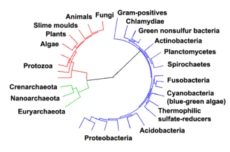
The relationships among the three domains are of central importance for understanding the origin of life. Most of the metabolic pathways, which are the object of the majority of an organism's genes, are common between Archaea and Bacteria, while most genes involved in genome expression are common between Archaea and Eukarya.[77] Within prokaryotes, archaeal cell structure is most similar to that of gram-positive bacteria, largely because both have a single lipid bilayer[78] and usually contain a thick sacculus (exoskeleton) of varying chemical composition.[79] In some phylogenetic trees based upon different gene/protein sequences of prokaryotic homologs, the archaeal homologs are more closely related to those of gram-positive bacteria.[78] Archaea and gram-positive bacteria also share conserved indels in a number of important proteins, such as Hsp70 and glutamine synthetase I;[78][80] but the phylogeny of these genes was interpreted to reveal interdomain gene transfer,[81][82] and might not reflect the organismal relationship(s).[83]
It has been proposed that the archaea evolved from gram-positive bacteria in response to antibiotic selection pressure.[78][80][84] This is suggested by the observation that archaea are resistant to a wide variety of antibiotics that are produced primarily by gram-positive bacteria,[78][80] and that these antibiotics act primarily on the genes that distinguish archaea from bacteria. The proposal is that the selective pressure towards resistance generated by the gram-positive antibiotics was eventually sufficient to cause extensive changes in many of the antibiotics' target genes, and that these strains represented the common ancestors of present-day Archaea.[84] The evolution of Archaea in response to antibiotic selection, or any other competitive selective pressure, could also explain their adaptation to extreme environments (such as high temperature or acidity) as the result of a search for unoccupied niches to escape from antibiotic-producing organisms;[84][85] Cavalier-Smith has made a similar suggestion.[86] This proposal is also supported by other work investigating protein structural relationships[87] and studies that suggest that gram-positive bacteria may constitute the earliest branching lineages within the prokaryotes.[88]

The evolutionary relationship between archaea and eukaryotes remains unclear. Aside from the similarities in cell structure and function that are discussed below, many genetic trees group the two.[90]
Complicating factors include claims that the relationship between eukaryotes and the archaeal phylum Thermoproteota is closer than the relationship between the "Euryarchaeota" and the phylum Thermoproteota[91] and the presence of archaea-like genes in certain bacteria, such as Thermotoga maritima, from horizontal gene transfer.[92] The standard hypothesis states that the ancestor of the eukaryotes diverged early from the Archaea,[93][94] and that eukaryotes arose through symbiogenesis, the fusion of an archaean and a eubacterium, which formed the mitochondria; this hypothesis explains the genetic similarities between the groups.[89] The eocyte hypothesis instead posits that Eukaryota emerged relatively late from the Archaea.[95]
A lineage of archaea discovered in 2015, Lokiarchaeum (of the proposed new phylum "Lokiarchaeota"), named for a hydrothermal vent called Loki's Castle in the Arctic Ocean, was found to be the most closely related to eukaryotes known at that time. It has been called a transitional organism between prokaryotes and eukaryotes.[96][97]
Several sister phyla of "Lokiarchaeota" have since been found ("Thorarchaeota", "Odinarchaeota", "Heimdallarchaeota"), all together comprising a newly proposed supergroup Asgard, which may appear as a sister taxon to Proteoarchaeota.[29][3][98]
Details of the relation of Asgard members and eukaryotes are still under consideration,[99] although, in January 2020, scientists reported that Candidatus Prometheoarchaeum syntrophicum, a type of Asgard archaea, may be a possible link between simple prokaryotic and complex eukaryotic microorganisms about two billion years ago.[100][101]
Individual archaea range from 0.1 micrometers (μm) to over 15 μm in diameter, and occur in various shapes, commonly as spheres, rods, spirals or plates.[102] Other morphologies in the Thermoproteota include irregularly shaped lobed cells in Sulfolobus, needle-like filaments that are less than half a micrometer in diameter in Thermofilum, and almost perfectly rectangular rods in Thermoproteus and Pyrobaculum.[103] Archaea in the genus Haloquadratum such as Haloquadratum walsbyi are flat, square specimens that live in hypersaline pools.[104] These unusual shapes are probably maintained by both their cell walls and a prokaryotic cytoskeleton. Proteins related to the cytoskeleton components of other organisms exist in archaea,[105] and filaments form within their cells,[106] but in contrast with other organisms, these cellular structures are poorly understood.[107] In Thermoplasma and Ferroplasma the lack of a cell wall means that the cells have irregular shapes, and can resemble amoebae.[108]
Some species form aggregates or filaments of cells up to 200 μm long.[102] These organisms can be prominent in biofilms.[109] Notably, aggregates of Thermococcus coalescens cells fuse together in culture, forming single giant cells.[110] Archaea in the genus Pyrodictium produce an elaborate multicell colony involving arrays of long, thin hollow tubes called cannulae that stick out from the cells' surfaces and connect them into a dense bush-like agglomeration.[111] The function of these cannulae is not settled, but they may allow communication or nutrient exchange with neighbors.[112] Multi-species colonies exist, such as the "string-of-pearls" community that was discovered in 2001 in a German swamp. Round whitish colonies of a novel Euryarchaeota species are spaced along thin filaments that can range up to 15 centimetres (5.9 in) long; these filaments are made of a particular bacteria species.[113]
Archaea and bacteria have generally similar cell structure, but cell composition and organization set the archaea apart. Like bacteria, archaea lack interior membranes and organelles.[64] Like bacteria, the cell membranes of archaea are usually bounded by a cell wall and they swim using one or more flagella.[114] Structurally, archaea are most similar to gram-positive bacteria. Most have a single plasma membrane and cell wall, and lack a periplasmic space; the exception to this general rule is Ignicoccus, which possess a particularly large periplasm that contains membrane-bound vesicles and is enclosed by an outer membrane.[115]
Most archaea (but not Thermoplasma and Ferroplasma) possess a cell wall.[108] In most archaea the wall is assembled from surface-layer proteins, which form an S-layer.[116] An S-layer is a rigid array of protein molecules that cover the outside of the cell (like chain mail).[117] This layer provides both chemical and physical protection, and can prevent macromolecules from contacting the cell membrane.[118] Unlike bacteria, archaea lack peptidoglycan in their cell walls.[119] Methanobacteriales do have cell walls containing pseudopeptidoglycan, which resembles eubacterial peptidoglycan in morphology, function, and physical structure, but pseudopeptidoglycan is distinct in chemical structure; it lacks D-amino acids and N-acetylmuramic acid, substituting the latter with N-Acetyltalosaminuronic acid.[118]
Archaeal flagella are known as archaella, that operate like bacterial flagella – their long stalks are driven by rotatory motors at the base. These motors are powered by a proton gradient across the membrane, but archaella are notably different in composition and development.[114] The two types of flagella evolved from different ancestors. The bacterial flagellum shares a common ancestor with the type III secretion system,[120][121] while archaeal flagella appear to have evolved from bacterial type IV pili.[122] In contrast with the bacterial flagellum, which is hollow and assembled by subunits moving up the central pore to the tip of the flagella, archaeal flagella are synthesized by adding subunits at the base.[123]

Archaeal membranes are made of molecules that are distinctly different from those in all other life forms, showing that archaea are related only distantly to bacteria and eukaryotes.[124] In all organisms, cell membranes are made of molecules known as phospholipids. These molecules possess both a polar part that dissolves in water (the phosphate "head"), and a "greasy" non-polar part that does not (the lipid tail). These dissimilar parts are connected by a glycerol moiety. In water, phospholipids cluster, with the heads facing the water and the tails facing away from it. The major structure in cell membranes is a double layer of these phospholipids, which is called a lipid bilayer.[125]
The phospholipids of archaea are unusual in four ways:
Archaea exhibit a great variety of chemical reactions in their metabolism and use many sources of energy. These reactions are classified into nutritional groups, depending on energy and carbon sources. Some archaea obtain energy from inorganic compounds such as sulfur or ammonia (they are chemotrophs). These include nitrifiers, methanogens and anaerobic methane oxidisers.[132] In these reactions one compound passes electrons to another (in a redox reaction), releasing energy to fuel the cell's activities. One compound acts as an electron donor and one as an electron acceptor. The energy released is used to generate adenosine triphosphate (ATP) through chemiosmosis, the same basic process that happens in the mitochondrion of eukaryotic cells.[133]
Other groups of archaea use sunlight as a source of energy (they are phototrophs), but oxygen–generating photosynthesis does not occur in any of these organisms.[133] Many basic metabolic pathways are shared among all forms of life; for example, archaea use a modified form of glycolysis (the Entner–Doudoroff pathway) and either a complete or partial citric acid cycle.[134] These similarities to other organisms probably reflect both early origins in the history of life and their high level of efficiency.[135]
Some Euryarchaeota are methanogens (archaea that produce methane as a result of metabolism) living in anaerobic environments, such as swamps. This form of metabolism evolved early, and it is even possible that the first free-living organism was a methanogen.[136] A common reaction involves the use of carbon dioxide as an electron acceptor to oxidize hydrogen. Methanogenesis involves a range of coenzymes that are unique to these archaea, such as coenzyme M and methanofuran.[137] Other organic compounds such as alcohols, acetic acid or formic acid are used as alternative electron acceptors by methanogens. These reactions are common in gut-dwelling archaea. Acetic acid is also broken down into methane and carbon dioxide directly, by acetotrophic archaea. These acetotrophs are archaea in the order Methanosarcinales, and are a major part of the communities of microorganisms that produce biogas.[138]

Other archaea use CO
2 in the atmosphere as a source of carbon, in a process called carbon fixation (they are autotrophs). This process involves either a highly modified form of the Calvin cycle[140] or another metabolic pathway called the 3-hydroxypropionate/ 4-hydroxybutyrate cycle.[141] The Thermoproteota also use the reverse Krebs cycle while the "Euryarchaeota" also use the reductive acetyl-CoA pathway.[142] Carbon fixation is powered by inorganic energy sources. No known archaea carry out photosynthesis[143] (Halobacterium is the only known phototroph archeon but it uses an alternative process to photosynthesis). Archaeal energy sources are extremely diverse, and range from the oxidation of ammonia by the Nitrosopumilales[144][145] to the oxidation of hydrogen sulfide or elemental sulfur by species of Sulfolobus, using either oxygen or metal ions as electron acceptors.[133]
Phototrophic archaea use light to produce chemical energy in the form of ATP. In the Halobacteria, light-activated ion pumps like bacteriorhodopsin and halorhodopsin generate ion gradients by pumping ions out of and into the cell across the plasma membrane. The energy stored in these electrochemical gradients is then converted into ATP by ATP synthase.[102] This process is a form of photophosphorylation. The ability of these light-driven pumps to move ions across membranes depends on light-driven changes in the structure of a retinol cofactor buried in the center of the protein.[146]
Archaea usually have a single circular chromosome,[147] with as many as 5,751,492 base pairs in Methanosarcina acetivorans,[148] the largest known archaeal genome. The tiny 490,885 base-pair genome of Nanoarchaeum equitans is one-tenth of this size and the smallest archaeal genome known; it is estimated to contain only 537 protein-encoding genes.[149] Smaller independent pieces of DNA, called plasmids, are also found in archaea. Plasmids may be transferred between cells by physical contact, in a process that may be similar to bacterial conjugation.[150][151]

Archaea are genetically distinct from bacteria and eukaryotes, with up to 15% of the proteins encoded by any one archaeal genome being unique to the domain, although most of these unique genes have no known function.[153] Of the remainder of the unique proteins that have an identified function, most belong to the Euryarchaeota and are involved in methanogenesis. The proteins that archaea, bacteria and eukaryotes share form a common core of cell function, relating mostly to transcription, translation, and nucleotide metabolism.[154] Other characteristic archaeal features are the organization of genes of related function – such as enzymes that catalyze steps in the same metabolic pathway into novel operons, and large differences in tRNA genes and their aminoacyl tRNA synthetases.[154]
Transcription in archaea more closely resembles eukaryotic than bacterial transcription, with the archaeal RNA polymerase being very close to its equivalent in eukaryotes,[147] while archaeal translation shows signs of both bacterial and eukaryotic equivalents.[155] Although archaea have only one type of RNA polymerase, its structure and function in transcription seems to be close to that of the eukaryotic RNA polymerase II, with similar protein assemblies (the general transcription factors) directing the binding of the RNA polymerase to a gene's promoter,[156] but other archaeal transcription factors are closer to those found in bacteria.[157] Post-transcriptional modification is simpler than in eukaryotes, since most archaeal genes lack introns, although there are many introns in their transfer RNA and ribosomal RNA genes,[158] and introns may occur in a few protein-encoding genes.[159][160]
Haloferax volcanii, an extreme halophilic archaeon, forms cytoplasmic bridges between cells that appear to be used for transfer of DNA from one cell to another in either direction.[161]
When the hyperthermophilic archaea Sulfolobus solfataricus[162] and Sulfolobus acidocaldarius[163] are exposed to DNA-damaging UV irradiation or to the agents bleomycin or mitomycin C, species-specific cellular aggregation is induced. Aggregation in S. solfataricus could not be induced by other physical stressors, such as pH or temperature shift,[162] suggesting that aggregation is induced specifically by DNA damage. Ajon et al.[163] showed that UV-induced cellular aggregation mediates chromosomal marker exchange with high frequency in S. acidocaldarius. Recombination rates exceeded those of uninduced cultures by up to three orders of magnitude. Frols et al.[162][164] and Ajon et al.[163] hypothesized that cellular aggregation enhances species-specific DNA transfer between Sulfolobus cells in order to provide increased repair of damaged DNA by means of homologous recombination. This response may be a primitive form of sexual interaction similar to the more well-studied bacterial transformation systems that are also associated with species-specific DNA transfer between cells leading to homologous recombinational repair of DNA damage.[165]
Archaea are the target of a number of viruses in a diverse virosphere distinct from bacterial and eukaryotic viruses. They have been organized into 15-18 DNA-based families so far, but multiple species remain un-isolated and await classification.[166][167][168] These families can be informally divided into two groups: archaea-specific and cosmopolitan. Archaeal-specific viruses target only archaean species and currently include 12 families. Numerous unique, previously unidentified viral structures have been observed in this group, including: bottle-shaped, spindle-shaped, coil-shaped, and droplet-shaped viruses.[167] While the reproductive cycles and genomic mechanisms of archaea-specific species may be similar to other viruses, they bear unique characteristics that were specifically developed due to the morphology of host cells they infect.[166] Their virus release mechanisms differ from that of other phages. Bacteriophages generally undergo either lytic pathways, lysogenic pathways, or (rarely) a mix of the two.[169] Most archaea-specific viral strains maintain a stable, somewhat lysogenic, relationship with their hosts – appearing as a chronic infection. This involves the gradual, and continuous, production and release of virions without killing the host cell.[170] Prangishyili (2013) noted that it has been hypothesized that tailed archaeal phages originated from bacteriophages capable of infecting haloarchaeal species. If the hypothesis is correct, it can be concluded that other double-stranded DNA viruses that make up the rest of the archaea-specific group are their own unique group in the global viral community. Krupovic et al. (2018) states that the high levels of horizontal gene transfer, rapid mutation rates in viral genomes, and lack of universal gene sequences have led researchers to perceive the evolutionary pathway of archaeal viruses as a network. The lack of similarities among phylogenetic markers in this network and the global virosphere, as well as external linkages to non-viral elements, may suggest that some species of archaea specific viruses evolved from non-viral mobile genetic elements (MGE).[167]
These viruses have been studied in most detail in thermophilics, particularly the orders Sulfolobales and Thermoproteales.[171] Two groups of single-stranded DNA viruses that infect archaea have been recently isolated. One group is exemplified by the Halorubrum pleomorphic virus 1 (Pleolipoviridae) infecting halophilic archaea,[172] and the other one by the Aeropyrum coil-shaped virus (Spiraviridae) infecting a hyperthermophilic (optimal growth at 90–95 °C) host.[173] Notably, the latter virus has the largest currently reported ssDNA genome. Defenses against these viruses may involve RNA interference from repetitive DNA sequences that are related to the genes of the viruses.[174][175]
Archaea reproduce asexually by binary or multiple fission, fragmentation, or budding; mitosis and meiosis do not occur, so if a species of archaea exists in more than one form, all have the same genetic material.[102] Cell division is controlled in a cell cycle; after the cell's chromosome is replicated and the two daughter chromosomes separate, the cell divides.[176] In the genus Sulfolobus, the cycle has characteristics that are similar to both bacterial and eukaryotic systems. The chromosomes replicate from multiple starting points (origins of replication) using DNA polymerases that resemble the equivalent eukaryotic enzymes.[177]
In Euryarchaeota the cell division protein FtsZ, which forms a contracting ring around the cell, and the components of the septum that is constructed across the center of the cell, are similar to their bacterial equivalents.[176] In cren-[178][179] and thaumarchaea,[180] the cell division machinery Cdv fulfills a similar role. This machinery is related to the eukaryotic ESCRT-III machinery which, while best known for its role in cell sorting, also has been seen to fulfill a role in separation between divided cell, suggesting an ancestral role in cell division.[181]
Both bacteria and eukaryotes, but not archaea, make spores.[182] Some species of Haloarchaea undergo phenotypic switching and grow as several different cell types, including thick-walled structures that are resistant to osmotic shock and allow the archaea to survive in water at low salt concentrations, but these are not reproductive structures and may instead help them reach new habitats.[183]
Quorum sensing was originally thought to not exist in Archaea, but recent studies have shown evidence of some species being able to perform cross-talk through quorum sensing. Other studies have shown syntrophic interactions between archaea and bacteria during biofilm growth. Although research is limited in archaeal quorum sensing, some studies have uncovered LuxR proteins in archaeal species, displaying similarities with bacteria LuxR, and ultimately allowing for the detection of small molecules that are used in high density communication. Similarly to bacteria, Archaea LuxR solos have shown to bind to AHLs (lactones) and non-AHLs ligans, which is a large part in performing intraspecies, interspecies, and interkingdom communication through quorum sensing.[184]

Archaea exist in a broad range of habitats, and are now recognized as a major part of global ecosystems,[18] and may represent about 20% of microbial cells in the oceans.[185] However, the first-discovered archaeans were extremophiles.[132] Indeed, some archaea survive high temperatures, often above 100 °C (212 °F), as found in geysers, black smokers, and oil wells. Other common habitats include very cold habitats and highly saline, acidic, or alkaline water, but archaea include mesophiles that grow in mild conditions, in swamps and marshland, sewage, the oceans, the intestinal tract of animals, and soils.[7][18] Similar to PGPR, Archaea are now considered as a source of plant growth promotion as well.[7]
Extremophile archaea are members of four main physiological groups. These are the halophiles, thermophiles, alkaliphiles, and acidophiles.[186] These groups are not comprehensive or phylum-specific, nor are they mutually exclusive, since some archaea belong to several groups. Nonetheless, they are a useful starting point for classification.[187]
Halophiles, including the genus Halobacterium, live in extremely saline environments such as salt lakes and outnumber their bacterial counterparts at salinities greater than 20–25%.[132] Thermophiles grow best at temperatures above 45 °C (113 °F), in places such as hot springs; hyperthermophilic archaea grow optimally at temperatures greater than 80 °C (176 °F).[188] The archaeal Methanopyrus kandleri Strain 116 can even reproduce at 122 °C (252 °F), the highest recorded temperature of any organism.[189]
Other archaea exist in very acidic or alkaline conditions.[186] For example, one of the most extreme archaean acidophiles is Picrophilus torridus, which grows at pH 0, which is equivalent to thriving in 1.2 molar sulfuric acid.[190]
This resistance to extreme environments has made archaea the focus of speculation about the possible properties of extraterrestrial life.[191] Some extremophile habitats are not dissimilar to those on Mars,[192] leading to the suggestion that viable microbes could be transferred between planets in meteorites.[193]
Recently, several studies have shown that archaea exist not only in mesophilic and thermophilic environments but are also present, sometimes in high numbers, at low temperatures as well. For example, archaea are common in cold oceanic environments such as polar seas.[194] Even more significant are the large numbers of archaea found throughout the world's oceans in non-extreme habitats among the plankton community (as part of the picoplankton).[195] Although these archaea can be present in extremely high numbers (up to 40% of the microbial biomass), almost none of these species have been isolated and studied in pure culture.[196] Consequently, our understanding of the role of archaea in ocean ecology is rudimentary, so their full influence on global biogeochemical cycles remains largely unexplored.[197] Some marine Thermoproteota are capable of nitrification, suggesting these organisms may affect the oceanic nitrogen cycle,[144] although these oceanic Thermoproteota may also use other sources of energy.[198]
Vast numbers of archaea are also found in the sediments that cover the sea floor, with these organisms making up the majority of living cells at depths over 1 meter below the ocean bottom.[199][200] It has been demonstrated that in all oceanic surface sediments (from 1000- to 10,000-m water depth), the impact of viral infection is higher on archaea than on bacteria and virus-induced lysis of archaea accounts for up to one-third of the total microbial biomass killed, resulting in the release of ~0.3 to 0.5 gigatons of carbon per year globally.[201]
Archaea recycle elements such as carbon, nitrogen, and sulfur through their various habitats.[202] Archaea carry out many steps in the nitrogen cycle. This includes both reactions that remove nitrogen from ecosystems (such as nitrate-based respiration and denitrification) as well as processes that introduce nitrogen (such as nitrate assimilation and nitrogen fixation).[203][204] Researchers recently discovered archaeal involvement in ammonia oxidation reactions. These reactions are particularly important in the oceans.[145][205] The archaea also appear crucial for ammonia oxidation in soils. They produce nitrite, which other microbes then oxidize to nitrate. Plants and other organisms consume the latter.[206]
In the sulfur cycle, archaea that grow by oxidizing sulfur compounds release this element from rocks, making it available to other organisms, but the archaea that do this, such as Sulfolobus, produce sulfuric acid as a waste product, and the growth of these organisms in abandoned mines can contribute to acid mine drainage and other environmental damage.[207]
In the carbon cycle, methanogen archaea remove hydrogen and play an important role in the decay of organic matter by the populations of microorganisms that act as decomposers in anaerobic ecosystems, such as sediments, marshes, and sewage-treatment works.[208]
The well-characterized interactions between archaea and other organisms are either mutual or commensal. There are no clear examples of known archaeal pathogens or parasites,[209][210] but some species of methanogens have been suggested to be involved in infections in the mouth,[211][212] and Nanoarchaeum equitans may be a parasite of another species of archaea, since it only survives and reproduces within the cells of the Crenarchaeon Ignicoccus hospitalis,[149] and appears to offer no benefit to its host.[213]
Mutualism is an interaction between individuals of different species that results in positive (beneficial) effects on per capita reproduction and/or survival of the interacting populations. One well-understood example of mutualism is the interaction between protozoa and methanogenic archaea in the digestive tracts of animals that digest cellulose, such as ruminants and termites.[214] In these anaerobic environments, protozoa break down plant cellulose to obtain energy. This process releases hydrogen as a waste product, but high levels of hydrogen reduce energy production. When methanogens convert hydrogen to methane, protozoa benefit from more energy.[215]
In anaerobic protozoa, such as Plagiopyla frontata, archaea reside inside the protozoa and consume hydrogen produced in their hydrogenosomes.[216][217] Archaea also associate with larger organisms. For example, the marine archaean Cenarchaeum symbiosum lives within (is an endosymbiont of) the sponge Axinella mexicana.[218]
Archaea can also be commensals, benefiting from an association without helping or harming the other organism. For example, the methanogen Methanobrevibacter smithii is by far the most common archaean in the human flora, making up about one in ten of all the prokaryotes in the human gut.[219] In termites and in humans, these methanogens may in fact be mutualists, interacting with other microbes in the gut to aid digestion.[220] Archaean communities also associate with a range of other organisms, such as on the surface of corals,[221] and in the region of soil that surrounds plant roots (the rhizosphere).[222][223]
Although Archaea do not have a historical reputation of being pathogens, Archaea are often found with similar genomes to more common pathogen like E. coli,[224] showing metabolic links and evolutionary history with today's pathogens. Archaea also has inconsistent detection within clinical studies because of the lack of categorization of Archaea into more specific species.[225]
Extremophile archaea, particularly those resistant either to heat or to extremes of acidity and alkalinity, are a source of enzymes that function under these harsh conditions.[226][227] These enzymes have found many uses. For example, thermostable DNA polymerases, such as the Pfu DNA polymerase from Pyrococcus furiosus, revolutionized molecular biology by allowing the polymerase chain reaction to be used in research as a simple and rapid technique for cloning DNA. In industry, amylases, galactosidases and pullulanases in other species of Pyrococcus that function at over 100 °C (212 °F) allow food processing at high temperatures, such as the production of low lactose milk and whey.[228] Enzymes from these thermophilic archaea also tend to be very stable in organic solvents, allowing their use in environmentally friendly processes in green chemistry that synthesize organic compounds.[227] This stability makes them easier to use in structural biology. Consequently, the counterparts of bacterial or eukaryotic enzymes from extremophile archaea are often used in structural studies.[229]
In contrast with the range of applications of archaean enzymes, the use of the organisms themselves in biotechnology is less developed. Methanogenic archaea are a vital part of sewage treatment, since they are part of the community of microorganisms that carry out anaerobic digestion and produce biogas.[230] In mineral processing, acidophilic archaea display promise for the extraction of metals from ores, including gold, cobalt and copper.[231]
Archaea host a new class of potentially useful antibiotics. A few of these archaeocins have been characterized, but hundreds more are believed to exist, especially within Haloarchaea and Sulfolobus. These compounds differ in structure from bacterial antibiotics, so they may have novel modes of action. In addition, they may allow the creation of new selectable markers for use in archaeal molecular biology.[232]
Archaea (/ɑːrˈkiːə/ (![]() listen) ar-KEE-ə; singular archaeon /ɑːrˈkiːən/ ar-KEE-ən) is a domain of single-celled organisms. These microorganisms lack cell nuclei and are therefore prokaryotes. Archaea were initially classified as bacteria, receiving the name archaebacteria (in the Archaebacteria kingdom), but this term has fallen out of use.
listen) ar-KEE-ə; singular archaeon /ɑːrˈkiːən/ ar-KEE-ən) is a domain of single-celled organisms. These microorganisms lack cell nuclei and are therefore prokaryotes. Archaea were initially classified as bacteria, receiving the name archaebacteria (in the Archaebacteria kingdom), but this term has fallen out of use.
Archaeal cells have unique properties separating them from the other two domains, Bacteria and Eukaryota. Archaea are further divided into multiple recognized phyla. Classification is difficult because most have not been isolated in a laboratory and have been detected only by their gene sequences in environmental samples. It is unknown if these are able to produce endospores.
Archaea and bacteria are generally similar in size and shape, although a few archaea have very different shapes, such as the flat, square cells of Haloquadratum walsbyi. Despite this morphological similarity to bacteria, archaea possess genes and several metabolic pathways that are more closely related to those of eukaryotes, notably for the enzymes involved in transcription and translation. Other aspects of archaeal biochemistry are unique, such as their reliance on ether lipids in their cell membranes, including archaeols. Archaea use more diverse energy sources than eukaryotes, ranging from organic compounds such as sugars, to ammonia, metal ions or even hydrogen gas. The salt-tolerant Haloarchaea use sunlight as an energy source, and other species of archaea fix carbon (autotrophy), but unlike plants and cyanobacteria, no known species of archaea does both. Archaea reproduce asexually by binary fission, fragmentation, or budding; unlike bacteria, no known species of Archaea form endospores. The first observed archaea were extremophiles, living in extreme environments such as hot springs and salt lakes with no other organisms. Improved molecular detection tools led to the discovery of archaea in almost every habitat, including soil, oceans, and marshlands. Archaea are particularly numerous in the oceans, and the archaea in plankton may be one of the most abundant groups of organisms on the planet.
Archaea are a major part of Earth's life. They are part of the microbiota of all organisms. In the human microbiome, they are important in the gut, mouth, and on the skin. Their morphological, metabolic, and geographical diversity permits them to play multiple ecological roles: carbon fixation; nitrogen cycling; organic compound turnover; and maintaining microbial symbiotic and syntrophic communities, for example.
No clear examples of archaeal pathogens or parasites are known. Instead they are often mutualists or commensals, such as the methanogens (methane-producing strains) that inhabit the gastrointestinal tract in humans and ruminants, where their vast numbers facilitate digestion. Methanogens are also used in biogas production and sewage treatment, and biotechnology exploits enzymes from extremophile archaea that can endure high temperatures and organic solvents.
Arkeoj aŭ arĥeoj (latine Archaea) estas biologia regno de prokariotaj estaĵoj. Arkeoj estas unuĉelaj kaj ne havas ĉelkernojn, tamen ili ne estas bakterioj, kvankam ili dekomence estis klasigitaj kiel bakterioj, kaj ricevis la nomon prabakterioj (latine archaebacteria, en la Prabakteria regno) - tiu klasigo eksmodiĝis post la studoj fare de Carl Woese.[1] Sur la genetika nivelo, ili estas pli similaj al eŭkariotoj kiel ni mem. Estis Carl Woese, kiu eltrovis la arĥeojn en 1977.
Arkiaj ĉeloj havas unikajn trajtojn kiuj apartigas ilin el la aliaj du vivoregnoj, nome Bakterioj kaj Eŭkariotoj. La arkioj estas krome subdividitaj en multaj agnoskitaj filumoj. Ties klasigo estas malfacile farebla ĉar la majoritato ankoraŭ ne estis izoligitaj en la laboratorioj kaj estis detektitaj nur pere de analizo de ties nukleataj acidoj en montraĵoj elprenitaj el ties medioj.
Arkioj kaj bakterioj estas ĝenerale similaj laŭ grando kaj formo, kvankam kelkaj arkioj havas formojn tre malsimilajn al tiuj de bakterioj, kiaj la ebenaj kaj kvadrat-formaj ĉeloj de Haloquadratum walsbyi.[2] Spite tiun morfologian similecon al la bakteriio, arkioj posedas genojn kaj kelkajn metabolajn sistemojn kiuj estas pli proksime rilataj al tiuj de eŭkariotoj, ĉefe la enzimoj kiuj funkcias en la genetikaj procezoj de transskrinado kaj tradukado. Aliaj aspektoj de arkia biokemio estas unikaj, kiaj ilia dependo el eteraj lipidoj en iliaj ĉelmembranoj, kiel ĉe la arkeoloj. Arkioj uzas pli da energifontoj ol la eŭkariotoj: tiuj gamas el organikaj komponaĵoj, kiel sukeroj, al amoniako, metalaj jonoj aŭ eĉ hidrogenhava gaso. Sal-toleraj arkioj (nome Haloarchaea aŭ Salarkioj) uzas sunlumon kiel energifonto, kaj aliaj specioj de arkioj fiksigas karbonon, sed malkiel ĉe plantoj kaj cianobakterioj, neniu konata specio de arkio plenumas ambaŭ procezojn. Arkioj reproduktiĝas nesekse pere de binara fisio, fragmentado, aŭ burĝonado; malkiel ĉe bakterioj kaj eŭkariotoj, neniu konata specio de arkio formas sporojn.
Multaj arkeoj estas ekstremofiloj, tio estas vivas en tre akraj medioj — ekzemple, varmegaj, salegaj, kaj acidaj lagoj. Inter tiuj estis registritaj dekomence termofontoj kaj sallagoj, sed ekde tiam ili troviĝis ankaŭ en ampleksa gamo de habitatoj, kiel grundoj, oceanoj kaj marĉoj. Ili estas ankaŭ parto de la homa mikrobaro, kiu troviĝas en kojlo, buŝo kaj haŭto.[3] Arkioj estas partikulare multnombraj en la oceanoj, kaj la arkioj de la planktono povas esti el la plej abundaj grupoj de organismoj sur la planedo. Arkioj estas la ĉefa parto de la surtera vivo kaj ili povas ludi gravajn rolojn kaj en la karbona kaj en la nitrogena cikloj.
Oni ne konas klarajn ekzemplojn de arkiaj patogenoj aŭ parazitoj, sed ili estas ofte mutualistoj aŭ kunmanĝantoj. Unu ekzemplo estas la metanogenoj kiuj loĝas en la digesta sistemo de homoj aŭ remaĉuloj, kie ilia multnombreco fakte helpas la digestadon. Metanogenoj estas uzataj ankaŭ en produktado de biogaso kaj en procezoj de akvopurigado, kaj bioteknologio ekspluatas enzimojn el ekstremofilaj arkioj kiuj povas rezisti altajn temperaturojn kaj organikajn solvigaĵojn. Laŭ nuna scio, la arkioj kaŭzas neniun ajn homan aŭ bestan malsanon — ili estas plej interesaj laŭ la vidpunkto de evoluo. Multaj sciencistoj nun kredas ke la komuna parencaro de ĉiuj eŭkariotoj estis simila al arkeoj.
Dum multo de la 20a jarcento, prokariotoj estis konsiderataj kiel aparta grupo de organismoj kaj klasigitaj baze sur sia biokemio, morfologio kaj metabolo. Por ekzemplo, mikrobiologoj klopodis klasigi mikroorganismojn baze sur la strukturoj de iliaj ĉelmuroj, ilia formoj kaj la substancojn kiujn ili konsumas.[4] En 1965, Emile Zuckerkandl kaj Linus Pauling[5] proponis anstataŭe uzi la sekvencojn de la genoj en diversaj prokariotoj por studi kiel ili estas rilataj unu al la alia. Tiu filogenetika alproksimiĝo estas la ĉefa metodo uzita nune.
Arkioj estis dekomence klasigitaj kiel aparta grupo de prokariotoj en 1977 fare de Carl Woese kaj George E. Fox baze sur la sekvencoj de ribosomaj genoj RRNA.[6] Tiuj du grupoj estis origine nomitaj Arkiobakterioj kaj Eŭbakterioj kaj estis termigitaj Urkingdoms fare de Woese kaj Fox; aliaj esploristoj traktis ilin kiel regnoj aŭ subgenroj. En tiu publikigo Woese kaj Fox prezentis la unuajn pruvarojn por la koncepto de arkiobakterioj - kiuj tiam enhavis nur la metanogenojn - kiel aparta "devenlinio":
Por emfazi tiun diferencon, Woese, Otto Kandler kaj Mark Wheelis poste proponis novan naturan sistemon de organismoj kun tri apartaj domanioj: nome Eŭkariotoj, Bakterioj kaj Arkioj,[7] en tio kio estas nune konata kiel "La Woese-a Revolucio". La historo de la esplorado pri Arkioj estas dokumenita detale fare de Jan Sapp.[8]
La vorto archaea de la scienca nomo derivas el la antikva greka ἀρχαῖα, signife "antikvaj aĵoj",[9] ĉar la unuaj reprezentantoj de la domanio Archaea estis metanogenoj kaj oni supozis, ke ilia metabolo spegulis la praatmosferon de la Tero kanj la antikveco de la organismoj, sed kiam aliaj novaj habitatoj estis studitaj, pliaj organismoj estis malkovritaj. Ankaŭ ekstremaj halofiloj[10] kaj hipertermofilaj mikroboj[11] estis inkluditaj en Archaea. Dum longe, archaea estis konsideritaj kiel ekstremofiloj kiuj ekzistas nur en ekstremaj habitatoj kiaj termofontoj kaj sallagoj. Fine de la 20a jarcento, archaea estis identigitaj ankaŭ en ne-ekstremaj medioj. Nuntempe, ili estas konata kiel granda kaj diversa grupo de organismoj kiuj estas amplekse distribuataj en naturo kaj estas oftaj en ĉiuj habitatoj.[12] Tiu nova alproksimiĝo al la gravo kaj ĉieeco de la arkioj venis el la uzado de polimeraza ĉen-reakcio (PĈR) por detekti prokariotojn el mediaj montraĵoj (kiel akvo aŭ grundo) per de la multobligo de iliaj ribosomaj genoj. Tio ebligas la detekton kaj identigon de organismoj kiuj ne estis produktitaj en la laboratorio.[13] [14]
Arkeoj aŭ arĥeoj (latine Archaea) estas biologia regno de prokariotaj estaĵoj. Arkeoj estas unuĉelaj kaj ne havas ĉelkernojn, tamen ili ne estas bakterioj, kvankam ili dekomence estis klasigitaj kiel bakterioj, kaj ricevis la nomon prabakterioj (latine archaebacteria, en la Prabakteria regno) - tiu klasigo eksmodiĝis post la studoj fare de Carl Woese. Sur la genetika nivelo, ili estas pli similaj al eŭkariotoj kiel ni mem. Estis Carl Woese, kiu eltrovis la arĥeojn en 1977.
Las arqueas (Archaea; et: del griego αρχαία [arjaía], «las antiguas»), a veces llamadas árqueas, son un gran grupo de microorganismos procariotas unicelulares que, al igual que las bacterias, no presentan núcleo ni orgánulos membranosos internos, pero son fundamentalmente diferentes a estas, de tal manera que conforman su propio dominio o reino.
En el pasado se agruparon en el antiguo reino Monera, y cuando fueron identificadas como grupo en 1977, recibieron el nombre de arqueobacterias (reino Archaebacteria), pero esta clasificación ya no se utiliza.[5] En realidad, las arqueas tienen una historia evolutiva independiente y muestran muchas diferencias bioquímicas y genéticas con las otras formas de vida, por lo que se clasificaron en un dominio separado dentro del sistema de tres dominios: Archaea, Bacteria y Eukarya.
Las arqueas son un dominio (y también un reino)[6] que se divide en múltiples filos. Los grupos Crenarchaeota y Euryarchaeota son los más estudiados. La clasificación de las arqueas todavía es difícil, porque la gran mayoría nunca se han estudiado en el laboratorio y solo se han detectado mediante análisis de sus ácidos nucleicos en muestras tomadas de diversos ambientes.
Las arqueas y bacterias son bastante similares en tamaño y forma, aunque algunas arqueas tienen formas muy inusuales, como las células aplanadas y cuadradas de Haloquadratum walsbyi.[7] A pesar de esta semejanza visual con las bacterias, las arqueas poseen genes y varias rutas metabólicas que son más cercanas a las de los eucariotas, en especial en las enzimas implicadas en la transcripción y la traducción. Otros aspectos de la bioquímica de las arqueas son únicos, como los éteres lipídicos de sus membranas celulares. Las arqueas explotan una variedad de recursos mucho mayores que los eucariotas, desde compuestos orgánicos comunes como los azúcares, hasta el uso de amoníaco,[8] iones de metales o incluso hidrógeno como nutrientes. Las arqueas tolerantes a la sal (las haloarqueas) utilizan la luz solar como fuente de energía, y otras especies de arqueas fijan carbono;[9] sin embargo, a diferencia de las plantas y las cianobacterias, no se conoce ninguna especie de arquea que sea capaz de ambas cosas. Las arqueas se reproducen asexualmente y se dividen por fisión binaria,[10] fragmentación o gemación; a diferencia de las bacterias y los eucariotas, no se conoce ninguna especie de arquea que forme esporas.[11]
Inicialmente, las arqueas eran consideradas todas metanógenas o extremófilas que vivían en ambientes hostiles tales como aguas termales y lagos salados, pero actualmente se sabe que están presentes en los más diversos hábitats, tales como el suelo, océanos, pantanos y en el colon humano. Las arqueas son especialmente numerosas en los océanos, y las que forman parte del plancton podrían ser uno de los grupos de organismos más abundantes del planeta. Actualmente se consideran una parte importante de la vida en la Tierra y podrían desempeñar un papel importante tanto en el ciclo del carbono como en el ciclo del nitrógeno. No se conocen ejemplos claros de arqueas patógenas o parásitas, pero suelen ser mutualistas o comensales. Son ejemplos las arqueas metanógenas que viven en el intestino de los humanos y los rumiantes, donde están presentes en grandes cantidades y contribuyen a digerir el alimento. Las arqueas tienen su importancia en la tecnología, hay metanógenos que son utilizados para producir biogás y como parte del proceso de depuración de aguas, y las enzimas de arqueas extremófilas son capaces de resistir temperaturas elevadas y disolventes orgánicos, siendo por ello utilizadas en biotecnología.
El grupo de arqueas que se ha estudiado desde más antiguo es el de las metanógenas. La metanogénesis fue descubierta en el lago Mayor de Italia en 1776, al observar en él el burbujeo de "aire combustible". En 1882 se observó que la producción de metano en el intestino de animales se debía a la presencia de microorganismos (Popoff, Tappeiner, y Hoppe-Seyler).[12]
En 1936, año que marcó el principio de la era moderna en el estudio de la metanogénesis, H.A Barker brindó las bases científicas para el estudio de su fisiología y logró desarrollar un medio de cultivo apropiado para el crecimiento de los metanógenos. En ese mismo año se identificaron los géneros Methanococcus y Methanosarcina.[13]
Las primeras arqueas extremófilas se encontraron en ambientes calientes. En 1970, Thomas D. Brock de la Universidad de Wisconsin descubrió a Thermoplasma, un arquea termoacidófila y en 1972 a Sulfolobus, una hipertermófila.[14] Brock se inició en 1969 en el campo de la biología de los hipertermófilos con el descubrimiento de la bacteria Thermus.
En 1977 se identifica a las arqueas como el grupo prokaryota más distante al descubrir que los metanógenos presentan una profunda divergencia con todas las bacterias estudiadas. Ese mismo año se propone la categoría de superreino para este grupo con el nombre de Archaebacteria. En 1978, el manual de Bergey le da la categoría de filo con el nombre de Mendosicutes y en 1984 divide al reino Procaryotae o Monera en 4 divisiones, agrupándolas en la división Mendosicutes.[15]
Las arqueas hipertermófilas se agruparon en 1984 bajo el nombre Eocyta, identificándolas como un grupo independiente de las entonces llamadas arqueobacterias (en referencia a los metanógenos) y las eubacterias, descubriéndose además que Eocyta era el grupo más cercano a las eucariotas.[16] La relación filogenética entre metanógenos e hipertermófilos hace que en 1990 se renombre a Eocyta como Crenarchaeota y a las metanógenas como Euryarchaeota, formando el nuevo grupo Archaea como parte del sistema de tres dominios.[17]
A principios del siglo XX, los prokaryotas se consideraban un único grupo de organismos y se clasificaban según su bioquímica, morfología y metabolismo. Por ejemplo, los microbiólogos intentaban clasificar los microorganismos según la estructura de su pared celular, su forma y las sustancias que consumían.[18] Sin embargo, en 1965 se propuso un nuevo sistema,[19] utilizando las secuencias genéticas de estos organismos para averiguar qué prokaryotas están realmente relacionadas entre sí. Este método, conocido como filogenia molecular, es el principal método utilizado desde entonces.
Las arqueas se clasificaron inicialmente en 1977 como un superreino separado de las bacterias, por Carl Woese y George E. Fox en árboles filogenéticos basados en las secuencias de genes de ARN ribosómico (ARNr).[20] Estos dos grupos se denominaron originalmente Eubacteria y Archaebacteria, lo que Woese y Fox denominaron "reinos originales". Woese argumentó que este grupo de prokaryotas es un tipo de vida fundamentalmente distinto. Para enfatizar esta diferencia, usaron el término dominio en 1990 y los rebautizaron Bacteria y Archaea.[17] El nombre científico Archaea proviene del griego antiguo ἀρχαῖα, que significa "los antiguos".[21] El término "arqueobacteria" proviene de la combinación de esta raíz y del término griego baktērion, que significa "pequeño bastón".
Originalmente, solo se clasificaron los metanógenos en este nuevo dominio, luego los considerados extremófilos que solo vivían en hábitats como aguas termales y lagos salados. A finales del siglo XX, los microbiólogos se dieron cuenta de que Archaea son un grupo grande y diverso de organismos ampliamente distribuidos en la naturaleza, y que son comunes en hábitats mucho menos extremos, como suelos y océanos.[22] Esta nueva toma de conciencia de la importancia y la omnipresencia de estos organismos vino del uso de la reacción en cadena de la polimerasa para detectar prokaryotas en muestras de agua o suelo a partir de, únicamente, sus ácidos nucleicos. Esto permite detectar e identificar organismos cuyo cultivo en el laboratorio es complejo.[23][24]
La clasificación de las arqueas, y de los procariotas en general, es un tema en constante fluctuación. Los sistemas actuales de clasificación intentan organizar las arqueas en grupos que comparten rasgos estructurales y antepasados comunes.[25] Estas clasificaciones se basan especialmente en el uso de secuencias de genes de ARN ribosómico para revelar las relaciones entre los organismos (análisis moleculares de ADN).[26] En la actualidad (2016) figuran cinco filos en LPSN (List of Prokaryotic names with Standing in Nomenclature, Lista de nombres de prokaryotas con pie en la nomenclatura). Estos son: Euryarchaeota, Crenarchaeota, Korarchaeota, Nanoarchaeota y Thaumarchaeota. La mayoría de especies de arqueas cultivables y bien investigadas son miembros de dos filos principales, Euryarchaeota y Crenarchaeota. A la peculiar especie Nanoarchaeum equitans, que fue descubierta en 2003, se le ha atribuido su propio filo, Nanoarchaeota.[27] El reciente candidato Korarchaeota contiene un número reducido de inusuales especies termófilas que comparten rasgos de los dos filos principales, pero que son más cercanas a Crenarchaeota.[28][29]
Los análisis genómicos de las muestras tomadas del medio ambiente han revelado un gran número de especies nuevas de arqueas que tienen una relación distante con cualquiera de los grupos conocidos. Por ejemplo, los nanoorganismos arqueobacterianos acidófilos de la mina Richmond (ARMAN), que fueron descubiertos en 2006[30] y están entre los organismos conocidos más pequeños.[31] Así, el número de filos candidatos en 2018 es de catorce.[4][32] Los 19 taxones candidatos conocidos de arqueas se agrupan en cuatro supergrupos, usualmente con rango de filo o superfilo: DPANN, Euryarchaeota, Thermoproteota o TACK y Asgardarchaeota o Asgard.
Las características de los filos son:
Una filogenia algo consensuada en el GTDB database y el Annotree es la siguiente:[34][35]
Archaea DPANN Nanoarchaeota Euryarchaeota Methanobacteriota Neoeuryarchaeota Thermoplasmatota Halobacteriota Proteoarchaeota Thermoproteota Nitrososphaeria Methanomethylicia Thermoproteia Asgardarchaeota + α─proteobacteriaOtros análisis filogenéticos han sugerido que DPANN como clado puede no ser monofilético y sería causado por la atracción de ramas largas.[36] Análisis filogenéticos sugirieron que DPANN pertenece a Euryarchaeota, estando el filo Nanohaloarchaeota totalmente separado del resto. El clado de DPANN sin Nanohaloarchaeota se ha nombrado "Micrarchaea".[37] Por esta razón una filogenia alternativa para DPANN es la siguiente:[38][39]
Archaea Euryarchaeota Methanobacteriota "Micrarchaea" Nanoarchaeota Neoeuryarchaeota Thermoplasmatota Halobacteriota Proteoarchaeota Thermoproteota Nitrososphaeria Methanomethylicia Thermoproteia Asgardarchaeota + α─proteobacteriaLa clasificación de las arqueas en especies también es controvertida. En biología, una especie es un grupo de organismos relacionados. Una definición de especie muy extendida entre los animales es un conjunto de organismos que pueden reproducirse entre ellos y que están reproductivamente aislados de otros grupos de organismos (es decir, no pueden reproducirse con otras especies).[40] Sin embargo, los esfuerzos por clasificar los prokaryotas, como las arqueas, en especies se complican debido a que son asexuales y que presentan un alto nivel de transferencia horizontal de genes entre linajes. Este tema es controvertido; por ejemplo, algunos datos sugieren que en arqueas como Ferroplasma, se pueden agrupar células individuales en poblaciones de genoma muy similar y que raramente transfieren genes a grupos más divergentes de células.[41] Algunos argumentan que estos grupos de células son análogos a especies. Por otra parte, estudios de Halorubrum descubrieron un intercambio genético significativo entre estas poblaciones.[42] Estos resultados han llevado a pensar que clasificar estos grupos de organismos como especies tendría poco sentido práctico.[43]
El conocimiento actual sobre la diversidad de las arqueas es fragmentario, y no se puede estimar con ningún tipo de precisión el número total de especies existentes.[26] Incluso se desconoce el número total de filos arqueobacterianos, de los cuales actualmente hay propuestos 16 y solo ocho tienen representantes que se han cultivado y estudiado directamente. Muchos de estos grupos hipotéticos son conocidos únicamente a partir de una sola secuencia de ARNr, lo que indica que la diversidad de estos organismos permanece completamente desconocida.[44] El problema de cómo estudiar y clasificar microbios no cultivados también se da en las bacterias.[45] Recientemente, y aunque el proyecto plantea las dificultades mencionadas anteriormente, el consorcio público GEBA (acrónimo en inglés de Genomic Enciclopedy of Bacteria and Archaea, Enciclopedia genómica de Bacteria y Archaea) está llevando a cabo la tarea de completar y anotar la mayor cantidad de genomas de estos dos dominios con el fin, entre otros, de llevar a cabo una clasificación basada en el genoma.[46]
Aunque se han encontrado probables fósiles de prokaryotas de casi 3770-4280 millones de años de antigüedad, la morfología de la mayoría de los prokaryotas y de sus fósiles no permite distinguir entre bacterias y arqueas.[50] En cambio, los "fósiles químicos" de lípidos característicos de las arqueas son más informativos, porque dichos compuestos no aparecen en otros organismos.[51] Algunas publicaciones sugieren que se encuentran lípidos característicos de arqueas o eucariotas, en sedimentos de hace 2700 millones de años;[52] pero estos datos fueron cuestionados.[53] Estos lípidos han sido detectados en rocas que datan del Precámbrico. Los restos más antiguos conocidos de lípidos de isopreno datan del cinturón de Isua, al oeste de Groenlandia, que incluyen sedimentos formados hace 3800 millones de años, siendo estos los más antiguos encontrados hasta la fecha.[54] El linaje de las arqueas podría ser el más antiguo de la Tierra.[55]
Woese consideraba que las bacterias, arqueas y eucariotas representan líneas separadas de descendencia que divergieron temprano en la evolución de colonias ancestrales de organismos.[56][57] Una posibilidad[57][58] es que esto ocurriera antes de la evolución de las células, cuando la falta de una membrana celular típica permitía una transferencia lateral de genes no restringida, y que el antepasado común de los tres dominios originó por fijación de un subconjunto específico de genes.[57][58] Es posible que el último antepasado común de las bacterias y arqueas fuera un termófilo, lo que presenta la posibilidad de que las temperaturas bajas sean "ambientes extremos" para las arqueas, y que los organismos que viven en ambientes más fríos aparecieran más tarde en la historia de la vida en la Tierra.[59] Como Archaea y Bacteria ya no están más relacionados entre sí que lo que son para los eucariotas, el término prokaryota solo tiene el significado de "no-eucariota", lo que limita su utilidad.[60]
Por su parte, Gupta propone que las arqueas evolucionaron a partir de bacterias grampositivas en respuesta a una presión selectiva ejercida por los antibióticos liberados por otras bacterias.[61][62][63] Esta idea está apoyada porque las arqueas son resistentes a una amplia variedad de antibióticos producidos principalmente por las grampositivas,[61][62] y estos antibióticos actúan principalmente sobre genes que distinguen las arqueas. Su propuesta es que la presión selectiva hacia la resistencia a los antibióticos generada por los antibióticos de las grampositivas fue finalmente suficiente para causar grandes cambios en muchos de los genes que eran el objetivo de los mismos, y que estas cepas de microorganismos representaban el ancestro común de las arqueas actuales.[63] La evolución de las arqueas en respuesta a la selección por antibióticos, o cualquier otra presión selectiva competitiva, también podría explicar su adaptación a ambientes extremos (tales como alta temperatura o acidez) como resultado de una búsqueda de nichos ecológicos desocupados para escapar de los organismos productores de antibióticos;[63][64] La propuesta de Gupta está apoyada también por otras investigaciones sobre la relación entre las proteínas estructurales[65] y por los estudios que sugieren que las bacterias grampositivas pueden constituir uno de los linajes que primero ramificaron en los prokaryotas.[66] Cavalier-Smith hizo una sugerencia similar, aunque considera que las bacterias gramnegativas son las más antiguas y que las grampositivas y las arqueas se originaron a partir de ellas por la pérdida de la membrana externa.[67] No obstante, hasta ahora no hay ningún análisis molecular que pueda respaldar estas teorías porque tanto las bacterias como arqueas forman grupos monofiléticos lo que indica que ambos son igualmente antiguos y evolucionaron del último antepasado común universal.
Se ha sugerido que el último antepasado común universal de bacterias y arqueas es un termófilo que vivió hace 4350 millones de años durante el eón Hádico.[48] La bifurcación entre arqueas y bacterias se produjo a mediados del Hádico, mientras que los eucariotas son más recientes y surgieron a finales del Paleoproterozoico. Las arqueas ultrapequeñas (DPANN) y el filo Euryarchaeota se separaron del resto de las arqueas a finales del Hádico y a comienzos del Arcaico. Proteoarchaeota que incluye los supergrupos Thermoproteota y Asgard se originaron a mediados del Arcaico.[68][48][49] La separación de Asgard y los eucariotas se calculó a finales del Arcaico (periodo Neoarcaico).[69]
La relación entre los tres dominios es de gran importancia para comprender el origen de la vida. La mayoría de las vías metabólicas, que implican la mayoría de los genes de un organismo, son comunes entre arqueas y bacterias, y la mayoría de los genes implicados en la expresión del genoma son comunes entre Archaea y Eukarya.[70] En los prokaryotas la estructura de la celda de las arqueas es muy similar a las bacterias grampositivas, principalmente porque ambas tienen una bicapa lipídica[61] y generalmente contiene un grueso sáculo de composición química variada.[71] En los árboles filogenéticos basados en las secuencias de diferentes genes/proteínas de homólogos procarióticos, los homólogos de arqueas están más cerca de los de las bacterias grampositivas.[61] Las arqueas y las bacterias grampositivas también comparten indeles en varias proteínas importantes, como la Hsp70 y la glutamina sintetasa I.[61][62][72]
La relación evolutiva entre las arqueas y los eucariotas es generalmente aceptada, aunque hay detalles que todavía se desconocen. Además de las similitudes en la estructura y las funciones celulares que serán discutidas más adelante, muchos árboles genéticos agrupan los dos linajes. La hipótesis principal es que el antepasado arqueano de las eucariotas divergió muy temprano[73][74] y que las eucariotas son el resultado de la fusión de esta arquea con una proteobacteria. Esto explicaría varias similitudes genéticas, pero resulta difícil explicar la estructura celular.[75] Un paso importante para la comprensión del origen arqueano de la primera célula eucariota fue el descubrimiento del clado TACK o filo Thermoproteota.[76] A continuación se descubrió la clase Lokiarchaeia, un linaje de arqueas que combina todas las características arqueanas compartidas con los eucariotas que previamente se encontraban distribuidas entre diferentes grupos de arqueas.[77] Finalmente se identificó un nuevo supergrupo o filode arqueas relacionadas con Lokiarchaeia que se denominó Asgardarchaeota o Asgard.[32]
Cruzando estos datos se obtiene un árbol filogenético que agrupa a varios grupos de arqueas con Eukarya (hipótesis del eocito) combinado con la simbiogénesis pre-eucariota,[78][32] lo que se puede resumir del siguiente modo:
Prokaryota Archaea ProteoarchaeotaThermoproteota (TACK)
Asgardarchaeota (Asgard) + α─proteobacteriaEn las hipótesis anteriores como la de Woese, argumentaron que las bacterias, las arqueas y las eucariotas representaban tres linajes evolutivos distintos que divergieron muchos millones de años atrás de un grupo de ancestral de organismos.[79][80] Otros argumentaron que las arqueas y eucariotas surgieron de un grupo de bacterias.[81] Cavalier-Smith propuso el clado Neomura para representar esta teoría; Neomura significa "paredes nuevas" y hace referencia a la teoría de que las arqueas y los eucariotas hayan derivado de bacterias que (entre otras adaptaciones) sustituirían las paredes de peptidoglicano por otras glucoproteínas. Según Woese, como arqueas y bacterias no estarían relacionadas más estrechamente unas con otras que con los eucariotas, se propuso que el término "prokaryota" no tendría sentido evolutivo auténtico y habría que desecharlo por completo.[60] Sin embargo, muchos biólogos evolutivos creen que en el sistema de tres dominios se exagera la diferencia entre las arqueas y las bacterias, y sostienen que la transición más drástica se produjo entre Prokaryota y Eucariota (sistema de dos imperios), este último de origen más reciente por eucariogénesis y como resultado de la fusión endosimbiótica de por lo menos dos prokaryotas: una arquea y una bacteria.
Las arqueas tienen medidas comprendidas entre 0,1 μm y más de 15 μm y se presentan en diversas formas, siendo comunes esferas, barras, espirales y placas.[10] El grupo Crenarchaeota incluye otras morfologías, como células lobuladas irregularmente en Sulfolobus, finos filamentos de menos de 0,5 μm de diámetro en Thermofilum y barras casi perfectamente rectangulares en Thermoproteus y Pyrobaculum.[82] Recientemente, se ha descubierto en piscinas hipersalinas una especie de forma cuadrada y plana (como un sello de correos) denominada Haloquadra walsbyi.[83] Estas formas inusuales probablemente se mantienen tanto por la pared celular como por un citoesqueleto procariótico, pero estas estructuras celulares, al contrario que en el caso de las bacterias, son poco conocidas.[84] En las células de las arqueas se han identificado proteínas relacionadas con los componentes del citoesqueleto,[85] así como filamentos.[86]
Algunas especies forman agregados o filamentos celulares de hasta 200 μm de longitud y pueden ser miembros importantes de las comunidades de microbios que conforman biopelículas.[10][87] Un ejemplo particularmente elaborado de colonias multicelulares lo constituyen las arqueas del género Pyrodictium. En este caso, las células se ordenan formando tubos largos, delgados y huecos, denominados cánulas, que se conectan y dan lugar a densas colonias ramificadas.[88] La función de estas cánulas se desconoce, pero pueden permitir que las células se comuniquen o intercambien nutrientes con sus vecinas.[89] También se pueden formar colonias por asociación entre especies diferentes. Por ejemplo, en una comunidad que fue descubierta en 2001 en un humedal alemán, había colonias blancas y redondas de una nueva especie de arquea del filo Euryarchaeota esparcidas a lo largo de filamentos delgados que pueden medir hasta 15 cm de largo; estos filamentos se componen de una especie particular de bacterias.[90]
Las arqueas son similares a las bacterias en su estructura celular general, pero la composición y organización de algunas de estas estructuras son muy diferentes. Como las bacterias, las arqueas carecen de membranas internas, de modo que sus células no contienen orgánulos.[60] También se parecen a las bacterias en que su membrana celular está habitualmente delimitada por una pared celular y en que nadan por medio de uno o más flagelos.[91] En su estructura general, las arqueas se parecen especialmente a las bacterias grampositivas, pues la mayoría tienen una única membrana plasmática y pared celular, y carecen de espacio periplasmático; la excepción de esta regla general es la arquea Ignicoccus, que tiene un espacio periplasmático particularmente grande que contiene vesículas limitadas por membranas, y que queda cerrado por una membrana exterior.[92]
La siguiente tabla describe algunas de las principales características que comparten las arqueas con otros dominios o que son únicas.[93] Muchas de estas características serán analizadas a continuación.
Las membranas arqueanas se componen de moléculas que difieren mucho de las que se encuentran en otras formas de vida, lo que es una prueba de que las arqueas solo tienen una relación distante con las bacterias y eucariotas.[95] En todos los organismos, las membranas celulares se componen de moléculas conocidas como fosfolípidos. Estas moléculas tienen una parte polar que se disuelve en el agua (la "cabeza" polar), y una parte "grasa" no polar que no se disuelve en el agua (la "cola" apolar). Estas partes diferentes quedan conectadas por glicerol. En el agua, los fosfolípidos se aglomeran, con las cabezas polares hacia el agua y las colas lipídicas no polares lejos de ella. Esto hace que se estructuren en capas. La estructura principal de la membrana celular es una doble capa de estos fosfolípidos, que recibe el nombre de bicapa lipídica.
Los fosfolípidos de las membranas arqueanas son inusuales en cuatro cosas. Primeramente, las bacterias tienen membranas compuestas principalmente de lípidos unidos con glicerol mediante enlaces éster, mientras que en las arqueas los lípidos se unen al glicerol mediante enlaces éter.[96] La diferencia entre estos dos tipos de fosfolípidos es el tipo de enlace que los une al glicerol. Los enlaces éter tienen una resistencia química superior a la de los enlaces éster, lo que podría contribuir a la capacidad de algunas arqueas de sobrevivir a temperaturas extremas o en ambientes muy ácidos o alcalinos.[97] Las bacterias y eucariotas también contienen algunos lípidos con enlaces éter, pero a diferencia de las arqueobacterias, estos lípidos no forman una parte importante de sus membranas.
En segundo lugar, los lípidos arqueanos son únicos porque la estereoquímica del grupo glicerol es la inversa de la que se observa en otros organismos. El grupo glicerol puede existir en dos formas que son la imagen especular la una de la otra, y que se pueden denominar formas "diestra" y "siniestra"; en lenguaje químico se les denomina enantiómeros. Del mismo modo que una mano derecha no entra fácilmente en un guante para la mano izquierda, una molécula de glicerol diestra generalmente no puede ser utilizada o creada por enzimas adaptados por la forma siniestra. Esto sugiere que las arqueas utilizan enzimas completamente diferentes para sintetizar sus fosfolípidos de los que utilizan las bacterias y eucariotas; como estas enzimas se desarrollaron muy al principio de la historia de la vida, esto sugiere a su vez que las arqueas se separaron muy pronto de los otros dos dominios.[95]
En tercer lugar, las colas lipídicas de los fosfolípidos de las arqueanos tienen una composición química diferente a las de otros organismos. Los lípidos arqueanos se basan en una cadena isoprenoide y son largas cadenas con múltiples ramas laterales y, a veces, incluso anillos de ciclopropano o ciclohexano.[98] Esto contrasta con los ácidos grasos que hay en las membranas de otros organismos, que tienen cadenas rectas sin ramificaciones ni anillos. Aunque los isoprenoides desempeñan un papel importante en la bioquímica de muchos organismos, solo las arqueas los utilizan para producir fosfolípidos. Estas cadenas ramificadas podrían ayudar a evitar que las membranas arqueobacterianas tengan fugas a altas temperaturas.[99]
Finalmente, en algunas arqueas la bicapa lipídica es sustituida por una única monocapa. De hecho, las arqueas fusionan las colas de dos moléculas fosfolipídicas independientes en una única molécula con dos cabezas polares, esta fusión podría hacer su membrana más rígida y más apta para resistir ambientes severos.[100] Por ejemplo, todos los lípidos de Ferroplasma son de este tipo, lo que se cree que ayuda a este organismo a sobrevivir en los medios extraordinariamente ácidos en que habita.
La mayoría de las arqueas tienen una pared celular, las excepciones son Thermoplasma y Ferroplasma.[101] En la mayoría de arqueas, la pared se compone de proteínas de superficie, que forman una capa S.[102] Una capa S es una agrupación rígida de moléculas proteínicas que cubren el exterior de la célula como una cota de malla.[103] Esta capa ofrece una protección química y física, y puede servir de barrera, impidiendo que entren en contacto macromoléculas con la membrana celular.[104] A diferencia de las bacterias, la mayoría de arqueas carecen de peptidoglicano en la pared celular.[105] La excepción es el pseudopeptidoglicano, que se encuentra en las archaeas metanógenas, pero este polímero es diferente del peptidoglicano bacteriano, ya que carece de aminoácidos y ácido N-acetilmurámico.[104]
Las arqueas también tienen flagelos, que funcionan de una manera parecida a los flagelos bacterianos —son largas colas que se mueven por motores rotatorios situados en la base de los flagelos—. Estos motores son impulsados por el gradiente de protones de la membrana. Sin embargo, los flagelos arqueobacterianos son notablemente diferentes en su composición y su desarrollo.[91] Cada tipo de flagelo evolucionó de un antepasado diferente, el flagelo bacteriano evolucionó de un sistema de secreción de tipo III, mientras que los flagelos arqueanos parecen haber evolucionado de los pili bacterianos de tipo IV.[106] A diferencia del flagelo bacteriano, que es un tubo vacío y que está formado por subunidades que se mueven por la cavidad central y luego se añaden a la punta del flagelo, los flagelos arqueanos se sintetizan mediante la adición de subunidades en su base.[107]
Las arqueas presentan una gran variedad de reacciones químicas en su metabolismo; siendo idénticas a las de los otros dominios, y utilizan muchas fuentes de energía diferentes. Estas formas de metabolismo se clasifican en grupos nutricionales, según la fuente de la energía y del carbono. Algunas arqueas obtienen la energía de compuestos inorgánicos como el azufre o el amoníaco (son litótrofas). Estas arqueas incluyen nitrificantes, metanógenos y oxidantes anaeróbicos de metano.[109] En estas reacciones, un compuesto pasa electrones al otro (en una reacción redox), liberando energía que es utilizada para alimentar las actividades de las células. Un compuesto actúa como donante de electrones y el otro como aceptor. Una característica común de todas estas reacciones es que la energía liberada es utilizada para generar adenosín trifosfato (ATP) mediante la quimiosmosis, que es el mismo proceso básico que tiene lugar en las mitocondrias de las células animales.[110]
Otros grupos de arqueas utilizan la luz solar como fuente de energía (son fotótrofas), como las algas, protistas y bacterias. Sin embargo, ninguno de estos organismos presenta una fotosíntesis generadora de oxígeno (fotosíntesis oxigénica), como las cianobacterias.[110] Muchas de las rutas metabólicas básicas son compartidas por todas las formas de vida, por ejemplo, las arqueas utilizan una forma modificada de la glucólisis (la ruta de Entner-Doudoroff), y un ciclo de Krebs completo o parcial.[111] Estas semejanzas con el resto de organismos probablemente reflejan tanto la evolución temprana de estas partes del metabolismo en la historia de la vida, como su alto nivel de eficiencia.[112]
Algunas Euryarchaeota son metanógenas y producen gas metano en ambientes anaeróbicos como pantanos. Este tipo de metabolismo evolucionó pronto, e incluso es posible que el primer organismo de vida libre fuera un metanógeno.[113] Una reacción típica de estos organismos implica el uso de dióxido de carbono como aceptor de electrones para oxidar hidrógeno. La metanogénesis implica una variedad de coenzimas que son únicos de estas arqueas, como la coenzima M o el metanofurano.[114] Otros compuestos orgánicos como alcoholes, ácido acético o ácido fórmico son utilizados como receptores de electrones por los metanógenos. Estas reacciones son habituales en las arqueas intestinales. El ácido acético también es descompuesto en metano y dióxido de carbono directamente, por arqueas acetótrofas. Estas acetótrofas pertenecen al orden Metanosarcinales, y son una parte importante de las comunidades de microorganismos productoras de biogás.[115]
Otras arqueas utilizan el CO2 de la atmósfera como fuente de carbono, en un proceso llamado fijación del carbono (son autótrofas). En las arqueas, este proceso implica o bien una forma muy modificada del ciclo de Calvin,[117] o una ruta metabólica recientemente descubierta conocida como ciclo del 3-hidroxipropionato/4-hidroxibutirato.[118] Las Crenarchaeota también utilizan el ciclo de Krebs inverso y las Euryarchaeota también utilizan la ruta reductora del acetil-CoA.[118] En estos organismos, la fijación del carbono es alimentada por fuentes inorgánicas de energía, en lugar de por la luz solar como en el caso en las plantas y cianobacterias. No se conocen arqueas que puedan llevar a cabo la fotosíntesis, que es cuando la luz es utilizada por los fotoautótrofos como fuente de energía, además de fuente de alimento para la fijación del dióxido de carbono.[119] Las fuentes de energía utilizadas por las arqueas para fijar el carbono son extremadamente diversas, y de la oxidación del amoníaco por parte de los Nitrosopumilales[120][121] hasta la oxidación de ácido sulfhídrico o azufre elemental por parte de Sulfolobus, utilizando oxígeno o iones metálicos como aceptor de electrones.[110]
Las arqueas fotótrofas utilizan luz para producir energía química en forma de ATP. En las haloarqueas hay bombas de iones que se activan por la luz, como la bacteriorodopsina y la halorodopsina, que generan gradientes de iones a través del bombeo de iones hacia el exterior de la célula a través de la membrana plasmática. La energía almacenada en estos gradientes electroquímicos es posteriormente convertida en ATP por la ATP sintasa.[10] Este proceso es una forma de fotofosforilación. La estructura y el funcionamiento de estas bombas activadas por la luz han sido estudiadas en gran detalle, lo que ha revelado que su capacidad de mover iones a través de las membranas depende en unos cambios producidos por la luz en la estructura de un cofactor de retinol en el centro de la proteína.[122]
Las arqueas, por lo general, tienen un único cromosoma circular al igual que las bacterias,[123] que varía en tamaño desde 5.751.492 pares de bases en Methanosarcina acetivorans,[124] el mayor genoma secuenciado hasta la fecha, hasta 490.885 pares de bases en Nanoarchaeum equitans, el genoma más pequeño conocido que puede contener solo 537 genes codificadores de proteínas. Las arqueas también pueden presentar plásmidos que se pueden propagar por contacto físico entre células, en un proceso que puede ser similar a la conjugación bacteriana.[125][126][127] Los plásmidos son cada vez más importantes como herramientas genéticas, pues permiten la realización de estudios genéticos en Archaea.[128]
Al igual que los bacteriófagos que infectan bacterias, existen virus que se replican en las arqueas. Esto incluye virus con formas ya conocidas en las bacterias y eucariotas y otros con una variedad de formas inusuales, como botellas, barras con un gancho o incluso lágrimas que no parecen tener relación evolutiva con otros virus, estos últimos han sido descritos en mayor detalle en los termófilos, en particular los órdenes Sulfolobales y Thermoproteales.[130][131] La defensa contra estos virus pueden implicar la interferencia de ARN por secuencias de ADN repetitivas en el genoma de las arqueas.[132][133]
Las arqueas son genéticamente distintas a otros organismos, con hasta un 15 % de proteínas exclusivas codificadas por el genoma de cualquiera arquea.[134] Los genes que son compartidos entre Archaea, Bacteria y Eukarya forman un núcleo de funciones de la célula, relacionados principalmente con la transcripción, traducción y metabolismo de nucleótidos.[135] La mayoría de los genes exclusivos de las arqueas no tienen una función conocida, pero de los que tienen una función identificada, la mayoría participan en la metanogénesis. Otros elementos característicos de los genomas de las arqueas son la organización de genes de función relacionada, tales como las enzimas que catalizan las etapas de la misma ruta metabólica, nuevos operones y grandes diferencias en genes ARNt y sus aminoacil ARNt sintetasas.[135]
La transcripción y traducción en Archaea son más similares a Eukarya que a Bacteria; por ejemplo, en las subunidades y secuencias de la ARN polimerasa II y en los ribosomas.[123] Las funciones e interacciones de la ARN polimerasa en la transcripción en Archaea también parece estar relacionada con la de Eukarya, con un similar ensamblado de proteínas (factores de transcripción genéricos) dirigiendo la unión de la ARN polimerasa a un promotor de gen. Sin embargo, muchos otros factores de transcripción en las arqueas son similares a los de bacterias.[136]
Las arqueas existen en una gran variedad de hábitats, son una parte importante de los ecosistemas globales,[22] y podrían representar hasta un 20 % del total de biomasa de la Tierra.[137] Gran cantidad de arqueas son extremófilas, y este tipo de hábitat fue visto históricamente como su nicho ecológico.[109] De hecho, algunas arqueas sobreviven a altas temperaturas, como la cepa 121 de Geogemma barossii, a menudo por encima de 100 °C, como las que hay en los géiseres, chimeneas mineralizadas, y pozos de petróleo. Otros viven en hábitats muy fríos, y otros en aguas altamente salinas, ácidas o alcalinas. Sin embargo, otras arqueas son mesófilas, ya que viven en condiciones mucho más suaves y húmedas como las alcantarillas, los océanos y el suelo.[22]
Las arqueas extremófilas son miembros de cuatro grupos fisiológicos principales. Son los halófilos, termófilos, alcalófilos y acidófilos.[138] Estos grupos no son incluyentes ni tienen una relación con el filo al que pertenece una determinada arquea, ni son mutuamente exclusivos, ya que algunas arqueas pertenecen a varios de estos grupos. Sin embargo, son útiles como punto de partida para la clasificación de estos organismos.
Los halófilos, incluyendo el género Halobacterium, viven en ambientes extremadamente salinos, como lagos salados, y empiezan a superar a sus homólogos bacterianos a salinidades superiores al 20-25 %.[109] Los termófilos prosperan a temperaturas por encima de 45 °C, en lugares como aguas termales; las arqueas hipertermófilas son los que prosperan en temperaturas superiores a 80 °C.[139] La cepa 116 de Methanopyrus kandleri crece a 122 °C, que es la temperatura más alta registrada en la que puede vivir un organismo.[140] Otras arqueas existen en condiciones muy ácidas o alcalinas.[138] Por ejemplo, uno de los acidófilos arquobacterianos más extremos es Picrophilus torridus, que crece a un pH 0, lo que equivale a prosperar en ácido sulfúrico con una concentración molar de 1,2.[141]
Esta resistencia a ambientes extremos ha convertido a las arqueas en el centro de especulación sobre las posibles propiedades de la vida extraterrestre.[142] Esta hipótesis eleva las probabilidades de que exista vida microbial en Marte,[143] e incluso se ha llegado a sugerir que microbios viables podrían viajar entre planetas en meteoritos.[144] Recientemente, varios estudios han demostrado que las arqueas no existen únicamente en medios mesófilos y termófilos, sino que también están presentes, a veces en altas cantidades, a temperaturas bajas. Por ejemplo, las arqueas son comunes en ambientes oceánicos fríos como los mares polares.[145] Aún son más significativas las grandes cantidades de arqueobacterias que viven en todo el mundo en la comunidad planctónica (como parte del picoplancton).[146] Aunque estas arqueas pueden estar presentes en cantidades extremadamente grandes (hasta un 40 % de la biomasa microbial), casi ninguna de estas especies ha sido aislada y estudiada en un cultivo puro.[147] Por consiguiente, la comprensión actual del papel de las arqueas en la ecología de los océanos es rudimentaria, por lo que su influencia completa sobre los ciclos biogeoquímicos globales continúa siendo desconocida.[148] Asimismo, un estudio reciente ha demostrado que un grupo de Crenarchaeota marinos son capaces de llevar a cabo la nitrificación, sugiriendo que estos organismos podrían ser importantes en el ciclo del nitrógeno oceánico.[149] También se encuentran grandes cantidades de arqueas en los sedimentos que cubren el fondo marino, y estos organismos forman la mayoría de células vivientes a profundidades de más de un metro dentro de este sedimento.[150][151]
Las arqueas reciclan elementos como carbono, nitrógeno y azufre de los diversos hábitats de cada ecosistema. Estas actividades son vitales para el funcionamiento normal de los ecosistemas, pero las arqueas pueden contribuir a incrementar los cambios causados por el hombre, e incluso pueden producir contaminación.
Las arqueas pueden llevar a cabo muchos de los pasos del ciclo del nitrógeno. Esto incluye tanto reacciones disimilatorias que eliminan nitrógeno de los ecosistemas (como la respiración basada en nitratos y desnitrificación), como procesos asimilatorios que introducen nitrógeno, como la asimilación de nitrato y la fijación de nitrógeno.[152] La impricación de las arqueas en las reacciones de oxidación de amoníaco se descubrió en 2007. Estas reacciones son particularmente importantes en los océanos.[120][153] Las arqueas son también importantes en la oxidación de amoníaco en el suelo. Producen nitritos, que son posteriormente otros microbios oxidadan en nitratos. Las plantas y otros organismos consumen este último.[154]
En el ciclo del azufre, las arqueas que crecen oxidando compuestos de azufre liberan este elemento de las rocas, haciendo que quede disponible para otros organismos. Sin embargo, las arqueas que hacen esto, como Sulfolobus producen ácido sulfúrico como producto residual, y el crecimiento de estos organismos en minas abandonadas puede contribuir a la formación de los líquidos del drenaje minero ácido.[8] y otros daños ambientales.[8]
En el ciclo del carbono, las arqueas metanógenas liberan hidrógeno y son importantes en la descomposición de la materia orgánica realizada por las poblaciones de microorganismos que actúan como descomponedores en los sistemas anaeróbicos, como depósitos de sedimentos, pantanos y en el tratamiento de aguas residuales.[155] Sin embargo, el metano es uno de los gases de efecto invernadero más abundantes en la atmósfera terrestre, y constituye el 18 % del total global.[156] Es 25 veces más potente como gas invernadero que el dióxido de carbono.[157] Los microorganismos metanógenos son la primera fuente de metano atmosférico, y son responsables de la mayoría de las emisiones de metano anuales mundiales.[158] Como consecuencia, estas arqueas contribuyen a las emisiones de gases invernadero globales y al calentamiento global.
Las interacciones bien definidas entre arqueas y otros organismos son o bien mutualistas o comensalistas. En 2007, todavía no se conocía ningún ejemplo claro de patógeno o parásito arqueobacteriano.[159][160] Sin embargo, se ha sugerido una relación entre la presencia de algunas especies de metanógenos y las infecciones en la boca,[161] y Nanoarchaeum equitans podría ser parásito, pues solo sobrevive y se reproduce dentro de las células del Crenarchaeota Ignicoccus hospitalis,[162] y parece no ofrecer ningún beneficio a su hospedador.[163]
Un ejemplo bien comprendido de mutualismo es la interacción entre los protozoos y arqueas metanógenas del sistema digestivo de animales que digieren celulosa, como los rumiantes y las termitas.[164] En estos ambientes anaeróbicos, los protozoos descomponen la celulosa del material vegetal para obtener energía. Este proceso libera hidrógeno como producto residual, pero los niveles altos de hidrógeno reducen la energía generada por esta reacción. Cuando los metanógenos convierten el hidrógeno en metano, los protozoos se benefician, ya que podrán obtener más energía de la descomposición de la celulosa.[165]
Estas asociaciones entre metanógenos y protozoos van un paso más allá en diversas especies de protozoos anaeróbicos, como Plagiopyla frontata, en cuyo caso, las arqueas viven en el protozoo y consumen el hidrógeno producido en sus hidrogenosomas.[166][167] Se están descubriendo asociaciones similares con organismos más grandes, con el descubrimiento de que la arquea marina Cenarchaeum symbiosum que vive dentro de la esponja Axinella mexicana.[168]
Las arqueas también pueden ser comensales, beneficiándose de una asociación sin ayudar o dañar al otro organismo. Por ejemplo, el metanógeno Methanobrevibacter smithii es de largo la arquea más común de la flora humana, representando aproximadamente un 10 % de todos los prokaryotas del intestino humano.[169] Como en el caso de las termitas, es posible que estos metanógenos sean en realidad mutualistas en los humanos, interactuando con otros microbios del intestino para facilitar la digestión de los alimentos.[170] También hay comunidades arqueobacterianas asociadas con una variedad de otros organismos, como en la superficie de corales,[171] y en la parte del suelo que rodea las raíces de las plantas (la rizosfera).[172][173]
Las arqueas se reproducen asexualmente por fisión binaria o múltiple, fragmentación o gemación. No se produce meiosis, de manera que si una especie de arquea existe en más de una forma, todas tienen el mismo número de cromosomas (tienen el mismo cariotipo).[10] La división celular está controlada como parte de un complejo ciclo celular, donde el cromosoma se replica, las copias se separan y luego la célula se divide.[174] Los detalles del ciclo celular solo han sido investigados en el género Sulfolobus, siendo similares a los de bacterias y eucariotas: los cromosomas se replican desde múltiples puntos de partida (origen de replicación) usando ADN polimerasas que son similares a las enzimas equivalentes eucarióticas.[175] Sin embargo, las proteínas que dirigen la división celular, como la proteína FtsZ que forma un anillo contráctil alrededor de la célula, parecen estar más relacionadas con sus equivalentes bacterianos.[174]
No se forman endosporas en ninguna especie de arquea,[176] aunque algunas especies de haloarqueas pueden alternar entre fenotipos y crecer como diferentes tipos de células, incluidas estructuras de paredes gruesas que son resistentes al choque osmótico y que les permiten sobrevivir a bajas concentraciones de sal.[177] No se trata de estructuras reproductivas, pero es posible que ayuden a estas especies a dispersarse en nuevos hábitats.
Las arqueas extremófilas, en particular las resistentes a las altas temperaturas o a los extremos de acidez y alcalinidad, son una importante fuente de enzimas que puede funcionar bajo estas duras condiciones.[178][179] Estas enzimas tienen una amplia gama de usos. Por ejemplo, las ADN polimerasas termoestables, como la ADN polimerasa Pfu de Pyrococcus furiosus, han revolucionado la biología molecular, al permitir el uso de la reacción en cadena de la polimerasa como método simple y rápido para la clonación del ADN. En la industria, las amilasas, galactosidasas y pululanasas de otras especies de Pyrococcus realizan su función a más de 100 °C, lo que permite la elaboración de alimentos a altas temperaturas, tales como leche baja en lactosa y suero de leche.[180] Las enzimas de estas arqueas termófilas también tienden a ser muy estables en solventes orgánicos, por lo que pueden utilizarse en una amplia gama de procesos respetuosos con el medio ambiente para la síntesis de compuestos orgánicos.[179]
En contraste con la amplia gama de aplicaciones de las enzimas, la utilización en biotecnología de los organismos en sí mismos es más reducida. Sin embargo, las arqueas metanógeneas son una parte vital del tratamiento de aguas residuales, realizando la digestión anaeróbica de los residuos y produciendo biogás.[181] Las arqueas acidófilas son también prometedores en minería para la extracción de metales como oro, cobalto y cobre.[182]
Una nueva clase de antibióticos potencialmente útiles se derivan de este grupo de organismos. Ocho de esas sustancias ya han sido caracterizadas, pero podría haber muchas más, especialmente en las haloarqueas. Estos compuestos son importantes porque tienen una estructura diferente a la de los antibióticos bacterianos, de manera que pueden tener un modo de acción diferente. Además, podrían permitir la creación de nuevos marcadores seleccionables para utilizarlos en la biología molecular arqueobacteriana. El descubrimiento de nuevas sustancias depende de la recuperación de estos organismos del medio ambiente y de su cultivo.[183]
|mes= (ayuda) |mes= (ayuda) |mes= (ayuda) |mes= (ayuda) |mes= (ayuda) |mes= (ayuda) |coautores= (ayuda) |mes= (ayuda) |mes= (ayuda) |mes= (ayuda) |mes= (ayuda) |mes= (ayuda) |coautores= (ayuda), excepto las partes que tienen notas. |mes= (ayuda) |mes= (ayuda) |mes= (ayuda) |mes= (ayuda) |mes= (ayuda) |mes= (ayuda) |mes= (ayuda) |mes= (ayuda) |mes= (ayuda) |mes= (ayuda) |mes= (ayuda) |mes= (ayuda) |mes= (ayuda) |mes= (ayuda) |mes= (ayuda) |mes= (ayuda) |mes= (ayuda) |mes= (ayuda) |mes= (ayuda) |mes= (ayuda) |mes= (ayuda) |mes= (ayuda) |mes= (ayuda) |mes= (ayuda) |mes= (ayuda) |mes= (ayuda) |mes= (ayuda) |mes= (ayuda) |mes= (ayuda) |mes= (ayuda) |mes= (ayuda) |mes= (ayuda) |mes= (ayuda) |coautores= (ayuda)
|coautores= (ayuda)
Las arqueas (Archaea; et: del griego αρχαία [arjaía], «las antiguas»), a veces llamadas árqueas, son un gran grupo de microorganismos procariotas unicelulares que, al igual que las bacterias, no presentan núcleo ni orgánulos membranosos internos, pero son fundamentalmente diferentes a estas, de tal manera que conforman su propio dominio o reino.
En el pasado se agruparon en el antiguo reino Monera, y cuando fueron identificadas como grupo en 1977, recibieron el nombre de arqueobacterias (reino Archaebacteria), pero esta clasificación ya no se utiliza. En realidad, las arqueas tienen una historia evolutiva independiente y muestran muchas diferencias bioquímicas y genéticas con las otras formas de vida, por lo que se clasificaron en un dominio separado dentro del sistema de tres dominios: Archaea, Bacteria y Eukarya.
Las arqueas son un dominio (y también un reino) que se divide en múltiples filos. Los grupos Crenarchaeota y Euryarchaeota son los más estudiados. La clasificación de las arqueas todavía es difícil, porque la gran mayoría nunca se han estudiado en el laboratorio y solo se han detectado mediante análisis de sus ácidos nucleicos en muestras tomadas de diversos ambientes.
Las arqueas y bacterias son bastante similares en tamaño y forma, aunque algunas arqueas tienen formas muy inusuales, como las células aplanadas y cuadradas de Haloquadratum walsbyi. A pesar de esta semejanza visual con las bacterias, las arqueas poseen genes y varias rutas metabólicas que son más cercanas a las de los eucariotas, en especial en las enzimas implicadas en la transcripción y la traducción. Otros aspectos de la bioquímica de las arqueas son únicos, como los éteres lipídicos de sus membranas celulares. Las arqueas explotan una variedad de recursos mucho mayores que los eucariotas, desde compuestos orgánicos comunes como los azúcares, hasta el uso de amoníaco, iones de metales o incluso hidrógeno como nutrientes. Las arqueas tolerantes a la sal (las haloarqueas) utilizan la luz solar como fuente de energía, y otras especies de arqueas fijan carbono; sin embargo, a diferencia de las plantas y las cianobacterias, no se conoce ninguna especie de arquea que sea capaz de ambas cosas. Las arqueas se reproducen asexualmente y se dividen por fisión binaria, fragmentación o gemación; a diferencia de las bacterias y los eucariotas, no se conoce ninguna especie de arquea que forme esporas.
Inicialmente, las arqueas eran consideradas todas metanógenas o extremófilas que vivían en ambientes hostiles tales como aguas termales y lagos salados, pero actualmente se sabe que están presentes en los más diversos hábitats, tales como el suelo, océanos, pantanos y en el colon humano. Las arqueas son especialmente numerosas en los océanos, y las que forman parte del plancton podrían ser uno de los grupos de organismos más abundantes del planeta. Actualmente se consideran una parte importante de la vida en la Tierra y podrían desempeñar un papel importante tanto en el ciclo del carbono como en el ciclo del nitrógeno. No se conocen ejemplos claros de arqueas patógenas o parásitas, pero suelen ser mutualistas o comensales. Son ejemplos las arqueas metanógenas que viven en el intestino de los humanos y los rumiantes, donde están presentes en grandes cantidades y contribuyen a digerir el alimento. Las arqueas tienen su importancia en la tecnología, hay metanógenos que son utilizados para producir biogás y como parte del proceso de depuración de aguas, y las enzimas de arqueas extremófilas son capaces de resistir temperaturas elevadas y disolventes orgánicos, siendo por ello utilizadas en biotecnología.
Arhed ehk ürgid (ladina keeles Archaea; on kasutatud ka nimetusi arhead, arhebakterid ehk ürgbakterid (Archaebacteria) ja metabakterid) on eluslooduse domeen ja ainuraksete mikroorganismide riik. Arhed on prokarüoodid, mis tähendab, et nende rakus puudub rakutuum ja teised membraaniga ümbritsetud organellid.
Esimesed arhede domeeni esindajad olid metanogeenid ja arvatakse, et selline metabolism peegeldab primitiivse Maa atmosfääri ja nende organismide iidsust.
Varem arvati arhed bakterite hulka ning kasutusel oli nimetus arhebakterid, kuid see klassifikatsioon on vananenud. Arhede rakkudel on unikaalsed omadused, mis eristavad neid bakteritest ja eukarüootidest.[1]
Esimestena avastatud ning siiani tuntuimad arhed elavad äärmuslikes elukeskkondades. Neid nimetatakse ekstremofiilideks. Näiteks termofiilsed ehk kuumalembesed arhed elavad kuumaveeallikates, kus veetemperatuur võib ulatuda 100°C. Halofiilsed arhed vajavad kõrget soolade kontsentratsiooni ning elavad ülisoolastes järvedes (näiteks Surnumeri), kus soolsus ulatub 300 promillini (maailmamere soolsus on 34...36 promilli).[2]
Mõned arhed elavad inimese sooles ja toodavad seal metaani, neid nimetatakse metanogeenideks.[3]
Veel 20. sajandi alguses arvati, et kõik prokarüoodid on ühtne grupp organisme. Selline vaade oli tingitud klassifitseerimiseks kasutatavatest meetoditest. Organisme grupeeriti nende biokeemilisi, morfoloogilisi ja metaboliitseid omadusi uurides.[4] Fülogeneetika arenedes hakati prokarüootide omavahelisi sugulussidemeid uurima nende geenijärjestusi võrreldes. 1977. aastal oli USA mikrobioloog Carl Woese esimene, kes klassifitseeris arhed eraldi rühmana. Tema pakkus välja ka uue süsteemi eluslooduse klassifitseerimiseks kolme domeeni: eukarüoodid, bakterid ja arhed.[5] Tänaseks teatakse, et arhed on mitmekülgne grupp organisme, kes on levinud väga erinevates elukeskkondades.
Arhede ja üldiselt prokarüootide klassifitseerimine on kiiresti arenev ning vastuoluline valdkond. Süstemaatikud üritavad grupeerida rühmadesse arhesid, millel on sarnane struktuur ja ühised eellased. Selline süstematiseerimine põhineb suurel osal ribosomaalse RNA järjestustel.[6] Enamik arhesid, mida on võimalik kultuuris kasvatada ning mis on hästi uuritud, kuuluvad ühte kahest peamisest hõimkonnast: Euryarchaeota ja Crenarchaeota. Teiste hõimkondade osas ei ole hetkel konsensust.[7]
Esimesed leitud arvatavasti prokarüootsete rakkude fossiilid on peaaegu 3,5 miljardit aastat vanad. Kuna enamikul prokarüootidel pole eristuvat morfoloogiat, ei ole võimalik määrata, kas need rakud kuulusid arhedele.[8] Selgemaid tõendeid on andnud keemilised fossiilid lipiididest, mis on iseloomulikud ainult arhedele.[9]
Woese väitis, et bakterid, arhed ja eukarüoodid esindavad erinevaid sugupuid, mis lahknesid varakult ühisest eellasorganismide kolooniast.[10][11]
Järgnev tabel võrdleb kolme domeeni tähtsamaid omadusi.[12]
Omadus Arhed Bakterid Eukarüoodid Rakumembraan Eeter-sidemetega lipiidid, pseudopeptidoglükaanid Ester-sidemetega lipiidid, peptidoglükaanid Ester-sidemetega lipiidid, erinevad struktuurid Geenistruktuurid Rõngaskromosoomid, translatsioon ja transkriptsioon
Arhed on lähemalt suguluses eukarüootidega, kuigi struktuurilt sarnanevad nad rohkem bakteritega.[13] Enne arhede väljakujunemist lahknesid ühisest eellasest esmalt bakterid ja seejärel eukarüoodid. Membraaniga ümbritsetud tuum tekkis evolutsioonis hetkel, kui arhede ja eukarüootide eellane oli bakterite eellasest juba lahknenud. [13]
Ainult arhedele omane tunnus on rohke eeter-sidemetega lipiidide esinemine nende rakumembraanides. Samuti ei teata teisi organisme, kes toodaks metabolismi produktina metaani.[14]
Arhed on tavaliselt kera-, pulga-, spiraali- või plaadikujulised. Nende tüüpiline läbimõõt on vahemikus 0,1 mikromeetrist (µm) kuni 15 µm.[15] Tuvastatud on ka erilise kujuga arhesid. Üheks selliseks on liik Haloquadratum walsbyi, kelle rakud esinevad lamedate ruudukujuliste plaatidena.[16] Arhede tsütoskeleti ehituse kohta teatakse võrdlemisi vähe. Mõned arhede liigid võivad moodustada agregaate või filamente, mille pikkus võib ulatuda 200 mikromeetrini. Sellised liigid on levinud biokiledes.[17] 2001. aastal leiti Saksamaalt soost arhede kogukond, mis koosnes mitmest eri liigist.[18]
Arhede ja bakterite rakkudel on palju sarnasusi. Neil mõlemal puuduvad rakusisesed membraanid ja organellid. Tüüpiliselt ümbritseb nii bakteri kui ka arhe rakku rakukest ja nad kasutavad liikumiseks vibureid. Ehituselt on arhed kõige sarnasemad grampositiivsete bakteritega. Enamusel on ainult üks plasmamembraan ning puudub periplasmaatiline ruum. Arhe rakke eristab bakterirakust nende koostis.[19]
Arhede seas on võimalik eristada kolme toitumistüüpi. Litotroofid kasutavad energiaallikana anorgaanilisi ühendeid, näiteks väävlit või ammoniaaki. Selle rühma ATP tootmise protsess sarnaneb eukarüootide mitokondrites toimuvaga.[20]
Arhede hulgas ei toimu fotosünteesi, kuid mõned arhed on siiski võimelised energiaallikana kasutama päikesevalgust. Neid nimetatakse fototroofideks ning nad toodavad ATP-d päikesevalgusest.[20]
Paljud metaboolsed rajad on iseloomulikud kõikidele elusorganismidele. See näitab ilmselt nii nende suurt efektiivsust kui ka varajast evolutsioonilist põlvnemist.[21]
Arhede metaboliitsed toitumistüübid Toitumistüüp Energiaallikas Süsiniku allikas Näited Fototroof päikesevalgus orgaaniline materjal Halobacterium Litotroof anorgaanilised ühendid orgaaniline materjal võiSuur hulk arhesid kasutab energiaallikana orgaanilisi komponente, neid nimetatakse organotroofideks. Nende orgaaniliste komponentide hulka kuuluvad näiteks alkoholid, äädikhape, sipelghape ja ammoniaak.[22]
Mõned arhed, kes elavad anaeroobses keskkonnas, näiteks soodes, on metanogeenid. See tähendab, et nad toodavad elutegevuse tulemusena metaani. Metanogeene leidub ainult arhede hulgas.[23]
Arhedel on tavaliselt üks rõngaskromosoom.[24] Arhedes leidub ka plasmiide ehk väikseid iseseisvaid DNA ühikuid. Arhede füüsilise kontakti käigus võivad plasmiidid liikuda ühest rakust teise.[25]
Arhesid võivad nakatada nii ühe- kui ka kaheahelalised DNA-viirused. Kaheahelalised viirused on täiesti iseseisev rühm viiruseid, millel on tihti omapärane kuju, sh pudelid, pisarad või konksuga vardad. Arhed erinevad geneetiliselt nii bakteritest kui ka eukarüootidest. Kuni 15% arhede genoomist on omane ainult arhedele. Kahjuks ei teata suurema osa nende unikaalsete geenide ülesandeid. Enim on välja selgitatud metaani tootmisega seotud geenide funktsioone. Mõnda valku leidub igas eluslooduse domeenis. Need valgud on suures osas seotud transkriptsiooni, translatsiooni ja nukleotiidide metabolismiga.[26][27]
Transkriptsioonil ja translatsioonil on ühiseid jooni nii vastavate bakteriaalsete kui ka eukarüootsete protsessidega. Arhede RNA polümeraas on väga sarnane eukarüootse vastega. Teised transkriptsiooni tegurid sarnanevad pigem bakterite omadega.[24]
Arhed paljunevad mittesuguliselt rakujagunemise, fragmentatsiooni või pungumise teel. Meioosi ei toimu ning tekkinud tütarorganismid omavad vanemaga identset geneetilist materjali.[28] Rakujagunemine toimub rakutsüklis. Esmalt toimub raku kromosoomi kahekordistumine, eralduvad kaks tütarkromosoomi ning rakk pooldub.[29] Arhede paljunemises on sarnaseid jooni nii eukarüootide kui ka bakterite paljunemisega.
Erinevalt bakteritest ja eukarüootidest ei moodusta arhed spoore. Mõnedele liikidele on siiski omane osmootsele rõhule vastupidavate vormide esinemine.[30]
Arhedesid esineb väga mitmekülgsetes elukeskkondades. Arvatakse, et arhed moodustavad kuni 20% ookeani mikroobidest. Esimesed tuvastatud arhed olid ekstremofiilid. Mõned liigid võivad elutseda väga kuumades keskkondades, näiteks geisrites, mustades suitsetajates ja nafta puurkaevudes. Arhed on levinud ka väga külmades paikades ning suure soolsusega ja tugevalt happelistes või aluselistes vetes. Hiljem on siiski leitud ka liike, kes elutsevad leebemates tingimustes, näiteks soodes, rabades, kanalisatsioonis, mullas ja loomade seedetraktis.[3] Ekstremofiilsed arhed jagatakse nelja füsioloogilisse rühma. Nendeks on soolalembesed, kuumalembesed, happelembesed ja aluselise keskkonna eelistajad. Esineb ka arhesid, kes kuuluvad mitmesse rühma.[3]
Erinevad arhed töötavad oma elutegevuse tagajärjel ja keskkonnast olenevalt ümber mitmesuguseid keemilisi elemente. Nende hulka kuuluvad süsinik, lämmastik, väävel. Osa arhede funktsioonidest on tähtsad ökosüsteemide normaalseks püsimiseks, kuid esineb ka arhede toodetud reostust.[31]
Molekulaarbioloogias on eriti kasulikuks osutunud ekstremofiilsed arhed, kellest eraldatud ensüümid on vastupidavad kõrgetele temperatuuridele ning äärmiselt happelistele või aluselistele keskkondadele.[32] Selliseid ensüüme on kasutatud näiteks uue lihtsa ja kiire DNA kloonimise meetodi loomiseks. Kuumataluvad ensüümid võimaldavad toota väikese laktoosisisaldusega piima ja vadakut. Kuna arhede ensüümid on tihti vastupidavamad, on bakterite ja eukarüootide ensüümide struktuuri uurimiseks lihtsam kasutada arhedelt pärit vasteid.[33] Biotehnoloogias kasutatakse lisaks ensüümidele ka organismide elutegevuse muid etappe. Metaani metaboliseerivad arhed osalevad reovee puhastamisel, kus nad toodavad anaeroobse seedimise tulemusena biogaasi.[34] Arhedest on eraldatud ka uus klass antibiootikume. Kahjuks on arhede kasutamine biotehnoloogias üsna vähe arenenud.[35]
Arhed ehk ürgid (ladina keeles Archaea; on kasutatud ka nimetusi arhead, arhebakterid ehk ürgbakterid (Archaebacteria) ja metabakterid) on eluslooduse domeen ja ainuraksete mikroorganismide riik. Arhed on prokarüoodid, mis tähendab, et nende rakus puudub rakutuum ja teised membraaniga ümbritsetud organellid.
Esimesed arhede domeeni esindajad olid metanogeenid ja arvatakse, et selline metabolism peegeldab primitiivse Maa atmosfääri ja nende organismide iidsust.
Varem arvati arhed bakterite hulka ning kasutusel oli nimetus arhebakterid, kuid see klassifikatsioon on vananenud. Arhede rakkudel on unikaalsed omadused, mis eristavad neid bakteritest ja eukarüootidest.
Esimestena avastatud ning siiani tuntuimad arhed elavad äärmuslikes elukeskkondades. Neid nimetatakse ekstremofiilideks. Näiteks termofiilsed ehk kuumalembesed arhed elavad kuumaveeallikates, kus veetemperatuur võib ulatuda 100°C. Halofiilsed arhed vajavad kõrget soolade kontsentratsiooni ning elavad ülisoolastes järvedes (näiteks Surnumeri), kus soolsus ulatub 300 promillini (maailmamere soolsus on 34...36 promilli).
Mõned arhed elavad inimese sooles ja toodavad seal metaani, neid nimetatakse metanogeenideks.
Arkeobakterioak edo Archaea mikrobio zelulabakarrak dira. Prokariotoak dira, bakterioen antzera. Hala ere, bakterioekin alderatuta desberdintasun handiak dauzkate, batik bat maila genetikoan. Izan ere, genetikoki arkeobakterioak hurbilago daude eukariotoengandik bakterioengandik baino.
1990 urtera arte arkeobakterioak beste bakterioekin batera sailkatzen ziren. Urte hartan, baina, mikrobiologoek beste Domeinu bat sortu zuten talde honentzat. Gaur egun, beraz, bizidun guztiak hiru domeinutan sailkatzen dira: Arkeobakterioak, Bakterioak eta Eukariotoak. Lehenengo biek Prokariotoa eremua osatzen dute.
Arkeobakterioak eta Eubakterioak (azken hauek, bakterio izenarekin deituak) Moneroen erreinuan sailkatzen ziren 1990 arte. Biak organismo prokariotoak direnez, ontzat jo da urte askotan sailkapen hori. Arkeobakterioak maila molekularrean hobeto ezagutzen direnetik banandu egin dira bakterioengandik, talde propio bat sortuz.
Mikroskopio optikoan aztertuta arkeobakterioek antz handia dute bakterioekin, ezin dira bereizi. Morfologia zein itxuraren aldetik ia berdinak dira. Maila genetikoan eta biokimikoan, ordea, alde handia dago talde batetik bestera [1]
Egiturari dagokionez bakterio eta arkeobakterioen arteko alderik handiena zelula horman eta zelula mintzaren osaketan dago. Zelula hormak, esaterako, arkeobakterioetan ez du mureina, pseudomureina izeneko molekula baizik. Zelula mintzean arkeobakterioek eter lipidikoak dituzte, eta gantz-azido eduki beharrean (glizerinari ester loturaren bidez lotzen direnak) lipido bereziak dituzte, eter loturaren bidez glizerinari lotzen direnak [2]. Bestalde, gai genetikoak eta oro har prozesu genetikoek eukariotoen zenbait ezaugarri izaten dituzte. Bizimoduaren aldetik arkeobakterio batzuk metanogenoak dira (behien digestio-aparatuan sortzen den metanoa arkeobakterioek ekoizten dute), eta asko muturreko baldintzak jasateko gai dira: asko anaerobioak, termofiloak edota halofiloak dira. Ezaugarri hauengatik hasieran pentsatu zen arkeobakterioak prokarioto primitiboenak izango zirela (izan ere honetatik dator izena). Hala ere azken urteotan zientzialariak konturatu dira uste baino askoz hedatuta daudela: eubakterioek okupatzen dituzten leku arruntetan ere hasi dira aurkitzen. Ozeanoan esaterako, arkeobakterio asko aurkitu egin dira, eta badakigu plaktonen ohiko mikroorganismoak direla ere. Posible da beraz, datorren urteotako aurkikuntzek mundu prokariotoari buruz dugun ikuspegia zeharo aldatzea. Izan ere ikerlari askoren ustez ezagutzen diren bakterio-motak batez ere laborategian kultiba daitezkeenak dira, baina azken estimazioen arabera bakterio horiek espezie prokariotiko guztietatik % 1 besterik ez litzateke izango.
Era berean, ezagutzen diren arkeobakterio asko ezin izan dira laborategian kultibatu, ez da orain arte mikrobio hauen kultura pururik lortu. Beren azido nukleikoen analisiaren bidez antzeman dira arkeobakterio gehienak.
Ekologiaren aldetik hainbat arkeobakterio muturreko habitatetan bizi dira; habitat oso latzak dira, gainontzeko bizidunek onartzen ez dituztenak: ur beroaren iturrietan (80º C-ko tenperaturetan), itsaso edo laku oso gazietan (gatzaren kontzentrazioa: %10-ekoa), ingurugiro oso anaerobioetan (zingira eta paduren hondoetan), gas sulfhidriko ugari duten tokietan (habitat hiper-azidoetan), etab.
Arkeobakterio extremofiloak hiru taldetan sailka daitezke bizi diren ingurugiroaren arabera: metanogenoak (habitat oso anaerobioetan daude, eta gas metanoa sortzen dute CO2 erreduzitzerakoan), halofiloak (gatz kontzentrazio altuak dituzten ingurugiroetan hazten dira) eta termoazidofiloak (tenperatura eta azidotasun handiko habitatetan egoten dira, azido sulfuriko duten iturri termaletan, adibidez).
Arkeobakterioek, bakterioen aldean, ez dute garrantzi klinikoa, patogenoak ez direlako (gaur arte ezagutzen direnak, bederen) [3] [4]. Gehienak komentsalismo edo mutualismo egiten bizi dira.
Arkeobakterioek garrantzi handia dute hainbat prozesu bioteknologikoetan: metanogeno batzuek biogasa sortzen dute araztegietan, hondakin-uren tratamenduan. Beste batzuen (archaea termofiloen) entzimak usu erabiltzen dira laborategi bioteknologikoetan, tenperatura altuetan lan egiten denean (DNA polimerasa termoegonkorrak, adibidez)
Archaea domeinuan bost filum daude: Crenarchaeota, Euryarchaeota, Korarchaeota, Nanoarchaeota eta Thaumarchaeota. Lehenengo biak dira hobekien ezagutzen direnak.
Arkeobakterioen sailkapena ez dago inondik inora finkatuta. Mikroorganismo horien multzoa oraindik ez da oso ongi ezagutzen, eta beraz urtez urte aldaketa berriak gertatzen dira. Biologia molekularraren garapenak eta espezie berrien aurkikuntzak aipatutako sailkapenean aldaketak ekarriko ditu etorkizun hurbilean.
Crenarchaeota filumean mikrobio hipertermofiloak daude. Besteak beste, Thermoproteus, Pyrolobus, Sulfolobus edo Pyrodictium. Filum honetako genero asko ozeanoetan ere bizi dira.
Euryarchaeota filumean mikrobio halofiloak daude: Halobacterium, Halococcus, Haloferax edo Natrialba, esaterako. Baita metanogenoak ere: Methanobacterium, Methanococcus, Methanobrevibacter edo Methanomicrobium. Termoazidofilo batzuk ere talde honetan sartzen dira: Thermoplasma, Thermococcus.... Hauetaz gain, talde honetako mikrobio ugarik planktonen osaketan parte hartzen dute.
Arkeobakterioak edo Archaea mikrobio zelulabakarrak dira. Prokariotoak dira, bakterioen antzera. Hala ere, bakterioekin alderatuta desberdintasun handiak dauzkate, batik bat maila genetikoan. Izan ere, genetikoki arkeobakterioak hurbilago daude eukariotoengandik bakterioengandik baino.
Arkeonit (ent. arkit) (Archaea) ovat yksisoluisia tumattomia mikro-organismeja, jotka muistuttavat ulkoisesti bakteereita.[1] Ne muodostavat yhden eliökunnan kolmesta domeenista eli pääryhmästä ja ovat yhdessä bakteerien kanssa tumattomia eliöitä eli prokaryootteja. Aluksi arkeoneja havaittiin vain äärimmäisissä olosuhteissa, kuten kuumissa lähteissä ja suolajärvissä, mutta sittemmin niitä on löydetty esimerkiksi myös eri maalajeista, marskimailta, valtameristä ja jopa ihmisen suolistosta.[2][3] Vaikka arkeonit muodostavat arviolta noin 20 prosenttia maapallon biomassasta, ainoat tällä hetkellä tunnetut niitä ravinnokseen käyttävät eläimet ovat syvällä meren pohjassa eläviä Dorvilleidae-heimon matoja.[4]
1900-luvun alkupuolella prokaryootit luokiteltiin vielä pääsääntöisesti yhdeksi eliöryhmäksi perustuen niiden biokemiallisiin ominaisuuksiin, anatomiaan ja aineenvaihduntaan. 1960-luvulta alkaen bakteereja alettiin luokitella perustuen niiden solurakenteeseen ja DNA- ja RNA-sekvenssien analyysiin. Geneettiseen informaatioon perustuva luokittelu, kladistiikka, onkin yhä vallalla oleva metodi eliöitä luokiteltaessa.[5]
1970-luvun loppupuolella Carl Woese ja George E. Fox tunnistivat arkeonit ensimmäistä kertaa erilliseksi ryhmäkseen rRNA-analyyseihin perustuen. Aiemmin kaikki eliöt oltiin luokiteltu kahteen domeeniin, eukaryootteihin ja prokaryootteihin, mutta Woesen ja Foxin tutkimusten seurauksena prokaryootit jaettiin kahdeksi domeeniksi: arkeoneiksi ja bakteereiksi.[5][6]
Tutkituimpien arkeonien keskeisimmät ryhmät fenotyypin mukaan ovat halofiilit, metanogeenit, termofiilit ja asidotermofiilit.[6] Taksonomisesti arkeonit jaetaan viiteen pääjaksoon, joskin pääjaksoja saattaa olla paljon useampiakin. Nykyisistä pääjaksoista tunnetuimpia ovat Crenarchaeota ja Euryarchaeota.[7] Viimeisin löydetty pääjakso on Thaumarchaeota.[8]
Kemiallisten fossiilien perusteella arkeoneja arveltiin olleen maapallolla jo 2,7 miljardia vuotta sitten, joskin tieto on sittemmin kyseenalaistettu tarpeeksi luotettavien fossiilisten jälkien puuttuessa.[9] Woese esitti niin arkeonien, bakteerien kuin eukaryoottienkin olevan erillisiä alkusolusta erkaantuneita kehityslinjoja,[10][11] mutta muutamat biologit ovat myöhemmin väittäneet arkeonien ja eukaryoottien polveutuvan ryhmästä bakteereja.[12][9] On mahdollista, että arkeonien ja bakteerien viimeisin yhteinen kantamuoto oli termofiili, mikä viittaisi matalien lämpötilojen olevan arkeonien kannalta äärimmäisiä ja kylmempiin olosuhteisiin sopeutuneiden arkeonien ilmaantuneen vasta myöhemmin.[9] Arkeonien ja virusten koevoluution arvellaan alkaneen noin kaksi miljardia vuotta sitten.[13]
Yksittäisten arkeonien koko vaihtelee välillä 0,1–15 μm.[14] Bakteerien tavoin arkeonit ovat muodoltaan yleensä pyöreitä, sauvamaisia, kierteisiä tai säiemäisiä. Viime aikoina on kuitenkin suolavesilammikosta löydetty jopa neliön muotoisia arkkeja.[15][16][17] Myös arkeonien ja bakteerien solutason rakenteessa on yhtäläisyyksiä. Niillä ei ole tumakotelon ympäröimää tumaa tai kalvorakenteisia soluelimiä, ja siten niiden geneettinen materiaalikin sijaitsee solulimassa yhtenä rengasmaisena kromosomina ja plasmideina. Kuten bakteereilla, myös osalla arkeista on liikkumiseen tarkoitettuja siimoja.[18] Arkeonien soluseinän rakenne kuitenkin eroaa bakteereista, sillä niillä ei ole soluseinässään mureiinia.[19] Arkeonien soluseinä onkin varsin uniikki, sillä sen pinnalla on proteiineista muodostunut jäykkä rakenne, joka suojaa solua vaativissa olosuhteissa.[18]
Arkeonien perimässä on eukaryoottien tavoin introneja ja histoniproteiineja.[19]
Itse arkeonien käyttö bioteknologiassa on vielä vähäistä, mutta joitakin sovelluksia on. Metanogeeniarkeonit ovat tärkeässä osassa jätevedenkäsittelyssä, koska ne kykenevät mädättämään ja tuottamaan biokaasua.[20] Lisäksi asidofiiliarkeonit vaikuttaisivat lupaavilta metallien, kuten kullan, koboltin ja kuparin, erotteluun malmista.[21]
Sen sijaan arkeonien tuottamia entsyymejä hyödynnetään enemmän. Etenkin kuumuutta tai äärimmäistä happamuutta ja emäksisyyttä kestäviä ekstremofiiliarkkeja käytetään lähteenä vaikeissakin olosuhteissa toimintakykynsä säilyttäville entsyymeille.[22][23] Esimerkiksi Pyrococcus furiosuksesta eristettyä hyvin kuumuutta kestävää Pfu DNA-polymeraasientsyymiä hyödynnetään PCR-tekniikassa. Teollisuudessa muista Pyrococcus-suvun arkeista eristettyjä entsyymejä käytetään muun muassa korkeaa lämpötilaa vaativassa ruoan prosessoinnissa, kuten tuotettaessa vähälaktoosista maitoa ja heraa.[24][20]
Yleistä
Luokittelu
Genomit
Arkeonit (ent. arkit) (Archaea) ovat yksisoluisia tumattomia mikro-organismeja, jotka muistuttavat ulkoisesti bakteereita. Ne muodostavat yhden eliökunnan kolmesta domeenista eli pääryhmästä ja ovat yhdessä bakteerien kanssa tumattomia eliöitä eli prokaryootteja. Aluksi arkeoneja havaittiin vain äärimmäisissä olosuhteissa, kuten kuumissa lähteissä ja suolajärvissä, mutta sittemmin niitä on löydetty esimerkiksi myös eri maalajeista, marskimailta, valtameristä ja jopa ihmisen suolistosta. Vaikka arkeonit muodostavat arviolta noin 20 prosenttia maapallon biomassasta, ainoat tällä hetkellä tunnetut niitä ravinnokseen käyttävät eläimet ovat syvällä meren pohjassa eläviä Dorvilleidae-heimon matoja.
Les archées aʁ.ke, ou Archaea (du grec ancien ἀρχαῖος, « originel, primitif »), anciennement appelés archéobactéries, sont des microorganismes unicellulaires procaryotes, c'est-à-dire des êtres vivants constitués d'une cellule unique qui ne comprend ni noyau ni organites, à l'instar des bactéries. D'apparence souvent semblable à ces dernières, les archées ont longtemps été considérées comme des bactéries extrêmophiles particulières, jusqu'à ce que les recherches phylogénétiques sur les procaryotes, commencées en 1965[2], aboutissent, avec les travaux de Carl Woese et George E. Fox[3], à la publication en 1977 d'un arbre phylogénétique fondé sur les séquences des gènes d'ARN ribosomique des organismes étudiés, arbre dans lequel les procaryotes étaient scindés en deux domaines distincts, celui des bactéries et celui des archées[4],[5]. On sait aujourd'hui que l'arbre des eucaryotes prend naissance parmi des archées d'Asgård. Ainsi les archées forment un clade avec les Eukaryota mais constituent un groupe paraphylétique situé à la base de ces derniers[6].
Du point de vue de leur génétique, leur biochimie et leur biologie moléculaire, les archées sont des organismes aussi différents des bactéries que des eucaryotes. Les enzymes réalisant la réplication de l'ADN, la transcription de l'ADN en ARN ainsi que la traduction de l'ARN messager en protéines chez les archées sont apparentées à celles des eucaryotes et non à celles des bactéries, de même que la présence d'histones dans le matériel génétique des archées rapproche ces dernières des eucaryotes et les distingue des bactéries. Par ailleurs, les gènes des archées possèdent des introns et leur ARN messager subit des modifications post-transcriptionnelles, ce qui est le cas également chez les eucaryotes mais pas chez les bactéries. D'autre part, certaines archées possèdent des voies métaboliques qui n'existent ni chez les bactéries ni chez les eucaryotes, comme la méthanogenèse chez les archées méthanogènes, tandis que les archées dans leur ensemble sont dépourvues d'acide gras synthase, contrairement à la fois aux bactéries et aux eucaryotes : elles font un usage très limité des acides gras, et leur membrane plasmique est constituée essentiellement d'étherlipides, à la différence des bactéries et des eucaryotes. Un autre trait propre aux archées est la présence chez certaines d'entre elles d'une paroi cellulaire constituée de pseudopeptidoglycane, ou pseudomuréine.
Les archées ont longtemps été vues comme des organismes essentiellement extrêmophiles présents notamment dans les sources hydrothermales océaniques, les sources chaudes volcaniques ou encore les lacs salés, mais on en a découvert depuis dans toute une variété de biotopes qui ne sont pas nécessairement extrêmes, tels que le sol, l'eau de mer, des marécages, la flore intestinale et orale[7] et même le nombril humain[8]. Les archées seraient particulièrement nombreuses dans les océans, et celles faisant partie du plancton constitueraient l'un des groupes d'organismes les plus abondants de la Terre. Les archées interviennent par ailleurs de façon non négligeable dans le cycle du carbone et le cycle de l'azote. On ne connaît pas vraiment d'exemple d'archée pathogène ou parasite, mais elles sont souvent mutualistes ou commensales. Les archées méthanogènes de l'intestin humain et des ruminants participent ainsi favorablement à la digestion.
La taille et la forme des archées sont généralement semblables à celles des bactéries, bien que certaines espèces d’archées présentent une forme inhabituelle, comme Haloquadratum walsbyi dont la cellule est plate et carrée. En dépit de ces similitudes visuelles avec les bactéries, les archées s’en distinguent par certains caractères biochimiques, comme la constitution de la membrane cellulaire. De plus, elles présentent des gènes et des voies métaboliques semblables à ceux rencontrés chez les eucaryotes, notamment les enzymes impliquées dans le mécanisme de réplication de l'ADN, la transcription et la traduction. Les archées utilisent une plus grande variété de sources d’énergie que les eucaryotes : composé organique comme les sucres, l’ammoniac, les ions métalliques et même l’hydrogène gazeux comme nutriments. Les Halobacteria utilisent la lumière solaire comme source d’énergie, et certaines espèces d’archées peuvent fixer le carbone. Cependant, il n’y a pas d’espèces d’archées connues capables de réaliser ces deux phénomènes, comme le font les plantes chez les eucaryotes et les cyanobactéries. Les archées se reproduisent de manière asexuée et se divisent par fission binaire, fragmentation ou bourgeonnement. Par opposition aux bactéries et aux eucaryotes, aucune espèce d’archées identifiée à ce jour n’est capable de former des spores.
Les archées sont extrêmement diversifiées. Certaines sont connues pour leur capacité à vivre dans des conditions extrêmes et occupent des niches écologiques qu'elles sont souvent seules à occuper (pH proche de 0, température supérieure à 100 °C, salinité élevée par exemple), mais il existe beaucoup d’archées vivant dans des biotopes plus courants et très variés comme le sol, les lacs, la mer ou l’intestin des animaux. Ces procaryotes sont maintenant ainsi reconnus comme une part majeure du vivant sur Terre, ils peuvent jouer un rôle dans le cycle du carbone et le cycle de l'azote. Il n’y a pas d’exemple clairement reconnu d’archées pathogènes ou parasites, mais il existe des espèces mutualistes ou commensales. Par exemple, les archées méthanogènes du tractus intestinal de l’homme et des ruminants participent à la digestion des aliments. Les archées ont également une importance en technologie, avec par exemple l’utilisation des méthanogènes pour produire des biogaz ou leur participation au traitement des eaux usées. Par ailleurs, les enzymes des archées extrêmophiles, résistantes aux températures élevées et aux solvants organiques, sont exploitées en biotechnologie[9].
Au début du XXe siècle, les procaryotes étaient considérés comme un seul groupe d'organismes et classés en fonction de leur biochimie, de leur morphologie et du métabolisme. Par exemple, les microbiologistes essayaient de classer les micro-organismes sur la base des structures de leurs parois cellulaires, leurs formes, et les substances qu'ils consomment. Cependant, une nouvelle approche a été proposée en 1965 qui permet d’étudier les liens de parentés entre les procaryotes en utilisant les séquences des gènes de ces organismes. Cette approche, connue sous le nom de la phylogénétique, est la méthode utilisée aujourd'hui.
Les archées ont d'abord été classées comme un groupe distinct des procaryotes en 1977 par Carl Woese (professeur à l'université de l'Illinois à Urbana aux États-Unis) et George E. Fox dans les arbres phylogénétiques fondés sur les séquences de l’ARN ribosomique 16S (ARNr) des gènes[3]. Ces deux groupes ont été initialement nommés les eubactéries et archaeobactéries et traités comme sous-règne ou règne. Woese fait valoir que ce groupe de procaryotes est fondamentalement différent des bactéries. Pour souligner cette différence, et pour insister sur le fait qu’ils composent, avec les eucaryotes, trois domaines bien distincts du vivant, ces deux domaines ont plus tard été renommés Archaea et Bacteria[10]. Le mot archées vient du grec ancien ἀρχαῖα, qui signifie « choses anciennes ». Pour leur part, d'autres comme Thomas Cavalier-Smith considéraient que les archées, alors appelées Archaebacteria, ne sont qu'un embranchement (phylum) des Unibacteria dans le règne des bactéries[11].
Dans un premier temps, seules les bactéries méthanogènes, isolées initialement par Carl Woese, ont été placées dans ce nouveau domaine et les archées ont été considérées comme des extrêmophiles qui n'existent que dans les habitats tels que les sources chaudes et les lacs salés : Woese découvre en 1979 les hyperhalophiles (genre Halobacterium) puis les thermoacidophiles (Thermoplasma acidophilum, Sulfolobus acidocaldarius), Karl Stetter isole en 1981 une archée anaérobie hyperthermophile, Pyrococcus furiosus. À la fin du XXe siècle, les microbiologistes se sont rendu compte que les archées sont en fait un grand groupe diversifié d'organismes qui sont très répandus dans la nature et qui sont communs dans une diversité d’habitats, tels que les sols et les océans[12]. Cette nouvelle appréciation de l’importance et de l'ubiquité des archées a été rendu possible grâce à la réaction en chaîne par polymérase pour détecter les procaryotes dans des échantillons d'eau ou de sol à partir de leurs acides nucléiques. Cela permet la détection et l'identification d'organismes qui ne peuvent pas être cultivés en laboratoire, ou dont la culture est difficile[13],[14].
Ces organismes ont longtemps été regroupés sous le terme générique de « procaryotes » avec les bactéries par opposition aux eucaryotes. Pour les différencier, les microbiologistes avaient élaboré un système de comparaison et de classification fondé sur de petites différences visibles au microscope, ainsi que sur des différences physiologiques (capacité à se développer sur un certain milieu par exemple).
Dès qu'il a été question d'élucider les relations généalogiques entre les différents procaryotes, les biologistes ont dû se rendre à l'évidence : les différences nutritionnelles et phénotypiques ne permettraient pas de classer correctement les différents organismes. Au cours des années 1970, les biologistes ont pris conscience de l'existence irremplaçable d'information, au cœur même des cellules des êtres vivants, permettant de déterminer la phylogénie, l'ADN. Le gène identifié dans une cellule est le variant d'un gène qui a existé il y a de très nombreuses années. La comparaison gène à gène entre deux organismes permet donc de mesurer le temps écoulé depuis la divergence à partir de l'ancêtre commun.
Carl Woese s'est rendu compte que l'ARN ribosomique (ou ARNr, une des molécules contenues dans la cellule) des organismes qu'il étudiait permettait de mettre en évidence l'existence de deux groupes clairement séparés : les bactéries et les archéobactéries. Plus précisément, les ARNr des archées sont en fait aussi différents des ARNr des bactéries que de ceux des eucaryotes. Woese en a conclu qu'il ne fallait plus uniquement séparer en deux grands groupes le monde du vivant, en fonction de la présence ou de l'absence d'un noyau, mais plutôt en trois domaines primitifs : les bactéries, les archées et les eucaryotes.
De nombreuses études confirment le caractère monophylétique de l'ensemble constitué des archées et des eucaryotes (mais pas des archées seules). Ces microorganismes ressemblent par leur forme aux bactéries, mais d'un point de vue moléculaire, si certains de leurs traits les rapprochent des bactéries, d'autres les rapprochent plutôt des eucaryotes. Il n'est donc pas possible de présenter les archées comme des ancêtres des bactéries.
Le classement des archées, et des procaryotes en général, est à la fois en évolution rapide et un domaine litigieux. Sur la base de critères uniquement métaboliques, les archées ont été divisées en quatre grands groupes selon qu'elles sont méthanogènes, halophiles, thermophiles ou sulfo-dépendantes.
Les systèmes de classifications actuels visent à organiser les archées en groupes d'organismes qui partagent des caractéristiques structurelles et des ancêtres communs[15]. Ces classifications s'appuient fortement sur l'usage de la séquence des gènes de l'ARN ribosomique pour révéler les relations entre les organismes (phylogénétique moléculaire)[16]. La plupart des archées cultivables sont membres de deux principaux embranchements : Euryarchaeota et Crenarchaeota. D'autres groupes ont été provisoirement créés. Par exemple, les espèces propres Nanoarchaeum equitans, qui ont été découvertes en 2003, ont été classées dans un nouveau phylum : Nanoarchaeota[17]. Un nouveau phylum, Korarchaeota, a également été proposé ; il contient un petit groupe d'espèces thermophiles inhabituelles qui partagent les caractéristiques des deux principaux embranchements, mais qui sont plus étroitement liées aux Crenarchaeota[18],[19]. Récemment mises en évidence, d'autres espèces d'archées, tels que les archaeal Richmond Mine acidophilic nanoorganisms (ARMAN), qui ont été découvertes en 2006, sont liées seulement de loin aux autres groupes antérieurement connus[20]. Le superphylum TACK a été proposé en 2011, pour regrouper les Thaumarchaeota, Aigarchaeota, Crenarchaeota et Korarchaeota [21].
L'archée Loki, identifiée en 2015 par son génome qualifié de Candidatus en nomenclature bactérienne, appartiendrait, du point de vue phylogénétique, à l'embranchement le plus proche des eucaryotes[22],[23]. De nouvelles lignées d'archées, apparentées à Loki, ont été identifiées dans les sédiments aquatiques par analyse métagénomique. Les archées Odin, Thor et Heimdall, formeraient, avec Loki, le super-embranchement Candidatus "Asgard", proposé en 2017 d'après le nom du royaume des dieux de la mythologie nordique[24]. Une autre étude du NCBI[25] indique que les archées d'Asgård et les Eukaryotes formeraient un clade monophylétique nommé Eukaryomorpha.
Selon LPSN[26]
et proposés[27]
D'après Tom A. Williams et al., 2017[28] et Castelle & Banfield, 2018[29] (DPANN) :
Neomura DPANNThermoplasma volcanium
Bien que les fossiles connus de cellules procaryotes aient été datés de près de 3,5 milliards d'années, la plupart des procaryotes n'ont pas de morphologies distinctives et les formes des fossiles ne peuvent pas être utilisées pour les identifier comme étant des archées[32]. Par contre, les fossiles chimiques, sous la forme des lipides caractéristiques des archées, donnent plus d'informations, car ces composés n’existent pas dans d'autres groupes d'organismes[33]. Certaines publications ont suggéré que des lipides fossiles provenant de procaryotes ou d’eucaryotes étaient présents dans les schistes datant de 2,7 milliards d'années[34]. Depuis, ces données ont toutefois été sujettes à question[35]. Ces lipides ont également été détectés dans des roches datant du Précambrien. Les plus anciennes traces connues de ces lipides isopréniques proviennent des roches de la formation d'Isua à l'ouest du Groenland, qui comprennent des sédiments formés il y a 3,8 milliards d'années et qui sont les plus anciens sur Terre[36].
Une fossilisation expérimentale est partie du principe que les premiers fossiles (> 3 Ga) se sont formés par silicification, c'est-à-dire via la précipitation de silice sur des structures cellulaires)[37]. On a fossilisé en laboratoire des souches différentes d'Archées (Methanocaldococcus jannaschii (en) et Pyrococcus abyssi) et de Bactéries (Chloroflexus aurantiacus (en) et Geobacillus (en) sp.) jugées proches des micro-organismes (thermophiles, anaérobies et autotrophes) qui ont colonisé la Terre primitive (et proches d'organismes qui auraient éventuellement pu avoir vécu sur la Planète Mars)[37]. Leur observation en microscopie électronique (MEB, MET, Cryo-MEB) a donné des indications morphologiques utiles pour le repérage de vrais fossiles anciens (à ne pas confondre avec des structures prébiotiques de type sphères submicrométriques, tubules et éléments filamenteux ou d'apparence fibreuses possiblement issus d'une simple chimie organique[38]) ; de même pour des analyses chimiques (GC, GC-MS, HPLC) ont apporté des données sur la dégradation/préservation de la matière organique durant ce processus de fossilisation par silicification[37]. Ce travail a confirmé que certains micro-organismes qui ne se silicifient pas : ainsi l'archée M. jannaschii s'est rapidement lysée alors que P. abyssi, Geobacillus sp. et C. aurantiacus se silicifiaient mais avec une intensité propre à chaque espèce. Certains de ces micro-organismes lors de la silicification tentent d'y survivre en produisant des EPS[Quoi ?] ou via un mécanisme de répulsion de la silice[37]. Les fossiles déjà découverts ne sont donc pas nécessairement représentatifs des espèces réellement présentes à l'époque (ni de leur nombre ou dominance)[37].
Woese propose que les bactéries, les archées et les eucaryotes représentent trois lignées séparées qui aurait divergé à partir d’une colonie d'organismes ancestrale[39],[40]. D’autres biologistes, comme Gupta ou Cavalier-Smith, cependant, ont proposé que les archées et les eucaryotes proviennent d'un groupe de bactéries[41],[42]. Il est possible que le dernier ancêtre commun des bactéries et des archées soit un organisme thermophile, ce qui soulève la possibilité que la vie soit apparue dans des conditions de températures élevées[43]. Cette hypothèse n’est toutefois pas approuvée par l’ensemble de la communauté scientifique[44],[45]. Par ailleurs, le fait que les archées sont plus proches des eucaryotes que des bactéries, conduit à penser le terme procaryote n'a pas de pertinence phylogénétique et devrait être rejeté[46].
La relation entre les archées et les eucaryotes reste un sujet d'importance. En plus des similitudes dans la structure cellulaire et les mécanismes biochimiques qui sont discutées ci-après, de nombreux arbres phylogénétiques groupent les archées et les eucaryotes ensemble. Toutefois, la découverte de gènes provenant d’archées dans le génome de certaines bactéries, telles que Thermotoga maritima, complique l'analyse des relations entre organismes, étant donné l’importance des transferts horizontaux de gènes[47].
Une théorie totalement différente, non basée sur les axiomes courants d'ancêtre commun d'une lignée et de différenciation arborescente, est la théorie endosymbiotique[48]. Selon celle-ci, les eucaryotes se sont développés à partir d’une fusion entre des bactéries et des archées, fusion elle-même découlant de l'évolution d'une relation symbiotique. Cette théorie est aujourd'hui largement acceptée en raison de la variété de faits connus qui la soutiennent. Au sein des archées, le groupe le plus proche des eucaryotes est le superphylum des archées d'Asgård (Asgardarchaeota)[49]. Leur génome code une série de protéines identiques ou similaires à des protéines qu'on pensait spécifiques des eucaryotes, et notamment l'actine qui forme le cytosquelette[50]. Les mitochondries proviendraient quant à elles de l'endosymbiose d'une rhodobactérie (une alpha-protéobactérie)[51].
Les archées ont généralement un seul chromosome circulaire. Le plus grand génome archéen séquencé à ce jour est celui de Methanosarcina acetivorans[52] avec 5 751 492 paires de bases alors que le génome de Nanoarchaeum equitans, le plus petit séquencé à ce jour[Quand ?] fait un dixième de cette taille avec seulement 490 885 paires de base. Il est estimé que le génome de Nanoarchaeum equitans comporte 537 gènes codant des protéines[53]. Les éléments extrachromosomiques, appelés plasmides sont également présents chez les archées. Ces plasmides peuvent être transférés entre les cellules par contact physique, dans un processus qui pourrait être similaire à la conjugaison bactérienne[54],[55].
La reproduction des archées a lieu de manière asexuée par division binaire, par fission multiple ou par fragmentation. La méiose ne se produit pas, tous les descendants ont le même matériel génétique. Après la réplication de l’ADN les chromosomes sont séparés et la cellule se divise[56]. Les détails du cycle cellulaire des archées ont fait l'objet de quelques études dans le genre Sulfolobus. Ce cycle a des caractères qui sont similaires à la fois des systèmes eucaryotes et bactériens. Selon les espèces d’archées, les chromosomes sont répliqués à partir de un ou plusieurs points de départ (origines de réplication) à l'aide d'ADN polymérases qui ressemblent aux enzymes équivalentes des eucaryotes[57]. Toutefois, les protéines de la division cellulaire, tels que la protéine FtsZ (Filamenting temperature-sensitive mutant Z), qui forme un anneau contractant autour de la cellule, et les composants de la cloison naissante dans le cœur de la cellule, sont similaires à leurs équivalents bactériens[56].
S’il existe des spores chez les bactéries et les eucaryotes, elles n’ont jamais été mises en évidence dans toutes les archées connues. Certaines espèces de Haloarchaea peuvent subir des modifications phénotypiques et croître avec différents types de cellules, incluant des parois épaisses. Ces structures qui sont résistantes aux chocs osmotiques (en) permettent aux archées de survivre dans l'eau à de faibles concentrations en sel, mais ce ne sont pas des structures de reproduction et elles ne peuvent aider à la dispersion dans de nouveaux habitats[58].
Les archées sont très diverses, aussi bien d'un point de vue morphologique que physiologique. Ce sont des êtres unicellulaires avec une taille variant entre 0,1 et 15 µm, mais certains se développent pour former des filaments ou des agrégats (filaments jusqu'à 200 µm). Elles peuvent être sphériques (coques), spirales, en forme de bâtonnet (bacilles), rectangulaires…
Elles font preuve d'une grande diversité de modes de reproduction, par fission binaire, bourgeonnement ou fragmentation.
D'un point de vue nutritionnel, elles se répartissent en de très nombreux groupes, depuis les chimiolithoautotrophes (tirant leur énergie de gradients chimiques d'origine non biologique) aux organotrophes.
D'un point de vue physiologique, elles peuvent être aérobies, anaérobies facultatives ou strictement anaérobies.
Les archées existent dans une large diversité d'habitats et sont une composante importante des écosystèmes de la planète[12]. Elles peuvent contribuer jusqu'à 20 % des cellules microbiennes dans les océans[59]. De nombreuses archées sont extrêmophiles, et les milieux extrêmes étaient initialement considérés comme leur niche écologique[60]. En effet, certaines archées vivent à des températures élevées, souvent supérieures à 100 °C, que l'on rencontre dans les geysers, les fumeurs noirs et des puits de pétrole. D'autres se trouvent dans des habitats très froids et d'autres en milieu très salé, acide ou dans l'eau alcaline. Toutefois, d'autres espèces d’archées sont mésophiles et poussent dans des conditions beaucoup plus douces, dans les marais, les eaux usées, les océans et les sols[12].
Les archées extrêmophiles sont membres des quatre principaux groupes physiologiques. Ce sont les halophiles, thermophiles, alcalophiles et acidophiles[61]. Ces groupes n’ont pas de lien avec leur embranchement dans la classification phylogénétique. Néanmoins, ils sont un point de départ utile pour la classification.
Les halophiles, par exemple le genre Halobacterium, vivent dans des environnements salins, tels que les lacs salés (Grand Lac Salé de l’Utah), le littoral marin, les marais salants, la mer Morte, avec des concentrations en sel jusqu'à 25 %. Les membres de l'ordre des Halobacteriales (Haloferax, Halobacterium, Halococcus, Halorubrum, Natrinema, Natronococcus…) sont des exemples d’archées halophiles. Elles ont souvent une pigmentation rouge à jaune à cause des caroténoïdes et sont responsables de la coloration de certains lacs (Lac Magadi au Kenya par exemple). Les thermophiles se développent mieux à des températures supérieures à 45 °C, dans des lieux tels que les sources d'eau chaude ; les archées hyperthermophiles sont définies comme celles qui se développent au mieux à une température supérieure à 80 °C[62]. Pyrococcus, Methanopyrus, Thermococcus, Sulfolobus, Pyrodictium sont des exemples d’archées hyperthermophiles. Pyrobaculum provient de réservoirs profonds de pétrole chaud. Pyrolobus fumarii est capable de se multiplier jusqu'à 113 °C. Une étude récente a montré que la souche 116 de Methanopyrus kandleri pousse à 122 °C, ce qui est la température la plus élevée enregistrée à laquelle un organisme est encore capable de se développer[63]. D’autres archées peuvent croître dans des conditions très acides ou alcalines[61]. Par exemple, l'une des archées acidophiles les plus extrêmes est Picrophilus torridus, qui croît à un pH de 0, ce qui équivaut à 1,2 mole d'acide sulfurique[64].
Des études plus récentes ont montré que les archées existent non seulement dans les environnements mésophile et thermophile, mais également à basse température, parfois en grand nombre. Ainsi, les archées sont communes dans les environnements océaniques froids tels que les mers polaires[65]. Les archées sont en fait présentes en grand nombre dans tous les océans du monde dans la communauté planctonique (dans le cadre du picoplancton)[66]. Bien que ces archées puissent représenter jusqu'à 40 % de la biomasse microbienne, presque aucune de ces espèces n’a été isolée et étudiée en culture pure[67]. Par conséquent, notre compréhension du rôle des archées dans l'écologie des océans est rudimentaire, de sorte que leur influence sur les cycles biogéochimiques mondiaux reste largement inexplorée[68]. Certaines Crenarchaeota marines sont capables de nitrification, suggérant que ces organismes jouent un rôle important dans le cycle de l'azote océanique[69], bien qu’elles puissent également utiliser d'autres sources énergétiques[70]. Un grand nombre d’archées sont également présentes dans les sédiments qui recouvrent le fond de la mer et constitueraient la majorité des cellules vivantes à des profondeurs de plus d'un mètre dans ces sédiments[71],[72]. Les archées méthanogènes (productrices de méthane) des marais sont responsables des gaz des marais (Poitevin par exemple). Beaucoup d’archées méthanogènes sont présentes dans le tube digestif des ruminants (Methanomicrobium, Methanosarcina), des termites ou des humains. Des études portant sur la faune nombrilienne (les micro-organismes vivant dans le nombril humain) ont démontré la présence d'archées à cet endroit[8].
Jusqu'à aujourd'hui, il n'y a pas de démonstration claire qu'il existe des archées pathogènes[73],[74], bien que des relations aient été proposées entre la présence d'archées méthanogènes et de maladies parodontales[75].
Bien qu’un grand nombre d’archées ne soient aujourd’hui pas cultivables en laboratoire, de nombreuses espèces peuvent être cultivées en utilisant des milieux de culture adaptés et en reproduisant au mieux les conditions environnementales de leurs habitats naturels. Les effets des archées présentes dans le nombril humain n'ont pas encore été étudiés[8].
Les archées sont semblables aux bactéries par beaucoup d’aspects de leur structure cellulaire et de leur métabolisme. Cependant, les mécanismes et les protéines impliqués dans les processus de réplication, de transcription et de traduction présentent des traits similaires à ceux rencontrés chez les eucaryotes. Les particularités des archées par rapport aux deux autres domaines du vivant (bactéries et eucaryotes) sont les suivantes :
Les archées aʁ.ke, ou Archaea (du grec ancien ἀρχαῖος, « originel, primitif »), anciennement appelés archéobactéries, sont des microorganismes unicellulaires procaryotes, c'est-à-dire des êtres vivants constitués d'une cellule unique qui ne comprend ni noyau ni organites, à l'instar des bactéries. D'apparence souvent semblable à ces dernières, les archées ont longtemps été considérées comme des bactéries extrêmophiles particulières, jusqu'à ce que les recherches phylogénétiques sur les procaryotes, commencées en 1965, aboutissent, avec les travaux de Carl Woese et George E. Fox, à la publication en 1977 d'un arbre phylogénétique fondé sur les séquences des gènes d'ARN ribosomique des organismes étudiés, arbre dans lequel les procaryotes étaient scindés en deux domaines distincts, celui des bactéries et celui des archées,. On sait aujourd'hui que l'arbre des eucaryotes prend naissance parmi des archées d'Asgård. Ainsi les archées forment un clade avec les Eukaryota mais constituent un groupe paraphylétique situé à la base de ces derniers.
Du point de vue de leur génétique, leur biochimie et leur biologie moléculaire, les archées sont des organismes aussi différents des bactéries que des eucaryotes. Les enzymes réalisant la réplication de l'ADN, la transcription de l'ADN en ARN ainsi que la traduction de l'ARN messager en protéines chez les archées sont apparentées à celles des eucaryotes et non à celles des bactéries, de même que la présence d'histones dans le matériel génétique des archées rapproche ces dernières des eucaryotes et les distingue des bactéries. Par ailleurs, les gènes des archées possèdent des introns et leur ARN messager subit des modifications post-transcriptionnelles, ce qui est le cas également chez les eucaryotes mais pas chez les bactéries. D'autre part, certaines archées possèdent des voies métaboliques qui n'existent ni chez les bactéries ni chez les eucaryotes, comme la méthanogenèse chez les archées méthanogènes, tandis que les archées dans leur ensemble sont dépourvues d'acide gras synthase, contrairement à la fois aux bactéries et aux eucaryotes : elles font un usage très limité des acides gras, et leur membrane plasmique est constituée essentiellement d'étherlipides, à la différence des bactéries et des eucaryotes. Un autre trait propre aux archées est la présence chez certaines d'entre elles d'une paroi cellulaire constituée de pseudopeptidoglycane, ou pseudomuréine.
Les archées ont longtemps été vues comme des organismes essentiellement extrêmophiles présents notamment dans les sources hydrothermales océaniques, les sources chaudes volcaniques ou encore les lacs salés, mais on en a découvert depuis dans toute une variété de biotopes qui ne sont pas nécessairement extrêmes, tels que le sol, l'eau de mer, des marécages, la flore intestinale et orale et même le nombril humain. Les archées seraient particulièrement nombreuses dans les océans, et celles faisant partie du plancton constitueraient l'un des groupes d'organismes les plus abondants de la Terre. Les archées interviennent par ailleurs de façon non négligeable dans le cycle du carbone et le cycle de l'azote. On ne connaît pas vraiment d'exemple d'archée pathogène ou parasite, mais elles sont souvent mutualistes ou commensales. Les archées méthanogènes de l'intestin humain et des ruminants participent ainsi favorablement à la digestion.
Is éard atá i gceist leis na haircéigh ná miocrorgánaigh aoncheallacha a bhfuil cuma na mbaictéar orthu ón taobh amuigh cé nach baictéir iad. Is prócaróit iad, rud a chiallaíonn nach bhfuil núicléas ceart ná orgánaidí eile iontu a bheadh á gcoinneáil le chéile ag seicní. Ríocht iontu féin iad na haircéigh, ar aon leibhéal leis na baictéir féin.
Is éard atá i gceist leis na haircéigh ná miocrorgánaigh aoncheallacha a bhfuil cuma na mbaictéar orthu ón taobh amuigh cé nach baictéir iad. Is prócaróit iad, rud a chiallaíonn nach bhfuil núicléas ceart ná orgánaidí eile iontu a bheadh á gcoinneáil le chéile ag seicní. Ríocht iontu féin iad na haircéigh, ar aon leibhéal leis na baictéir féin.
As arqueas (Archaea, do grego ἀρχαῖα 'os antigos') son un grupo de microorganismos unicelulares de morfoloxía procariótica (sen núcleo nin, en xeral, orgánulos membranosos internos), que forman un dos tres grandes dominios dos seres vivos, e que son diferentes das bacterias.
No pasado as arqueas foran clasificadas coas bacterias como procariotas encadradas no antigo reino Monera e recibían o nome de arquebacterias, pero esta clasificación xa non se utiliza.[1] En realidade, as arqueas teñen unha historia evolutiva independente e mostran moitas diferenzas na súa bioquímica coas outras formas de vida, polo que foron clasificadas nun dominio separado dentro do sistema de tres dominios: Archaea, Bacteria e Eukaryota.[2]
As Archaea divídense en cinco filos recoñecidos, pero pénsase que poden haber máis. Destes grupos, os Crenarchaeota e os Euryarchaeota son os máis intensamente estudados. A clasificación das arqueas é aínda difícil, porque a gran maioría nunca foron estudadas no laboratorio e só foron detectadas por análises dos seus ácidos nucleicos en mostras tomadas do ambiente.[3][4]
As arqueas e as bacterias son bastante similares en tamaño e forma, aínda que unhas poucas arqueas teñen formas moi pouco comúns, como as células de forma cadrada aplanadas de Haloquadratum walsbyi[5]. Malia a súa semellanza en aspecto coas bacterias, as arqueas posúen xenes e varias rutas metabólicas que son máis parecidas aos dos eucariotas, como os encimas implicados na transcrición xenética e na tradución de proteínas. Outros aspectos da bioquímica das arqueas son tamén exclusivos, como o feito de presentaren enlaces éter nos seus lípidos da membrana plasmática.[6] As arqueas utilizan unha variedade de fontes de enerxía moito maior ca os eucariotas, que van desde compostos orgánicos correntes como azucres, a amoníaco, ións metálicos ou mesmo gas hidróxeno. As arqueas tolerantes ao sal (as haloarqueas) utilizan a luz do sol como fonte de enerxía, e outras especies de arqueas fixan o carbono; pero, a diferenza de plantas e cianobacterias, ningunha especie de arqueas pode facer as dúas cousas. As arqueas reprodúcense asexualmente por fisión binaria, fragmentación ou xemación, pero, a diferenza de bacterias e eucariotas, ningunha forma esporas.[7]
Inicialmente, as arqueas eran consideradas todas extremófilas que vivían en ambientes con duras condicións, como fontes termais e lagos salgados, pero desde entón atopáronse arqueas nos máis diversos hábitats, como o solo, océanos, pantanos e no colon humano.[8] As arqueas son especialmente numerosas nos océanos, e as arqueas do plancto poden ser un dos grupos de seres vivos máis abundantes do planeta. As Archaea son hoxe recoñecidas como unha parte fundamental da vida na Terra, xa que xogan un papel nos ciclos do carbono e do nitróxeno. Non existen exemplos claros de arqueas patóxenas ou parasitas, pero si de mutualistas ou comensais. Un exemplo son os metanóxenos que habitan nos intestinos de humanos e ruminantes; nestes últimos a súa grande abundancia facilita a dixestión. Os metanóxenos utilízanse para a produción de biogás[9] e no tratamento de augas residuais,[10] e certos encimas de arqueas, que poden resistir altas temperaturas e solventes orgánicos, utilízanse en biotecnoloxía.
O grupo de arqueas que primeiro se estudou foi o das metanóxenas. A metanoxénese foi descuberta no lago Maior de Italia en 1776, ao observar nel o burbullar de "aire combustible". En 1882 observouse que a produción de metano no intestino de animais debíase á presenza de microorganismos (Popoff, Tappeiner e Hoppe-Seyler).[11]
En 1936, ano que marcou o principio da era moderna no estudo da metanoxénese, H.A Barker brindou as bases científicas para o estudo da súa fisioloxía e logrou desenvolver un medio de cultivo apropiado para o crecemento dos metanóxenos. Nese ano identificáronse os xéneros Methanococcus e Methanosarcina.[12]
As primeiras arqueas extremófilas encontráronse en ambientes quentes. En 1970, Thomas D. Brock, da Universidade de Wisconsin, descubriu a Thermoplasma, unha arquea termoacidófila e en 1972 a Sulfolobus, unha hipertermófila.[13] Brock iniciárase en 1969 no campo da bioloxía dos hipertermófilos co descubrimento de Thermus aquaticus, que non é unha arquea senón unha bacteria.
En 1977 identifícase ás arqueas como o grupo procariota máis distante ao descubrir que os metanóxenos presentan unha profunda diverxencia con todas as bacterias estudadas. Ese mesmo ano proponse a categoría de superreino para este grupo co nome de Archaebacteria. En 1978, o manual de Bergey dálle a categoría de filo co nome de Mendosicutes e en 1984 divide o reino Procaryotae ou Monera en 4 divisións, agrupando as Archaebacteria na división Mendosicutes.[14]
As arqueas hipertermófilas foron agrupadas en 1984 co nome de Eocyta, identificándoas como un grupo independente das entón chamadas arqueobacterias (en referencia aos metanóxenos) e as eubacterias, descubríndose ademais que Eocyta era o grupo máis achegado aos eucariontes.[15] A relación filoxenética entre metanóxenos e hipertermófilos fai que en 1990 se renomee a Eocyta como Crenarchaeota e ás metanóxenas como Euryarchaeota, formando o novo grupo Archaea como parte do sistema de tres dominios.[2]
Durante a maior parte do século XX os procariotas considerábanse como un único grupo de organismos e clasificábanse segundo as súas características bioquímicas, morfoloxía e metabolismo. Por exemplo, os microbiólogos clasificaban os microorganismos segundo a estrutura da súa parede celular, forma e substancias que consumían.[16] Porén, en 1965 propúxose un novo enfoque,[17] usar as secuencias dos xenes destes organismos para coñecer as relacións entre os distintos grupos de procariotas. Este enfoque, coñecido como filoxenética, é o principal método utilizado dende entón.
As Archaea foron ao primeiro clasificadas como un grupo separado de procariotas en 1977 por Carl Woese e George E. Fox en árbores filoxenéticas que estaban baseadas na comparación das secuencias dos xenes dos ARN ribosómicos.[18] Estes dous grupos foron nomeados entón Archaebacteria e Eubacteria e tratados como reinos ou subreinos, que Woese e Fox denominaron Urkingdoms. Woese consideraba que este grupo de procariotas era unha forma de vida fundamentalmente diferente das outras e para salientar esa diferenza, os dous dominios procarióticos foron despois redenominados Archaea e Bacteria.[19]
Inicialmente, só se situaron no novo dominio os metanóxenos, e as arqueas víanse como extremófilos que só vivían en fontes termais e lagos salgados. Ao final do século XX, os microbiólogos decatáronse de que as arqueas formaban un grupo amplo e diverso extensamente distribuído na natureza e común en hábitats moito menos extremos, como solos e océanos.[8] Esta nova apreciación da importancia e ubicuidade das arqueas chegou co uso da reacción en cadea da polimerase para detectar a presenza de procariotas nas mostras de auga e solo baseándose só nos seus ácidos nucleicos. Isto permitiu a detección e identificación de organismos que non se cultivaran no laboratorio.[3][4]
A clasificación das arqueas, e dos procariotas en xeral, é un campo rapidamente cambiante e controvertido. Os sistemas de clasificación actuais pretenden organizar as arqueas en grupos de organismos que comparten características estruturais e devanceiros comúns.[20] Estas clasificacións dependen principalmente da comparación das secuencias dos xenes do ARNr para revelar as relacións entre os distintos grupos (filoxenia molecular).[21] A maioría das especies cultivadas e ben investigadas de arqueas son membros de dous filos principais, Euryarchaeota e Crenarchaeota. Foron propostos ademais outros grupos para especies peculiares como Nanoarchaeum equitans, descuberta en 2003, que se clasificou no seu propio filo, o das Nanoarchaeota.[22] Tamén se propuxo o novo filo Korarchaeota, que contén un pequeno grupo de especies termófilas que comparten características dos dous filos principais de arqueas, pero que está máis relacionado cos Crenarchaeota.[23][24] Outras especies de arqueas están só lonxanamente relacionadas cos outros grupos, como os organismos ARMAN (Archaeal Richmond Mine Acidophilic Nanoorganisms, Nanoorganismos Acidófilos Arqueanos da Mina Richmond), que foron descubertos en 2006[25] e son uns dos organismos máis pequenos coñecidos.[26]
O filo proposto en 2008 dos Thaumarchaeota figura tamén na LPSN (List of Prokaryotic names with Standing in Nomenclature[27]). Conforme se codifiquen máis xenomas de arqueas é probable que o número de grupos aumente, e tamén será necesario comprobar os grupos controvertidos. Por exemplo, en 2011 propúxose o novo grupo dos Aigarchaeota.
As características dos filos son:
Un estudo realizado en 2013 baseado en 38 xenes marcadores, organiza a filoxenia arqueana en supergrupos, incluíndo novos filos candidatos do seguinte xeito:[29]
Archaea TACK [30] DPANN [31]A clasificación das arqueas en especies é tamén controvertida. En Bioloxía defínese especie como un grupo de organismos relacionados. O criterio de que se poden reproducir entre si e non con outros, que se pode aplicar con relativa facilidade a animais ou plantas, non é de ningunha axuda coas arqueas, que se reproducen asexualmente.[7]
Ademais, as Archaea mostran un alto grao de transferencia horizontal de xenes entre distintas liñaxes. Algúns investigadores suxiren que os individuos poden agruparse en poboacións similares a especies cando se observa unha gran similitude dos seus xenomas e unha pouco frecuente transferencia de xenes a/de células que teñan xenomas menos relacionados, como no caso do xénero Ferroplasma.[32] Por outra parte, estudos do xénero Halorubrum si atoparon unha transferencia de xenes significativa a/doutras poboacións menos relacionadas, o que limita a aplicabilidade deste criterio.[33] Unha segunda preocupación é en que medida esas designacións de especies teñen un significado práctico.[34]
Os coñecementos que se teñen da súa diversidade xenética son fragmentarios e o número total de especies de arqueas non se pode estimar con precisión.[21] A estimación dos posibles filos que podería haber (case todos sen describir) é de entre 18 e 23, dos cales só 8 teñen representantes que fosen cultivados e estudados directamente (na LPSN figuran 5 filos). Moitos destes grupos hipotéticos só se coñecen por unha soa secuencia de ARNr, o que indica que a diversidade entre estes organismos permanece pouco clara.[35] Entre as bacterias tamén hai moitos microbios non cultivados con implicacións similares para a súa caracterización.[36]
Aínda que se encontraron probables fósiles de procariotas de case 3.500 millóns de anos de antigüidade, a morfoloxía da maioría dos procariotas e dos seus fósiles non permite distinguir entre bacterias e arqueas.[37] Porén, os fósiles químicos de lípidos característicos das arqueas son máis informativos, porque ditos compostos non aparecen noutros organismos.[38] Algunhas publicacións suxiren que se encontran lípidos característicos de arqueas ou eucariotas en lousas de hai 2.700 millóns de anos;[39] pero estes datos foron cuestionados.[40] Tales lípidos foron tamén detectados en fósiles do Precámbrico. A máis antiga destas trazas procede do distrito de Isua, ao oeste de Groenlandia, onde se encontran os sedimentos máis antigos da Terra, formados hai 3.800 millóns de anos.[41] A liñaxe das arqueas pode ser a máis antiga da Terra.[42]
Woese consideraba que as bacterias, arqueas e eucariotas representan liñas separadas de descendencia que diverxeron cedo na evolución a partir de colonias de organismos ancestrais.[43][44] Unha posibilidade[44][45] é que isto ocorrese antes da evolución das células, cando a falta dunha membrana celular típica permitía unha transferencia lateral de xenes non restrinxida, e que o antepasado común dos tres dominios se orixinou por fixación dun subconxunto específico de xenes.[44][45] É posible que o último devanceiro común das bacterias e arqueas fose un termófilo, o que formula a posibilidade de que os ambientes de baixa temperatura fosen un "ambiente extremo" para as arqueas, e que os organismos que vivían en ambientes máis fríos non aparecesen ata máis tarde.[46] Como Archaea e Bacteria non están máis relacionados entre si ca o que o están cos eucariotas, ao termo procariota só lle queda o significado de 'non eucariota', o que limita a súa utilidade.[47]
A seguinte táboa describe algunhas das características principais que as arqueas comparten cos outros dominios ou que lles son exclusivas.[49] Moitas destas características serán discutidas máis abaixo.
A relación entre os tres dominios é de grande importancia para comprender a orixe da vida. A maioría das vías metabólicas, que implican a maioría dos xenes dun organismo, son comúns entre Archaea e Bacteria, e a maioría dos xenes implicados na expresión do xenoma son comúns entre Archaea e Eukarya.[57] Nos procariotas, a estrutura da célula das arqueas é moi similar á das bacterias grampositivas, principalmente porque ambas as dúas teñen unha bicapa lipídica[58] e xeralmente contén un groso sáculo de variada composición química.[59] Nas árbores filoxenéticas baseadas nas secuencias de diferentes xenes/proteínas de homólogos procarióticos, os homólogos de arqueas están máis próximos aos das bacterias grampositivas.[58] As arqueas e as bacterias grampositivas tamén comparten indeis en varias proteínas importantes, como a Hsp70 e a glutamina sintetase I.[58][60][61]
R. S. Gupta propuxo que as arqueas evolucionaron de bacterias grampositivas en resposta a unha presión selectiva exercida por antibióticos liberados por outras bacterias.[58][60][62] Esta idea está apoiada en que as arqueas son resistentes a unha ampla variedade de antibióticos producidos principalmente por bacterias grampositivas,[58][60] e en que estes antibióticos actúan principalmente sobre xenes que distinguen as arqueas das bacterias. A súa proposta é que a presión selectiva cara á resistencia xerada polos antibióticos das grampositivas foi finalmente suficiente para causar grandes cambios en moitos dos xenes que eran o obxectivo dos antibióticos, e que estas cepas de microorganismos representaban o devanceiro común das Archaea actuais.[62] A evolución das Archaea en resposta á selección por antibióticos, ou calquera outra presión selectiva competitiva, podería tamén explicar a súa adaptación a ambientes extremos (como a alta temperatura ou acidez) como resultado dunha procura de nichos ecolóxicos desocupados para escapar dos organismos produtores de antibióticos;[62][63] Cavalier-Smith fixo unha suxestión similar.[64] A proposta de Gupta está tamén apoiada por outros traballos de investigación sobre as relacións entre proteínas estruturais[65] e por estudos que suxiren que as bacterias grampositivas poden constituír unha das liñaxes que primeiro se ramificaron nos procariotas.[66]
As relacións evolutivas entre arqueas e eucariotas non están claras. Á parte das semellanzas na estrutura e funcións celulares que serán discutidas máis abaixo, moitas árbores xenéticas agrupan as dúas liñaxes.
As complicacións aumentan porque algúns opinan que as relacións entre os eucariotas e o filo arqueano das Crenarchaeota son máis próximas ca as relacións entre os filos arqueanos de Euryarchaeota e Crenarchaeota,[67] e pola presenza de xenes de tipo arqueano en certas bacterias, como Thermotoga maritima, debido a transferencia horizontal de xenes.[68] A hipótese principal é que o antepasado dos eucariotas diverxeu moi cedo das Archaea,[69][70] e que os eucariotas se orixinaron pola fusión dunha arquea e unha eubacteria, que deron lugar ao núcleo e ao citoplasma, respectivamente; isto explicaría varias semellanzas xenéticas pero atopa dificultades para explicar a estrutura celular.[71] Finalmente, outra hipótese establece que Eukaryota se relaciona con Archaea pola proximidade de ambos co superfilo TACK (siglas de catro filos arqueanos).[72] O descubrimento do clado TACK é fundamental para a comprensión da orixe das arqueas e da primeira célula eucariota. Nas diferentes árbores filoxenéticas elaboradas Archaea estaría relacionada con Eukaryota de diversas maneiras. Cruzando estes datos obtense un resultado que desbotaría a árbore dos tres dominios, apoiando a hipótese do eocito e á simbioxénese preeucariota e deduciríase a presenza do superfilo que agrupa a varios filos arqueanos con Eukaryota baixo as siglas TACK,[73] o que, segundo as árbores filoxenéticas dos partidarios desta hipótese, pode resumirse do seguinte modo:
Prokaryota Archaea TACK + α─proteobacteriaEn hipóteses anteriores como a de Woese, argumentouse que as bacterias, arqueas e eucariotas representaban tres liñaxes evolutivas distintas que diverxeran hai moitos millóns de anos dun grupo ancestral de organismos.[74][75] Outros argumentaron que as arqueas e eucariotas xurdiron dun grupo de bacterias.[76] Cavalier-Smith propuxo o clado Neomura para representar esta teoría; Neomura significa 'paredes novas' e fai referencia á teoría de que as arqueas e os eucariotas terían derivado de bacterias que (entre outras adaptacións) substituíran as paredes de peptidoglicano por outras de glicoproteínas. Segundo Woese, como arqueas e bacterias non estarían relacionadas máis estreitamente unhas con outras que cos eucariontes, propúxose que o termo "procariota" non tería sentido evolutivo auténtico e que habería que desbotalo por completo.[47] Porén, moitos biólogos evolutivos consideran que no sistema de tres dominios se esaxerou a diferenza entre arqueas e bacterias, e sosteñen que a transición máis drástica se produciu entre Prokaryota e Eukaryota (sistema de dous imperios), este último de orixe máis recente por eucarioxénese e como resultado da fusión endosimbiótica de polo menos dous procariontes: unha arquea e unha bacteria.
O tamaño das arqueas vai de 0,1 a 15 μm de diámetro, e a forma pode ser moi variada, normalmente esférica, bacilar, espiral ou plana.[77] Outras morfoloxías que aparecen nos Crenarchaeota son células lobuladas irregulares en Sulfolobus, filamentos con forma de agulla de menos dun micrómetro de diámetro en Thermofilum, e bacilos case perfectamente rectangulares en Thermoproteus e Pyrobaculum.[78] Haloquadratum walsbyi é unha arquea plana cadrada que vive en pozas hipersalinas.[79] Estas formas inusuais mantéñense probablemente grazas ás súas paredes celulares e ao citoesqueleto procariótico. Nas arqueas existen proteínas relacionadas co citoesqueleto doutros organismos,[80] e forman filamentos dentro das células,[81] pero a diferenza deles, estas estruturas celulares das arqueas non son ben coñecidas.[82] En Thermoplasma e Ferroplasma a falta de parede celular implica que a célula ten formas irregulares e pode parecer unha ameba.[83]
Algunhas especies forman agregados ou filamentos de células de ata 200 μm de longo.[77] Estes organismos poden ser abondosos nos biofilmes.[84] Hai que salientar que agregados de células de Thermococcus coalescens se fusionan en cultivo, formando unha soa célula xigante.[85] As arqueas do xénero Pyrodictium producen unha elaborada colonia multicelular na que hai un conxunto de tubos ocos longos e finos chamados cánulas que sobresaen da superficie das células e conéctanse formando unha aglomeración de aspecto arbustivo.[86] A función destas cánulas non está establecida, pero poden permitir a comunicación ou intercambio de nutrientes coas células veciñas.[87] Tamén existen colonias formadas por membros de varias especies distintas, como a comunidade de "colar de perlas" descuberta en 2001 nun pantano alemán. Nestas comunidades, colonias arredondadas branquechas dunha nova especie de Euryarchaeota están mesturadas con filamentos finos de ata 15 cm de longo formados por unha determinada especie de bacteria.[88]
As arqueas e as bacterias teñen xeralmente unha estrutura celular similar, pero a composición e organización da célula diferenza ás arqueas. Igual que as bacterias, as arqueas carecen de membranas internas e orgánulos membranosos.[47] Igual que as bacterias, as membranas plasmáticas das arqueas están normalmente rodeadas por unha parede celular e nadan utilizando un ou máis flaxelos.[89] Estruturalmente, as arqueas son máis similares ás bacterias grampositivas. A maioría teñen unha única membrana celular e parede celular, e carecen de espazo periplásmico (ou periplasma); a excepción a esta regra xeral é Ignicoccus, que posúe un periplasma especialmente grande que contén vesículas rodeadas de membrana e está encerrado dentro dunha membrana externa.[90]
As membrana arqueanas están formadas por moléculas que difiren moito das que se encontran noutras formas de vida, o que indica que as arqueas están só emparentadas moi de lonxe con bacterias e eucariotas.[91] En todos os organismos as membranas celulares están feitas de moléculas chamadas fosfolípidos. Estas moléculas son anfipáticas, xa que posúen unha parte polar ou hidrófila que ten afinidade pola auga (a cabeza fosfato), e unha parte apolar ou hidrófoba "graxa" que non ten esa afinidade (a cola lipídica). Estas dúas partes están conectadas por unha molécula de glicerol. Na auga, os fosfolípidos agrúpanse, colocando as súas cabezas polares en contacto coa auga e as partes apolares en dirección contraria. A principal estrutura das membranas celulares é unha bicapa lipídica formada por estes fosfolípidos.
Estes fosfolípidos son inusuais por catro razóns:
A maioría das arqueas (agás Thermoplasma e Ferroplasma) posúen parede celular.[83] Na maioría das arqueas a parede está unida a proteínas dunha capa superficial, que forman a chamada capa S.[97] Unha capa S é un armazón proteico ríxido que cobre exteriormente a célula (como unha cota de malla).[98] Esta capa proporciónalles protección química e física, e pode impedir que macromoléculas externas contacten coa membrana celular.[99] A diferenza das bacterias, as arqueas non teñen peptidoglicano na parede celular.[100] As Methanobacteriales teñen parede celular que contén pseudopeptidoglicano, o cal lembra o peptidoglicano das eubacterias en morfoloxía, función e estrutura física, pero é distinto en estrutura química, xa que lle faltan os D-aminoácidos e o ácido N-acetilmurámico.[99]
Os flaxelos arqueanos funcionan como os bacterianos, é dicir, ao seu longo talo móvese por motores rotatorios situados na base. Porén, os flaxelos arqueanos son completamente diferentes en composición e desenvolvemento.[89] Os dous tipos de flaxelos evolucionaron de diferentes antepasados. O flaxelo bacteriano ten homoloxías co sistema de secreción de tipo III,[101][102] mentres que o flaxelo arqueano parece que evolucionou do pili bacteriano tipo IV.[103] A diferenza do flaxelo bacteriano, que é oco e está formado por subunidades que flúen polo oco central ata o extremo, onde se ensamblan, os flaxelos arqueanos sintetízanse engadindo subunidades na base.[104]
As arqueas mostran unha gran variedade de reaccións químicas no seu metabolismo e usan moitas fontes de enerxía. Estas reaccións clasifícanse dependendo das fontes de enerxía e carbono. Algunhas arqueas obteñen enerxía de compostos inorgánicos como o xofre e o amoníaco (son litótrofos). Entre eles están as arqueas nitrificantes, metanóxenas e oxidantes anaeróbicas do metano.[105] Nestas reaccións un composto pasa electróns a outro nunha reacción redox, liberando enerxía, que será consumida nas actividades celulares. Un composto actúa como doante de electróns e o outro como aceptor de electróns. A enerxía liberada xera adenosín trifosfato (ATP) por medio de quimiosmose, polo mesmo proceso básico que ocorre nas mitocondrias das células eucarióticas.[106]
Outros grupos de arqueas usan a luz como fonte de enerxía (son fotótrofos). Porén, nestes organismos non ten lugar a fotosíntese xeradora de osíxeno.[106] Moitas rutas metabólicas básicas son compartidas por todas as formas de vida; por exemplo, as arqueas utilizan unha forma modificada da glicólise e presentan un ciclo do ácido cítrico completo ou parcial.[52] Estas similitudes con outros organismos reflicten probablemente que son organismos que apareceron moi cedo na historia da vida e que teñen un alto grao de eficiencia.[107]
Algunhas Euryarchaeota son metanóxenas e viven en ambientes anaeróbicos como os pantanos. Esta forma de metabolismo evolucionou moi cedo, e é posible que os primeiros organismos vivos de vida libre fosen metanóxenos.[108] Unha reacción común implica o uso de dióxido de carbono como aceptor de electróns para oxidar hidróxeno. A metanoxénese implica o uso dun conxunto de coencimas que é exclusivo destas arqueas, como o coencima M e o metanofurano.[109] Como alternativa os metanóxenos poden usar outros compostos orgánicos como alcohois, ácido acético ou ácido fórmico como aceptores de electróns. Estas reaccións son comúns nas arqueas que viven no tracto dixestivo de animais. O ácido acético tamén o poden degradar directamente a metano e dióxido de carbono as arqueas acetotróficas. Estes acetótrofos son arqueas da orde Methanosarcinales, e constitúen a maior parte das comunidades microbianas que producen biogás.[110]
Outras arqueas utilizan o CO2 atmosférico como fonte de carbono, nun proceso chamado fixación do carbono (son autótrofos). Este proceso pode utilizar unha forma moi modificada do ciclo de Calvin[112] ou ben unha vía metabólica descuberta en 2007 chamada ciclo do 3-hidroxipropionato/4-hidroxibutirato.[113] As Crenarchaeota tamén usan o ciclo de Krebs inverso, e as Euryarchaeota usan a vía do acetil-CoA redutiva.[114] A fixación do carbono está impulsada por fontes de enerxía inorgánicas. Ningunha arquea realiza unha fotosíntese como a das plantas.[115] As fontes de enerxía das arqueas son moi diversas, e van desde a oxidación do amoníaco nas Nitrosopumilales[116][117] á oxidación do sulfuro de hidróxeno ou do xofre elemental por especies do xénero Sulfolobus, utilizando oxíxeno ou ións metálicos como aceptores de electróns.[106]
As arqueas fotótrofas utilizan a luz para producir enerxía química en forma de ATP. As Halobacteria teñen bombas iónicas activadas pola luz, como a bacteriorrodopsina e a halorrodopsina, e xeran gradientes iónicos bombeando ións a través da membrana plasmática. A enerxía almacenada nestes gradientes electroquímicos é despois convertida en ATP pola ATP sintase.[77] Este proceso é unha forma de fotofosforilación. A capacidade destas bombas impulsadas pola luz de moveren ións a través das membranas depende de cambios causados pola luz na estrutura do cofactor retinol situado no centro da proteína.[118]
As arqueas xeralmente teñen un só cromosoma circular,[119] cun tamaño que vai desde 5.751.492 pares de bases en Methanosarcina acetivorans,[120] que é o meirande, ata só 490.885 pares de bases (10 veces menos) en Nanoarchaeum equitans, que é o menor entre as arqueas e que se estima que contén só 537 xenes codificantes de proteínas.[121] As arqueas tamén teñen pequenos círculos independentes máis pequenos de ADN chamados plásmidos. Os plásmidos poden ser transferidos dunha célula a outra por contacto físico, nun proceso que pode ser similar á conxugación bacteriana.[122][123]
As arqueas poden ser infectadas por virus de ADN bicatenario que non están relacionados con ningún outro tipo de virus e teñen varias formas pouco usuais, como forma de botella, bastóns ganchudos ou bágoa.[125] Estes virus foron estudados en maior detalle en termófilos das ordes Sulfolobales e Thermoproteales.[126] En 2009 identificouse un virus de ADN monocatenario que infectaba arqueas halófilas.[127] As defensas contra estes virus poden ser secuencias de ADN repetitivo do xenoma arqueano, que son homólogas con xenes dos virus, e que funcionan dun modo similar á interferencia de ARN.[128][129]
As arqueas son xeneticamente distintas das bacterias e dos eucariotas. Ata o 15% das proteínas codificadas polo xenoma arqueano son exclusivas dese dominio de seres vivos, pero a maioría deses xenes non teñen función coñecida.[130] Dos poucos dos que se coñece a función, a maioría son de euriarqueotas e están implicados na metanoxénese. As proteínas que comparten as arqueas coas bacterias e eucariotas forman un núcleo común para as funcións celulares básicas, e están relacionadas principalmente coa transcrición xenética, tradución de proteínas e metabolismo de nucleótidos.[131] Outras características tipicamente arqueanas son a organización dos xenes de funcións relacionadas como encimas que catalizan reaccións dunha mesma ruta metabólica agrupados en novos operóns, e grandes diferenzas nos xenes dos ARNt e das aminoacil ARNt sintetases.[131]
A transcrición e tradución nas arqueas é máis parecida á dos eucariotas ca á das bacterias, xa que as ARN polimerase e ribosomas de arqueas están moi próximos aos seus equivalentes en eucariotas.[119] Aínda que as arqueas só teñen un tipo de ARN polimerase, a súa estrutura e función na transcrición parece estar próxima á da ARN polimerase II eucariótica, cunha ensamblaxe similar de proteínas (os factores de transcrición xerais) que dirixen a unión da ARN polimerase ao promotor do xene.[132] Porén, outros factores de transcrición de arqueas son parecidos aos das bacterias.[133] A modificación postranscricional é máis simple ca nos eucariotas, xa que a maior parte dos xenes de arqueas carecen de intróns, pero teñen moitos intróns nos seus xenes dos ARNt e ARNr,[134] e hai tamén intróns nuns poucos xenes codificantes de proteínas.[135][136]
Haloferax volcanii (antes chamada Halobacterium volcanii), unha arquea halófila extrema, forma pontes citoplasmáticas entre células que parecen utilizarse para a transferencia de ADN dunha célula a outra en ambas as direccións.[137]
Cando as arqueas hipertermófilas Sulfolobus solfataricus[138] e Sulfolobus acidocaldarius[139] son expostas a axentes que producen danos no ADN como radiación ultravioleta, bleomicina ou mitomicina C, indúcese unha agregación celular específica de especie. A agregación en S. solfataricus non pode ser inducida por outros estresantes físicos, como os cambios de pH e temperatura,[138] o que suxire que a agregación está inducida especificamente polos danos no ADN. Ajon et al.[139] mostraron que a agregación celular inducida pola luz UV vai acompañada do intercambio de marcadores cromosómicos con alta frecuencia en S. acidocaldarius. As proporcións de recombinación exceden as de cultivos non inducidos en ata tres ordes de magnitude. Frols et al.[138][140] e Ajon et al.[139] formularon a hipótese de que a agregación celular potencia a transferencia de ADN específica de especie entre células de Sulfolobus para proporcionar un incremento na reparación dos danos no ADN por medio da recombinación homóloga. Esta resposta pode ser unha forma primitiva de interacción sexual similar aos sistemas de transformación bacterianos mellor estudados que están tamén asociados coa transferencia de ADN específica de especie entre células, que orixina unha reparación recombinacional homóloga dos danos no ADN.[141]
As arqueas reprodúcense asexualmente por fisión binaria ou múltiple, fragmentación ou xemación; non hai meiose, de modo que se unha arquea existe en máis dunha forma fenotípica todas elas teñen o mesmo material xenético.[77] A división celular ten lugar despois de que o cromosoma se replica e os dous cromosomas fillos se separan.[142] Só se investigaron os detalles no xénero Sulfolobus, pero viuse que o seu ciclo celular ten características similares tanto ao das bacterias coma ao eucariótico. Os cromosomas replícanse empezando desde múltiples puntos (orixes de replicación) utilizando ADN polimerases parecidas aos equivalentes eucarióticos.[143] Porén, as proteínas que dirixen a división celular, como a proteína FtsZ, que forma un anel de contracción arredor da célula, e os compoñentes do septo que dividirá ás dúas células fillas, son similares aos equivalentes bacterianos.[142]
As bacterias e os eucariotas, pero non as arqueas, forman esporas.[144] Algunhas especies de Haloarchaea sofren cambios fenotípicos e crecen como células de varios tipos diferentes, como estruturas de paredes grosas resistentes aos choques osmóticos, que posibilitan que as arqueas poidan vivir en ambientes con baixas concentracións salinas, pero que non son estruturas reprodutivas senón que poden servir para colonizar novos hábitats.[145]
As arqueas viven nunha gran variedade de hábitats, e son unha parte importante dos ecosistemas globais,[8] e poden supoñer ata o 20% das células no océano.[146] As primeiras arqueas que se descubriron eran extremófilas.[105] Algunhas poden vivir a altas temperaturas, a miúdo por riba dos 100 °C, en géyseres, chemineas negras submarinas e pozos de petróleo. Outros hábitats comúns son ambientes acuáticos moi fríos ou moi salinos, ácidos ou alcalinos. Porén, entre as arqueas tamén hai moitas especies mesófilas que crecen en condicións suaves, en pantanos, auga residual, océanos e solos.[8]
As arqueas extremófilas poden pertencer a catro grupos fisiolóxicos principais. Son os halófilos, termófilos, alcalófilos e acidófilos.[147] Estes grupos non pertencen a filos específicos, e non son mutuamente exclusivos dentro dun filo, xa que algunhas arqueas destes tipos pertencen a varios grupos, pero agrupalas así é un útil punto de comezo para a clasificación.
Os halófilos, como o xénero Halobacterium, viven en ambientes extremadamente salinos, como lagos salgados, e superan ás bacterias cando a salinidade supera o 20-25%.[105] As termófilas crecen mellor a temperaturas superiores a 45 °C, en lugares como fontes termais; as hipertermófilas crecen optimamente a temperaturas superiores a 80 °C.[148] A arquea Methanopyrus kandleri cepa 116 crece a 122 °C, que é a máxima temperatura rexistrada para un organismo vivo.[149]
Outras arqueas viven en condicións moi ácidas ou moi alcalinas.[147] Por exemplo, unha das arqueas acidófilas máis extremas é Picrophilus torridus, que crece a pH 0, o que equivale a vivir nunha concentración de ácido sulfúrico 1,2 molar.[150]
Esta resistencia a ambientes extremos fixo que as arqueas fosen o foco de especulacións sobre as posibles propiedades da vida extraterrestre.[151] Algúns hábitats extremófilos non son moi distintos dos que existen en Marte,[152] o que levou a suxerir que algúns microbios viables poderían ser transferidos dun planeta a outro por meteoritos (panspermia).[153]
No ano 2001, varios estudos encontraron que as arqueas viven non só en ambientes mesófilos e termófilos senón tamén, e ás veces con grande abundancia, a baixas temperaturas. Por exemplo, as arqueas son comúns en ambientes oceánicos fríos como os mares polares.[154] Aínda máis significativa é a gran cantidade de arqueas que viven nos océanos en condicións non extremas formando parte do plancto (como parte do picoplancto).[155] Aínda que estas arqueas poden estar presentes en enorme número (ata o 40% da biomasa microbiana), case ningunha destas especies foi illada e estudada nun cultivo puro.[156] En consecuencia, a nosa comprensión do papel das arqueas na ecoloxía dos océanos é rudimentaria, polo que a súa influencia na bioxeoquímica global está en gran medida inexplorada.[157] Algunhas Crenarchaeota mariñas poden facer a nitrificación, o que suxire que estes organismos poden afectar o ciclo do nitróxeno oceánico,[158] aínda que estas Crenarchaeota oceánicas poden utilizar outras fontes de enerxía.[159] Gran número de arqueas atópanse nos sedimentos que cobren o leito do mar, e estes organismos son a maioría dos que viven a 1 metro de profundidade nos sedimentos oceánicos.[160][161]
As arqueas reciclan elementos como o carbono, o nitróxeno e o xofre nos seus diversos hábitats. Estas actividades son vitais para o funcionamento normal dos ecosistemas, pero as arqueas poden contribuír a acrecentar cambios causados polo ser humano e mesmo poden producir contaminación.
As arqueas poden levar a cabo moitos dos pasos do ciclo do nitróxeno. Isto inclúe tanto reaccións que eliminan o nitróxeno dos ecosistemas, como a respiración baseada no nitrato e a desnitrificación, coma procesos que introducen nitróxeno, como a asimilación do nitrato e a fixación do nitróxeno.[162][163] A implicación das arqueas nas reaccións de oxidación do amoníaco descubriuse no ano 2007. Estas reaccións son especialmente importantes nos océanos.[164][165] As arqueas son tamén importantes na oxidación do amoníaco nos solos. Producen nitrito, que outros microbios oxidan a nitrato. As plantas e outros organismos consomen este último.[166]
No ciclo do xofre, as arqueas que crecen oxidando compostos de xofre liberan este elemento das rochas, facendo que quede dispoñible para outros organismos. Porén, as arqueas que fan isto, como Sulfolobus, producen ácido sulfúrico como produto residual, e o crecemento destes organismos en minas abandonadas pode contribuír á formación dos líquidos da drenaxe ácida de minas e outros danos ambientais.[167]
No ciclo do carbono, as arqueas metanóxenas eliminan hidróxeno e son importantes na descomposición da materia orgánica realizada polas poboacións de microorganismos que actúan como descompoñedores nos sistemas anaeróbicos, como depósitos de sedimentos, pantanos e no tratamento de augas residuais.[10] Porén, o metano é un dos gases invernadoiro máis abundantes na atmosfera terrestre, e constitúe o 18% do total global.[168] É 25 veces máis potente como gas invernadoiro ca o dióxido de carbono.[169] Os microorganismos metanóxenos son a primeira fonte de metano atmosférico, e son responsables da maioría das emisións de metano anuais mundiais.[170] Como consecuencia, estas arqueas contribúen ás emisións de gases invernadoiro globais e ao quecemento global.
As interaccións ben caracterizadas entre as arqueas e outros organismos poden ser do tipo do mutualismo ou do comensalismo. Non hai claros exemplos de arqueas coñecidas como patóxenas ou parasitas.[171][172] Porén, propúxose unha relación entre determinadas arqueas metanóxenas e a periodontite,[173][174] e Nanoarchaeum equitans pode ser parasita doutras especies de arqueas, xa que só sobrevive e se reproduce en asociación coa crenarqueota Ignicoccus hospitalis,[175] e non parece que lle proporcione beneficios ao seu hóspede.[176] A diferenza do caso anterior, os Nanoorganismos Acidófilos Arqueanos da Mina Richmond (ARMAN) ocasionalmente conectan con outras células de arqueas nos biofilmes das drenaxes ácidas das minas.[177] A natureza desta relación é descoñecida, pero é distinta da Nanoarchaeum-Ignicoccus en que as superdiminutas células ARMAN vense normalmente vivindo independentemente das células das Thermoplasmatales coas que viven.
Un exemplo ben coñecido de mutualismo é a interacción que establecen cos ruminantes e as térmites os protozoos e arqueas metanóxenas que viven nos seus tractos dixestivos e lles axudan a dixerir a celulosa.[178] Nestes ambientes anaeróbicos, os protozoos degradan a celulosa da materia vexetal para obter enerxía. Este proceso libera hidróxeno como produto residual, pero os altos niveis de hidróxeno reducen a produción de enerxía. Despois os metanóxenos converten o hidróxeno en metano, e os protozoos benefícianse de máis enerxía.[179]
En protozoos anaeróbicos como Plagiopyla frontata, as arqueas viven dentro dos protozoos e consomen o hidróxeno producido nos seus hidroxenosomas.[180][181] As arqueas tamén se asocian con organismos meirandes. Por exemplo, a arquea mariña Cenarchaeum symbiosum vive dentro (como endosimbionte) dunha esponxa do xénero Axinella.[182]
As arqueas poden ser tamén comensais que se benefician dunha asociación sen danar nin beneficiar o outro organismo. Por exemplo, o metanóxeno Methanobrevibacter smithii é con diferenza a arquea máis común na flora intestinal humana, e supón a décima parte de todos os procariotas do tracto dixestivo humano.[183] En térmites e humanos, estes metanóxenos poden ser mutualistas que interaccionan con outros microbios no tracto dixestivo para axudaren nas funcións do aparato dixestivo.[184] As comunidades arqueanas tamén se asocian con outros organismos, como fan os que viven na superficie do coral,[185] e na rexión do solo que rodea as raíces das plantas (rizosfera).[186][187]
As arqueas extremófilas, especialmente as resistentes ás altas temperaturas ou a grande acidez ou alcalinidade, son unha fonte de encimas que poden funcionar nesas duras condicións e que teñen moitas aplicacións.[188][189] Por exemplo, as ADN polimerases termoestables, como a ADN polimerase Pfu de Pyrococcus furiosus, revolucionou a bioloxía molecular ao permitir utilizar a reacción en cadea da polimerase como unha técnica simple para o clonado do ADN. Na industria, as amilases, galactosidases e pululanases doutras especies de Pyrococcus, que funcionan por riba dos 100 °C, permiten o procesamento de alimentos a altas temperaturas, como na produción de leite e soro lácteo baixos en lactosa.[190] Encimas destas arqueas termófilas tamén adoitan a ser moi estables en solventes orgánicos, permitindo o seu uso en procesos inocuos para o ambiente da chamada química verde para sintetizar compostos orgánicos.[189] Esta estabilidade fainos máis fáciles de usar en bioloxía estrutural. En consecuencia os encimas equivalentes aos bacterianos e eucarióticos das arqueas extremófilas son a miúdo utilizados en estudos estruturais.[191]
A diferenza da gran gama de aplicacións que teñen os encimas arqueanos, o uso das propias células das arqueas en biotecnoloxía está menos desenvolvido. As arqueas metanóxenas son unha parte vital dos procesos de tratamento de augas residuais, xa que forman parte da comunidade de microorganismos que levan a cabo a dixestión anaeróbica e producen biogás.[9] No procesamento de minerais, as arqueas acidófilas son moi prometedoras para a extracción de metais de minerais como ouro, cobalto e cobre.[192]
As arqueas producen un novo tipo de antibióticos potencialmente útiles. Algunhas destas arqueocinas xa foron caracterizadas, pero pénsase que deben de existir centos máis, especialmente nas haloarqueas e Sulfolobus.[193] Estes compostos difiren en estrutura dos antibióticos bacterianos, polo que poden ter novas formas de acción. Ademais, poden permitir a creación de novos marcadores seleccionables para o seu uso en bioloxía molecular arqueana.[194]

As arqueas (Archaea, do grego ἀρχαῖα 'os antigos') son un grupo de microorganismos unicelulares de morfoloxía procariótica (sen núcleo nin, en xeral, orgánulos membranosos internos), que forman un dos tres grandes dominios dos seres vivos, e que son diferentes das bacterias.
No pasado as arqueas foran clasificadas coas bacterias como procariotas encadradas no antigo reino Monera e recibían o nome de arquebacterias, pero esta clasificación xa non se utiliza. En realidade, as arqueas teñen unha historia evolutiva independente e mostran moitas diferenzas na súa bioquímica coas outras formas de vida, polo que foron clasificadas nun dominio separado dentro do sistema de tres dominios: Archaea, Bacteria e Eukaryota.
As Archaea divídense en cinco filos recoñecidos, pero pénsase que poden haber máis. Destes grupos, os Crenarchaeota e os Euryarchaeota son os máis intensamente estudados. A clasificación das arqueas é aínda difícil, porque a gran maioría nunca foron estudadas no laboratorio e só foron detectadas por análises dos seus ácidos nucleicos en mostras tomadas do ambiente.
As arqueas e as bacterias son bastante similares en tamaño e forma, aínda que unhas poucas arqueas teñen formas moi pouco comúns, como as células de forma cadrada aplanadas de Haloquadratum walsbyi. Malia a súa semellanza en aspecto coas bacterias, as arqueas posúen xenes e varias rutas metabólicas que son máis parecidas aos dos eucariotas, como os encimas implicados na transcrición xenética e na tradución de proteínas. Outros aspectos da bioquímica das arqueas son tamén exclusivos, como o feito de presentaren enlaces éter nos seus lípidos da membrana plasmática. As arqueas utilizan unha variedade de fontes de enerxía moito maior ca os eucariotas, que van desde compostos orgánicos correntes como azucres, a amoníaco, ións metálicos ou mesmo gas hidróxeno. As arqueas tolerantes ao sal (as haloarqueas) utilizan a luz do sol como fonte de enerxía, e outras especies de arqueas fixan o carbono; pero, a diferenza de plantas e cianobacterias, ningunha especie de arqueas pode facer as dúas cousas. As arqueas reprodúcense asexualmente por fisión binaria, fragmentación ou xemación, pero, a diferenza de bacterias e eucariotas, ningunha forma esporas.
Inicialmente, as arqueas eran consideradas todas extremófilas que vivían en ambientes con duras condicións, como fontes termais e lagos salgados, pero desde entón atopáronse arqueas nos máis diversos hábitats, como o solo, océanos, pantanos e no colon humano. As arqueas son especialmente numerosas nos océanos, e as arqueas do plancto poden ser un dos grupos de seres vivos máis abundantes do planeta. As Archaea son hoxe recoñecidas como unha parte fundamental da vida na Terra, xa que xogan un papel nos ciclos do carbono e do nitróxeno. Non existen exemplos claros de arqueas patóxenas ou parasitas, pero si de mutualistas ou comensais. Un exemplo son os metanóxenos que habitan nos intestinos de humanos e ruminantes; nestes últimos a súa grande abundancia facilita a dixestión. Os metanóxenos utilízanse para a produción de biogás e no tratamento de augas residuais, e certos encimas de arqueas, que poden resistir altas temperaturas e solventes orgánicos, utilízanse en biotecnoloxía.
Arheje (lat. Archaea) (starogrčki ἀρχαῖος, archaĩos "prastar", "prvobitan") ranije nazivane i Archaebakterijama ili prabakterijama tvore, pored bakterija i eukariota jednu od tri domene u koje se dijele sva živa bića kod kojih postoji stanica.
Mikroskopski su jednostanični organizmi. [1]
Pronađene su 1977. godine. Prvo su bile klasificirane kao bakterije, a kasnijim genskim i biokemijskim analizama ustanovilo se da su arheje srodnije eukariotima. To su otkrili Woese i suradnici.[1] Na njihov su prijedlog 1996. godine uvrštene u novu domenu, koja je taksonomski razina iznad kraljevstva, pa je cijeli živi svijet re-klasificiran u tri domene: bakterije, eukarioti i arheje. Do danas je opisano oko 250 vrsta arheja koje su svrstane dva koljena: Euryarchaeota i Crenoarchaeota.[1]
To su jednostanični organizmi s uglavnom zatvorenom molekulom DNK (nazvanom i kromosom).
Veći dio osobina stanične strukture i metabolizma Archaea liči na druge prokariote. No, njihova genetska transkripcija i translacija - dva osnovna procesa molekularne biologije - pokazuju tipične osobine bakterija, ali su vrlo slični onima, koji se sreću kod eukariota. Osobine koje arheje razlikuju bitno od prokariota su nepostojanje peptidoglikana u staničnoj stijenci te DNK namotana na histone. Ono što ih pak razlikuje od eukariota jest postojanje plazmida u citoplazmi i prije navedena stanična stijenka.[2]
Druge osobine ih opet svrstavaju u zasebnu skupinu. Za razliku od većine bakterija, imaju jednostruku staničnu membranu. Pored toga, dok su stanične membrane bakterija i eukariota građene pretežno od lipida koji su glicerolni esteri, membrane archaea su građene od lipida koji su glicerolni eteri. Te razlike su možda rezultat prilagodbe dijela archaea ekstremnim uvjetima u određenim staništima. Archaee imaju bičeve, ali oni se svojim sastavom i razvojem bitno razlikuju od na oko sličnih bičeva koje imaju bakterije.
Većina archaea su ekstremofili, što znači da su prilagođeni životu u ekstremnim biotopima. Mnoge archaee mogu živjeti pri vrlo visokim temperaturama (više od 80°C, hipertermofil), vrlo niskoj i vrlo visokoj pH vrijednosti (acidofil odnosno alkalofil) ili vrlo visokoj koncentraciji soli (halofil). Pokazalo se da mogu živjeti na mjestima za koje se mislilo da život tamo ne može postojati.[1]
Zbog te ekstremofilnosti ekološko značenje archaea je do nedugo smatrano relativno malim. Ustanovilo se da postoje i arheje umjerenih staništa, mezofilne arheje. [1] Zadnjih se godina korištenjem novih molekularnih metoda spoznalo, da su archaee veliki dio živih bića u relativno hladnoj morskoj vodi, u tlu i u slatkoj vodi. U određenim dijelovima oceana neke archaee čine i do 90% svih živih bića. Ukupno se procjenjuje da u oceanima postoji oko 1,3 x 1028 archaea i 3,1 x 1028 bakterija.
Arheje imaju enzime koji funkcioniraju na visokim temperaturama, zbog čega ih se istražuje kako bi se ti enzimi mogli komercijalno primijeniti. Izolirani arhejski enzimi mogli bi se dodati u detergente i enzimi koji pretvaraju kukuruzni škrob u dekstrine. Također se arheje može primijeniti u ekološke svrhe, za čišćenje kontaminiranih mjesta poput naftnih mrlja u oceanima.[1]
Arheje (lat. Archaea) (starogrčki ἀρχαῖος, archaĩos "prastar", "prvobitan") ranije nazivane i Archaebakterijama ili prabakterijama tvore, pored bakterija i eukariota jednu od tri domene u koje se dijele sva živa bića kod kojih postoji stanica.
Mikroskopski su jednostanični organizmi.
Arkea atau archaea (tunggal archaeon) merupakan satu domain atau kerajaan mikroorganisme bersel satu. Mikroba-mikroba ini adalah prokariota, yang berarti arkea tidak memiliki inti sel atau organel yang dibatasi membran lainnya di dalam selnya. Arkea awalnya diklasifikasikan sebagai bakteri dan diberi nama archaebacteria (di kerajaan Archaebacteria), tetapi klasifikasi ini sudah usang.[5]
Sel arkea memiliki sifat unik yang memisahkan arkea dari dua domain kehidupan yang lain, Bacteria dan Eukaryota. Arkea dibagi lagi menjadi beberapa filum yang diakui. Klasifikasi sulit karena mayoritas belum diteliti di laboratorium dan hanya terdeteksi oleh analisis asam nukleat arkea dalam sampel dari lingkungannya.
Arkea dan bakteri umumnya memiliki ukuran dan bentuk yang serupa, meskipun beberapa arkea memiliki bentuk yang sangat aneh, seperti sel-sel datar dan berbentuk persegi dari Haloquadratum walsbyi.[6] Meskipun terdapat kesamaan morfologi antara arkea dengan bakteri, arkea memiliki gen-gen dan beberapa jalur metabolik yang lebih serupa dengan yang dimiliki eukariota, terutama enzim yang terlibat dalam transkripsi dan translasi. Salah satu aspek lain dari biokimia arkea yang unik adalah ketergantungan arkea pada lipid eter di membran sel mereka. Arkea menggunakan lebih banyak sumber energi dari eukariota: berkisar dari senyawa organik seperti gula, amonia, ion logam atau bahkan gas hidrogen. Arkea toleran garam (Haloarchaea) menggunakan sinar matahari sebagai sumber energi, dan spesies lain dari arkea memfiksasi karbon; namun, tidak seperti tumbuhan dan cyanobacteria, tidak ada arkea yang melakukan semuanya. Arkea bereproduksi secara aseksual dengan pembelahan biner, fragmentasi, atau tunas; tidak seperti bakteri dan eukariota, tidak ada spesies yang diketahui membentuk spora.
Arkea awalnya dipandang sebagai ekstremofil yang tinggal di lingkungan yang keras, seperti mata air panas dan danau garam, tetapi arkea telah ditemukan di berbagai habitat, termasuk tanah, lautan, dan rawa. Arkea juga merupakan bagian dari mikrobiota dari semua organisme, dan dalam mikrobiota manusia arkea berperan penting di usus besar, rongga mulut, dan kulit.[7] Arkea sangat banyak di lautan, dan arkea dalam bentuk plankton mungkin adalah salah satu kelompok organisme yang paling banyak di planet ini. Arkea adalah bagian utama dari kehidupan bumi dan mungkin memainkan peran dalam siklus karbon dan siklus nitrogen. Tidak ada contoh yang jelas dari patogen atau parasit arkea yang dikenal, tetapi arkea seringkali mutualis atau komensal. Salah satu contoh adalah metanogen yang hidup di saluran pencernaan manusia dan ruminansia, di mana jumlahnya yang besar membantu pencernaan. Metanogen digunakan dalam produksi biogas dan pengolahan limbah, dan enzim dari arkea ekstremofil yang dapat bertahan pada suhu tinggi dan pelarut organik dimanfaatkan dalam bioteknologi.
Pada sebagian besar abad ke-20, prokariota dianggap sebagai satu kelompok organisme dan diklasifikasikan berdasarkan biokimia, morfologi dan metabolisme mereka. Misalnya, mikrobiolog mencoba mengklasifikasikan mikroorganisme berdasarkan struktur dinding sel mereka, bentuk mereka, dan zat-zat yang mereka konsumsi.[8] Pada tahun 1965, Linus Pauling dan Emile Zuckerland[9] mengusulkan untuk menggunakan urutan gen dalam berbagai prokariota untuk mengetahui bagaimana mereka berhubungan satu sama lain. Pendekatan ini, yang dikenal sebagai filogenetik, adalah metode utama yang digunakan saat ini.
Arkea – yang diketahui pada saat itu hanya metanogen – pertama kali diklasifikasikan sebagai kelompok yang terpisah dari prokariota pada tahun 1977 oleh Carl Woese dan George E. Fox pada pohon filogenetik berdasarkan urutan dari gen RNA ribosomal (rRNA).[10] Kedua kelompok awalnya bernama Archaebacteria dan Eubacteria dan diperlakukan sebagai kerajaan atau upakerajaan, yang oleh Woese dan Fox disebut Urkingdoms. Woese berpendapat bahwa kelompok ini adalah semacam prokariota yang secara fundamental berbeda dari kehidupan yang lain. Untuk menekankan perbedaan ini, Woese kemudian mengusulkan sistem alami baru organisme dengan tiga Domain terpisah: Eukarya, Bacteria dan Archaea,[1] dalam apa yang sekarang dikenal sebagai "Revolusi Woesian".
Kata arkea berasal dari bahasa Yunani Kuno ἀρχαῖα, yang berarti "hal-hal kuno",[11] karena wakil pertama dari domain Archaea adalah metanogen dan diasumsikan bahwa metabolisme mereka mencerminkan suasana primitif bumi dan organisme kuno. Untuk waktu yang lama, arkea terlihat sebagai ekstremofil yang hanya ada di habitat ekstrem seperti mata air panas dan danau garam. Namun, seiring habitat baru dipelajari, organisme yang lebih ditemukan. Halofilik ekstrem[12] dan hipertermofilik mikroba[13] juga termasuk dalam Archaea. Pada akhir abad ke-20, arkea telah diidentifikasi dalam lingkungan non-ekstrem juga. Hari ini, mereka dikenal sebagai kelompok besar dan beragam organisme yang tersebar luas di alam dan yang umum di semua habitat.[14] Apresiasi baru tentang pentingnya dan keberadaan arkea berasal dari penggunaan reaksi berantai polimerase (polymerase chain reaction, PCR) untuk mendeteksi prokariota dari sampel lingkungan (seperti air atau tanah) dengan melipatgandakan gen ribosom mereka. Hal ini memungkinkan deteksi dan identifikasi organisme yang belum dibudidayakan di laboratorium.[15][16]
Klasifikasi arkea, dan prokariota pada umumnya, adalah bidang yang bergerak cepat dan diperdebatkan. Sistem klasifikasi saat ini bertujuan untuk mengatur arkea ke dalam kelompok organisme yang berbagi fitur struktural dan nenek moyang yang sama.[18] Klasifikasi ini sangat bergantung pada penggunaan urutan gen RNA ribosomal untuk mengungkapkan hubungan antara organisme (filogenetik molekuler).[19] Sebagian besar spesies yang dapat dikultur dan diteliti dengan baik dari arkea adalah anggota dari dua filum utama, Euryarchaeota dan Crenarchaeota. Kelompok-kelompok lain telah dibuat secara tentatif. Misalnya, spesies aneh Nanoarchaeum equitans, yang ditemukan pada tahun 2003, telah diberikan filum sendiri, Nanoarchaeota.[20] Sebuah filum baru Korarchaeota juga telah diusulkan. Ini berisi sekelompok kecil spesies termofilik biasa yang berbagi fitur dari kedua filum utama, tetapi yang paling erat terkait dengan crenarchaeota.[21][22] Spesies baru terdeteksi lain dari arkea hanya jauh terkait dengan salah satu kelompok ini, seperti Archaeal Richmond Mine acidophilic nanoorganisms (ARMAN), yang ditemukan pada tahun 2006[23] dan beberapa organisme terkecil.[24]
Sebuah superfilum – TACK – telah diusulkan yang mencakup Thaumarchaeota, Aigarchaeota, Crenarchaeota, dan Korarchaeota.[25] Superfilum ini mungkin terkait dengan asal usul eukariota. Baru-baru ini, superfilum Asgard telah dinamai dan diajukan sebagai lebih dekat dengan eukariota asli dan merupakan kelompok saudari dari TACK.[26]
Klasifikasi arkea menjadi spesies juga kontroversial. Biologi mendefinisikan spesies sebagai kelompok organisme terkait. Kriteria pembiakan eksklusif yang familiar (organisme yang dapat berkembang biak dengan satu sama lain tetapi tidak dengan yang lain) tidak membantu di sini karena arkea bereproduksi secara aseksual.[27]
Arkea menunjukkan transfer gen horizontal yang tinggi antara garis keturunan. Beberapa peneliti menyarankan bahwa individu dapat dikelompokkan menjadi populasi seperti-spesies dengan genom yang sangat mirip dan jarang mentransfer gen ke/dari sel dengan genom kurang terkait, seperti dalam genus Ferroplasma[28] Di sisi lain, studi di Halorubrum menemukan transfer genetik signifikan ke/dari populasi yang kurang terkait, membatasi penerapan kriteria ini.[29] Kekhawatiran kedua adalah sejauh mana sebutan spesies memiliki arti praktis.[30]
Pengetahuan terkini tentang keragaman genetik tidak lengkap dan jumlah spesies arkea tidak dapat diperkirakan dengan akurat.[19] Perkiraan jumlah filum berkisar antara 18-23, yang hanya 8 memiliki perwakilan yang telah dibudidayakan dan dipelajari secara langsung. Banyak dari kelompok-kelompok hipotesis dikenal dari urutan rRNA tunggal, menunjukkan bahwa perbedaan di antara organisme ini tetap tidak jelas.[31] Bakteri juga mengandung banyak mikroba yang belum dikultur dengan implikasi yang sama untuk karakterisasi.[32]
Umur Bumi adalah sekitar 4,54 miliar tahun.[34][35][36] Bukti ilmiah menunjukkan bahwa kehidupan dimulai di Bumi setidaknya 3,5 miliar tahun yang lalu.[37][38] Bukti-bukti awal untuk kehidupan di Bumi adalah grafit ditemukan biogenik di batuan metasedimentary 3,7 miliar tahun ditemukan di Greenland Barat[39] dan fosil tikar mikroba yang ditemukan di batu pasir setua 3,48 miliar tahun ditemukan di Australia Barat.[40][41] Pada tahun 2015, "sisa-sisa kehidupan biotik" ditemukan di batuan berumur 4,1 miliar tahun di Australia Barat.[42][43] Seorang peneliti yang tidak terkait, S. Blair Hedges, menulis di sebuah email tentang studi itu berkata, "Jika kehidupan muncul relatif cepat di Bumi ... kehidupan mungkin umum di alam semesta."[42]
Meskipun fosil sel prokariotik kemungkinan setua hampir 3,5 miliar tahun yang lalu, sebagian besar prokariota tidak memiliki morfologi dan bentuk fosil khas tidak dapat digunakan untuk mengidentifikasi mereka sebagai arkea.[44] Sebaliknya, fosil kimia lipid yang unik lebih informatif karena senyawa tersebut tidak ada pada organisme lain.[45] Beberapa publikasi menyatakan bahwa sisa-sisa lipid arkea atau eukariota yang hadir dalam serpih berasal dari 2,7 miliar tahun yang lalu;[46] data tersebut dipertanyakan.[47] Lipid tersebut juga telah terdeteksi di batuan yang bahkan lebih tua dari Greenland barat. Jejak tertua berasal dari distrik Isua, termasuk sedimen tertua yang dikenal di bumi, terbentuk 3,8 miliar tahun yang lalu.[48] Silsilah arkea mungkin yang paling kuno yang ada di Bumi.[49]
Woese berpendapat bahwa bakteri, arkea, dan eukariota merupakan garis keturunan yang terpisah yang menyimpang awal dari koloni leluhur organisme.[50][51] Salah satu kemungkinan[51][52] adalah bahwa ini terjadi sebelum evolusi sel, saat tidak adanya membran sel memperbolehkan transfer gen lateral yang tak terbatas, dan bahwa nenek moyang dari tiga domain muncul dengan fiksasi subset spesifik gen.[51][52] Ada kemungkinan bahwa nenek moyang terakhir dari bakteri dan arkea adalah termofil, yang meningkatkan kemungkinan bahwa suhu yang lebih rendah adalah "lingkungan yang ekstrem" dalam hal arkea, dan organisme yang hidup di lingkungan dingin muncul hanya setelah itu.[53] Karena Archaea dan Bacteria tidak lebih terkait satu sama lain daripada mereka dengan eukariota, arti yang selamat dari istilah prokariota adalah "bukan eukariota", membatasi nilai dari istilah tersebut.[54]
Tabel berikut membandingkan beberapa karakteristik utama dari tiga domain, untuk menggambarkan persamaan dan perbedaan mereka.[55] Banyak dari karakteristik ini juga dibahas di bawah.
Properti Archaea Bacteria Eukarya Membran sel Lipid dengan ikatan eter, pseudopeptidoglikan Lipid dengan ikatan ester, peptidoglikan Lipid dengan ikatan ester, struktur bermacam-macam Struktur gen Kromosom melingkar, translasi dan transkripsi hampir sama dengan Eukarya Kromosom melingkar, translasi and transkripsi unik Kromosom linear yang jumlahnya lebih dari satu, translasi dan transkripsi hampir sama dengan Archaea Struktur internal sel Tidak ada organel dibatasi membran atau nukleus Tidak ada organel dibatasi membran atau nukleus Organel dibatasi membran dan nukleus Metabolisme[56] Bermacam-macam, dengan metanogenesis unik untuk Archaea Bermacam-macam, termasuk fotosintesis, respirasi aerobik dan anaerobik, fermentasi, dan autotrofi Fotosintesis dan respirasi seluler Reproduksi Reproduksi aseksual, transfer gen horizontal Reproduksi aseksual, transfer gen horizontal Reproduksi seksual dan aseksualArchaea dipisahkan sebagai domain ketiga karena perbedaan besar dalam struktur RNA ribosomal mereka. Molekul RNA yang telah disekuensing, yang dikenal sebagai 16s rRNA, hadir di semua organisme dan selalu memiliki fungsi penting yang sama, produksi protein. Karena fungsi ini jadi pusat kehidupan, organisme dengan mutasi dari 16s rRNA yang tidak mungkin untuk bertahan hidup, menyebabkan stabilitas besar dalam struktur nukleotida ini selama beberapa generasi. 16s rRNA juga cukup besar untuk menyimpan informasi organisme tertentu, tetapi cukup kecil untuk diurutkan dalam jumlah dikelola waktu. Pada tahun 1977, Carl Woese, seorang ahli mikrobiologi mempelajari urutan genetik organisme, mengembangkan metode sekuensing baru yang melibatkan membelah RNA menjadi fragmen yang bisa diurutkan dan dibandingkan dengan fragmen lain dari organisme lain.[57] Semakin mirip pola antara spesies-spesies, organisme-organismenya adalah lebih dekat.[58]
Woese menggunakan metode perbandingan rRNA barunya untuk mengkategorikan dan mengkontraskan organisme yang berbeda. Dia mensekuensing berbagai spesies yang berbeda dan terjadi pada kelompok metanogen yang memiliki pola yang sangat berbeda daripada prokariota atau eukariota yang dikenal.[57] Metanogen ini jauh lebih mirip satu sama lain daripada mereka dengan organisme lain yang disekuensing, yang mengarahkan Woese untuk mengusulkan domain baru Archaea.[57] Salah satu hasil yang menarik dari eksperimen adalah bahwa Archaea lebih mirip dengan eukariota daripada prokariota, meskipun mereka lebih mirip dengan prokariota dalam struktur.[59] Hal ini menyebabkan kesimpulan bahwa Archaea dan Eukarya berbagi nenek moyang yang sama lebih baru daripada Eukarya dan Bacteria pada umumnya.[59] Perkembangan inti terjadi setelah perpecahan antara Bakteri dan nenek moyang yang sama ini.[59] Meskipun Archaea prokariotik, mereka lebih erat terkait dengan Eukarya dan dengan demikian tidak dapat ditempatkan dalam salah satu domain, Bacteria atau Eukarya.[1]
Salah satu properti yang unik untuk Archaea adalah penggunaan berlimpah lipid dengan ikatan eter di membran sel mereka. Ikatan eter lebih stabil secara kimiawi dari ikatan ester ditemukan dalam Bacteria dan Eukarya, yang mungkin menjadi faktor yang berkontribusi terhadap kemampuan banyak Archaea untuk bertahan di lingkungan ekstrem yang menempatkan stres berat pada membran sel, seperti panas ekstrem dan salinitas. Fitur lain yang unik dari Archaea adalah bahwa tidak ada organisme lain yang dikenal mampu melakukan metanogenesis (produksi metabolis metana). Archaea metanogen memainkan peran penting dalam ekosistem dengan organisme yang memperoleh energi dari oksidasi metana, kebanyakan dari mereka bakteri, karena mereka sering menjadi sumber utama metana di lingkungan tersebut dan dapat berperan sebagai produsen primer. Metanogen juga memainkan peran penting dalam siklus karbon, memecah karbon organik menjadi metana, yang juga merupakan gas rumah kaca utama.[60]
Hubungan antara tiga domain adalah sangat penting untuk memahami asal usul kehidupan. Sebagian besar jalur metabolik, yang mencakup sebagian besar gen suatu organisme, yang umum antara Archaea dan Bacteria, sementara sebagian besar gen yang terlibat dalam ekspresi genom yang umum antara Archaea dan Eukarya.[61] Di antara prokariota, struktur sel arkea paling mirip dengan bakteri gram positif, terutama karena keduanya memiliki lipid bilayer tunggal[62] dan biasanya mengandung sakulus tebal dari berbagai komposisi kimia.[63] Dalam beberapa pohon filogenetik berdasarkan urutan gen/protein yang berbeda dari homolognya prokariotik, homolog arkea lebih erat terkait dengan homolog dari bakteri gram positif.[62] Arkea dan bakteri gram positif juga berbagi indel yang dilestarikan dalam sejumlah protein penting, seperti Hsp70 dan glutamin sintetase I;[62][64] namun, filogeni dari gen ini ditafsirkan untuk mengungkapkan transfer gen interdomain,[65][66] dan mungkin tidak mencerminkan hubungan organisme.
Telah diusulkan bahwa arkea berevolusi dari bakteri gram positif dalam menanggapi tekanan seleksi antibiotik.[62][64][67] Ini disarankan oleh pengamatan bahwa arkea tahan terhadap berbagai antibiotik yang terutama dihasilkan oleh bakteri gram positif,[62][64] dan bahwa antibiotik terutama bekerja pada gen yang membedakan arkea dari bakteri. Proposal adalah bahwa tekanan selektif terhadap resistensi yang dihasilkan oleh antibiotik gram positif akhirnya cukup untuk menyebabkan perubahan yang luas dalam banyak gen target antibiotik, dan bahwa strain ini mewakili nenek moyang masa kini Archaea.[67] Evolusi Archaea dalam menanggapi pemilihan antibiotik, atau tekanan selektif kompetitif lain, juga bisa menjelaskan adaptasi mereka untuk lingkungan yang ekstrem (seperti suhu tinggi atau keasaman) sebagai hasil dari pencarian ceruk kosong untuk melarikan diri dari organisme yang memproduksi antibiotik;[67][68] Cavalier-Smith telah membuat usulan yang sama.[69] Usulan ini juga didukung oleh kerja lainnya menyelidiki hubungan protein struktural[70] dan studi yang menunjukkan bahwa bakteri gram positif mungkin merupakan awal garis keturunan bercabang dalam prokariota.[71]
Hubungan evolusi antara arkea dan eukariota masih belum jelas. Selain dari kesamaan dalam struktur dan fungsi sel yang dibahas di bawah, banyak pohon genetik mengelompokkan keduanya.
Faktor yang memperrumit termasuk klaim bahwa hubungan antara eukariota dan filum arkea Crenarchaeota lebih dekat daripada hubungan antara Euryarchaeota dan filum Crenarchaeota[72] dan adanya gen seperti-arkea pada bakteri tertentu, seperti Thermotoga maritima, dari transfer gen horizontal.[73] Hipotesis standar menyatakan bahwa nenek moyang eukariota berpisah awal dari Archaea,[74][75] dan bahwa eukariota muncul melalui fusi dari arkea dan bakteri, yang menjadi inti dan sitoplasma; ini menjelaskan berbagai kesamaan genetik tetapi kesulitan dalam menjelaskan struktur sel.[76] Sebuah hipotesis alternatif, hipotesis eosit, berpendapat bahwa Eukaryota muncul relatif terlambat dari Archaea.[77]
Sebuah garis keturunan arkea yang ditemukan pada 2015, Lokiarchaeum (dari filum baru Lokiarchaeota), yang dinamai berdasarkan sebuah ventilasi hidrotermal yang disebut Kastil Loki di Samudra Arktik, telah ditemukan sebagai kelompok yang paling berkerabat dengan eukariota. Organisme ini telah disebut organisme peralihan antara prokariota dan eukariota.[78][79]
Sampai saat ini, beberapa filum saudara dari "Lokiarchaeota" telah ditemukan ("Thorarchaeota", "Odinarchaeota", "Heimdallarchaeota"), semuanya membentuk sebuah supergrup Asgard yang baru diajukan, yang mungkin merupakan takson saudara dari TACK.[4][26][80] Rincian dari hubungan anggota Asgard dan eukariota masih dalam pertimbangan.
Berbagai arkea individu dari 0,1 mikrometer (μm) sampai lebih dari 15 μm dengan diameter, dan terjadi dalam berbagai bentuk, umumnya sebagai bola, batang, spiral atau piring.[81] Morfologi lain di Crenarchaeota termasuk sel berlobus yang bentuknya tidak teratur di Sulfolobus, filamen seperti-jarum yang kurang dari setengah mikrometer diameter di Thermofilum, dan batang hampir sempurna persegi panjang di Thermoproteus dan Pyrobaculum.[82] Haloquadratum walsbyi adalah arkea persegi yang datar, yang hidup di kolam hypersaline.[83] Bentuk yang tidak biasa ini mungkin dipertahankan baik oleh dinding sel mereka dan sitoskeleton prokariotik. Protein yang terkait dengan komponen sitoskeleton organisme lain ada di arkea,[84] dan filamen terbentuk dalam sel mereka,[85] tetapi berbeda dengan organisme lain, struktur seluler ini kurang dipahami.[86] Dalam Thermoplasma dan Ferroplasma ketiadaan dinding sel berarti bahwa sel memiliki bentuk yang tidak teratur, dan dapat menyerupai ameba.[87]
Beberapa spesies membentuk agregat atau filamen sel hingga panjang 200 μm.[81] Organisme ini bisa menonjol dalam biofilm.[88] Khususnya, agregat sel Thermococcus coalescens menyatu bersama dalam kultur, membentuk sel-sel raksasa tunggal.[89] Arkea di genus Pyrodictium menghasilkan koloni multisel rumit yang melibatkan jajaran panjang, tabung berongga tipis yang disebut kanula yang menonjol dari permukaan sel dan menghubungkan mereka ke dalam aglomerasi padat seperti-semak.[90] Fungsi dari kanula ini tidak diselesaikan, tetapi mereka dapat memungkinkan komunikasi atau pertukaran nutrisi dengan tetangga.[91] Koloni multi-spesies yang ada, seperti komunitas "string-of-pearls" yang ditemukan pada tahun 2001 di sebuah rawa Jerman. Koloni putaran keputihan dari novel spesies Euryarchaeota spasi sepanjang filamen tipis yang dapat berkisar hingga 15 sentimeter (5,9 in) panjang; filamen ini terbuat dari spesies bakteri tertentu.[92]
Arkea dan bakteri memiliki struktur sel yang umumnya sama, tetapi komposisi dan organisasi sel memisahkan arkea. Seperti bakteri, arkea tidak memiliki membran interior dan organel.[54] Seperti bakteri, membran sel arkea biasanya dibatasi oleh dinding sel dan mereka berenang menggunakan satu atau lebih flagela.[93] Secara struktural, arkea paling mirip dengan bakteri gram-positif. Kebanyakan memiliki membran plasma dan dinding sel tunggal, dan tidak ada ruang periplasmik; pengecualian untuk aturan umum ini adalah Ignicoccus, yang memiliki sebuah periplasma sangat besar yang berisi vesikel bermembran dan tertutup oleh membran luar.[94]
Kebanyakan arkea (tapi tidak Thermoplasma dan Ferroplasma) memiliki dinding sel.[87] Dalam kebanyakan arkea dinding dirakit dari protein permukaan-lapisan, yang merupakan lapisan-S.[95] Sebuah S-layer adalah array kaku protein molekul yang menutupi bagian luar sel (seperti zirah rantai).[96] Lapisan ini menyediakan baik perlindungan kimia dan fisik, dan dapat mencegah makromolekul mengalami kontak dengan membran sel.[97] Tidak seperti bakteri, arkea tidak memiliki peptidoglikan di dinding sel mereka.[98] Methanobacteriales memiliki dinding sel yang mengandung pseudopeptidoglikan, yang menyerupai peptidoglikan eubacteria dalam morfologi, fungsi, dan struktur fisik, tetapi pseudopeptidoglikan berbeda dalam struktur kimia; tidak memiliki asam amino-D dan asam N-asetilmuramat.[97]
Flagela arkea beroperasi seperti flagela bakteri—mereka tangkai panjang digerakkan oleh motor berputar di dasar. Motor ini didukung oleh gradien proton melintasi membran. Namun, flagela arkea terutama berbeda dalam komposisi dan pengembangan.[93] Kedua jenis flagela berevolusi dari nenek moyang yang berbeda. Flagela bakteri memiliki satu nenek moyang dengan sistem sekresi tipe III,[99][100] sementara flagela arkea tampaknya telah berevolusi dari tipe IV pili bakteri.[101] Berbeda dengan flagela bakteri, yang berongga dan dirakit oleh subunit bergerak naik pori pusat ke ujung flagela tersebut, flagela arkea disintesis dengan menambahkan subunit di dasar.[102]
Membran arkea terbuat dari molekul yang sangat berbeda dari molekul di bentuk kehidupan lain, menunjukkan arkea yang terkait jauh dari bakteri dan eukariota.[103] Dalam semua organisme, membran sel yang terbuat dari molekul yang dikenal sebagai fosfolipid. Molekul-molekul ini memiliki kedua bagian kutub yang larut dalam air ("kepala" fosfat), dan bagian non-polar "berminyak" yang tidak larut dalam air (ekor lipid). Bagian-bagian yang berbeda dihubungkan oleh gugus gliserol. Dalam air, fosfolipid mengelompok, dengan kepala menghadap air dan ekor menghadap jauh dari air. Struktur utama dalam membran sel adalah lapisan ganda fosfolipid ini, yang disebut lipid bilayer.
Fosfolipid ini tidak biasa dalam beberapa hal:
Arkea menunjukkan berbagai macam reaksi kimia dalam metabolisme mereka dan menggunakan banyak sumber energi. Reaksi-reaksi ini diklasifikasikan ke dalam kelompok nutrisi, tergantung pada sumber-sumber energi dan karbon. Beberapa arkea memperoleh energi dari senyawa anorganik seperti belerang atau amoniak (mereka litotrof). Ini termasuk nitrifier, metanogen dan oksidan metana anaerob.[110] Dalam reaksi ini satu senyawa memberikan elektron kepada senyawa yang lain (dalam reaksi redoks), melepaskan energi untuk bahan bakar aktivitas sel. Satu senyawa bertindak sebagai donor elektron dan satu sebagai akseptor elektron. Energi yang dilepaskan menghasilkan adenosina trifosfat (ATP) melalui kemiosmosis, dalam proses dasar yang sama yang terjadi di mitokondria sel eukariotik.[111]
Kelompok lain dari arkea menggunakan sinar matahari sebagai sumber energi (mereka fototrof). Namun, fotosintesis yang menghasilkan oksigen tidak terjadi dalam organisme-organisme ini.[111] Banyak jalur metabolik dasar dibagi antara semua bentuk kehidupan; misalnya, arkea menggunakan bentuk modifikasi dari glikolisis (jalur Entner-Doudoroff) dan baik siklus asam sitrat lengkap atau parsial.[112] Kesamaan dengan organisme lain ini mungkin mencerminkan baik asal usul awal dalam sejarah hidup dan tingkat efisiensi yang tinggi.[113]
Tipe nutrisional dalam metabolisme arkea Tipe nutrisional Sumber energi Sumber karbon Contoh Fototrof Cahaya matahari Senyawa organik Halobacterium Litotrof Senyawa anorganik Senyawa organik atau fiksasi karbon Ferroglobus, Methanobacteria atau Pyrolobus Organotrof Senyawa organik Senyawa organik atau fiksasi karbon Pyrococcus, Sulfolobus atau MethanosarcinalesBeberapa euryarchaeota adalah metanogen yang hidup di lingkungan anaerobik, seperti rawa-rawa. Bentuk metabolisme ini berkembang awal, dan bahkan mungkin bahwa organisme hidup bebas pertama adalah metanogen.[114] Reaksi umum melibatkan penggunaan karbon dioksida sebagai akseptor elektron untuk mengoksidasi hidrogen. Metanogenesis melibatkan berbagai koenzim yang unik untuk arkea ini, seperti koenzim M dan metanofuran.[115] Senyawa organik lain seperti alkohol, asam asetat atau asam format yang digunakan sebagai alternatif akseptor elektron oleh metanogen. Reaksi-reaksi ini sering terjadi pada arkea yang tinggal di usus. Asam asetat juga dipecah menjadi metana dan karbon dioksida secara langsung, oleh arkea asetotrofik. Asetotrof ini arkea dalam ordo Methanosarcinales, dan merupakan bagian utama dari komunitas mikroorganisme yang menghasilkan biogas.[116]
Arkea lainnya menggunakan CO2 di atmosfer sebagai sumber karbon, dalam proses yang disebut fiksasi karbon (mereka autotrof). Proses ini melibatkan baik bentuk yang sangat termodifikasi dari siklus Calvin[118] atau jalur metabolik yang baru ditemukan disebut siklus 3-hidroksipropionat/4-hidroksibutirat.[119] Crenarchaeota juga menggunakan siklus Krebs terbalik sementara euryarchaeota juga menggunakan jalur reduktif asetil-KoA.[120] Fiksasi karbon ini didukung oleh sumber energi anorganik. Tidak ada arkea yang diketahui melakukan fotosintesis.[121] Sumber energi arkea sangat beragam, dan berkisar dari oksidasi amonia oleh Nitrosopumilales[122][123] sampai oksidasi hidrogen sulfida atau unsur belerang oleh spesies Sulfolobus, baik menggunakan oksigen atau ion logam sebagai akseptor elektron.[111]
Arkea fototrofik menggunakan cahaya untuk menghasilkan energi kimia dalam bentuk ATP. Dalam Halobacteria, pompa ion diaktifkan-cahaya seperti bakteriorodopsin dan halorodopsin menghasilkan gradien ion dengan memompa ion keluar dari sel melintasi membran plasma. Energi yang tersimpan dalam gradien elektrokimia kemudian diubah menjadi ATP oleh ATP sintase.[81] Proses ini merupakan bentuk fotofosforilasi. Kemampuan pompa didorong-cahaya ini untuk menggerakkan ion melintasi membran tergantung pada perubahan didorong-cahaya dalam struktur kofaktor retinol yang ada di tengah protein.[124]
Arkea biasanya memiliki kromosom melingkar tunggal,[125] ukuran yang mungkin sebesar 5.751.492 pasangan basa di Methanosarcina acetivorans,[126] genom arkea terbesar yang diketahui. Sepersepuluh dari ukuran ini adalah genom kecil 490.885 pasangan basa Nanoarchaeum equitans, genom arkea terkecil dikenal; itu diperkirakan hanya berisi 537 gen pengkode-protein.[127] Potongan independen kecil dari DNA, yang disebut plasmid, juga ditemukan dalam arkea. Plasmid dapat ditransfer antara sel dengan kontak fisik, dalam proses yang mungkin mirip dengan konjugasi bakteri.[128][129]
Arkea dapat terinfeksi oleh virus DNA untai ganda yang tidak berhubungan dengan bentuk lain dari virus dan memiliki berbagai bentuk yang tidak biasa, termasuk botol, batang bengkok, atau air mata.[131] Virus ini telah dipelajari di sebagian besar rinci dalam termofil, khususnya ordo Sulfolobales dan Thermoproteales.[132] Dua kelompok virus DNA untai tunggal yang menginfeksi arkea telah baru-baru ini diisolasi. Satu kelompok dicontohkan oleh virus pleomorfik Halorubrum 1 ("Pleolipoviridae") menginfeksi arkea halofilik[133] dan yang lainnya oleh Aeropyrum coil berbentuk virus ("Spiraviridae") menginfeksi inang hipertermofilik (pertumbuhan optimal pada 90-95 °C).[134] Khususnya, virus yang terakhir memiliki terbesar saat ini dilaporkan ssDNA genom. Pertahanan terhadap virus ini mungkin melibatkan interferensi RNA dari urutan DNA repetitif yang terkait dengan gen dari virus.[135][136]
Arkea secara genetik berbeda dari bakteri dan eukariota, dengan sampai 15% dari protein yang dikodekan oleh setiap genom arkea yang unik untuk domain, meskipun sebagian besar gen yang unik tidak memiliki fungsi yang diketahui.[137] Dari sisa protein yang unik yang memiliki fungsi diidentifikasi, sebagian besar milik Euryarchaea dan terlibat dalam metanogenesis. Protein yang sama-sama dimiliki arkea, bakteri dan eukariota membentuk inti umum fungsi sel, yang berkaitan terutama untuk transkripsi, translasi, dan metabolisme nukleotida.[138] Fitur karakteristik arkea lainnya adalah organisasi gen terkait fungsi—seperti enzim yang mengkatalisis langkah dalam jalur metabolik yang sama ke dalam operon baru, dan perbedaan yang besar dalam gen tRNA dan tRNA sintetase aminoasilnya.[138]
Transkripsi di arkea lebih menyerupai transkripsi eukariotik daripada transkripsi bakteri, dengan RNA polimerase arkea yang sangat dekat dengan ekuivalen dalam eukariota;[125] sementara translasi arkea menunjukkan tanda-tanda ekuivalen baik bakteri dan eukariota.[139] Meskipun arkea hanya memiliki satu jenis RNA polimerase, struktur dan fungsi dalam transkripsi tampaknya dekat dengan RNA polimerase II eukariotik, dengan kumpulan protein yang sama (faktor transkripsi umum) mengarahkan pengikatan RNA polimerase ke promotor gen.[140] Namun, faktor transkripsi arkea lainnya lebih dekat dengan yang ditemukan pada bakteri.[141] Modifikasi setelah-transkripsi lebih sederhana daripada eukariota, karena sebagian besar gen arkea tidak memiliki intron, meskipun ada banyak intron pada gen RNA transfer dan RNA ribosomal mereka,[142] dan intron mungkin ada pada beberapa gen pengkode-protein.[143][144]
Halobacterium volcanii, sebuah arkea halofilik ekstrem, membentuk jembatan sitoplasma antara sel-sel yang sepertinya akan digunakan untuk transfer DNA dari satu sel ke sel lain di kedua arah.[145]
Ketika arkea hipertermofilik Sulfolobus solfataricus[146] dan Sulfolobus acidocaldarius[147] dikenai agen perusak DNA radiasi UV, bleomisin atau mitomisin C, agregasi seluler spesies-spesifik diinduksi. Agregasi di S. solfataricus tidak dapat diinduksi oleh stres fisik lainnya, seperti pH atau perubahan suhu,[146] menunjukkan bahwa agregasi diinduksi secara khusus oleh kerusakan DNA. Ajon et al.[147] menunjukkan bahwa agregasi seluler yang diinduksi UV memediasi pertukaran penanda kromosom dengan frekuensi tinggi di S. acidocaldarius. Tingkat rekombinasi melebihi kultur yang tidak diinduksi hingga tiga lipat. Frols et al.[146][148] dan Ajon et al.[147] berhipotesis bahwa agregasi seluler meningkatkan transfer DNA spesifik spesies antara sel-sel Sulfolobus dalam rangka memberikan peningkatan perbaikan DNA yang rusak dengan cara rekombinasi homolog. Tanggapan ini mungkin merupakan bentuk primitif dari interaksi seksual yang mirip dengan sistem transformasi bakteri yang dipelajari dengan lebih baik yang juga terkait dengan transfer DNA spesifik spesies antara sel-sel yang mengarah ke perbaikan rekombinasi homolog dari kerusakan DNA.[149]
Arkea bereproduksi secara aseksual dengan fisi biner atau multipel, fragmentasi, atau tunas; meiosis tidak terjadi, jadi jika spesies arkea ada di lebih dari satu bentuk, semua memiliki materi genetik yang sama.[81] Pembelahan sel dikendalikan dalam siklus sel; setelah kromosom sel direplikasi dan dua kromosom anak terpisah, sel membelah.[150] Dalam genus Sulfolobus, siklus memiliki karakteristik yang mirip dengan baik sistem bakteri dan eukariotik. Kromosom mereplikasi dari beberapa titik-awal (origo replikasi) menggunakan DNA polimerase yang menyerupai enzim eukariotik yang setara.[151]
Dalam euryarchaea protein pembelahan sel FtsZ, yang membentuk cincin kontraktor di sekitar sel, dan komponen septum yang dibangun di tengah sel, mirip dengan protein yang setara pada bakteri.[150] Di cren-[152][153] dan thaumarchaea,[154] mesin pembelahan sel CDV memenuhi peran serupa. Mesin ini terkait dengan mesin eukariotik ESCRT-III yang, sementara paling dikenal untuk perannya dalam pemilahan sel, juga telah terlihat untuk memenuhi peran dalam pemisahan antara sel yang membelah, menunjukkan peran leluhur dalam pembelahan sel.
Baik bakteri dan eukariota, tetapi tidak arkea, membuat spora.[155] Beberapa spesies Haloarchaea menjalani peralihan fenotipik dan tumbuh sebagai beberapa jenis sel yang berbeda, termasuk struktur berdinding tebal yang tahan terhadap guncangan osmotik dan memungkinkan arkea untuk bertahan hidup dalam air pada konsentrasi garam rendah, tetapi ini bukanlah struktur reproduksi dan dapat membantu mereka mencapai habitat baru.[156]
Arkea ada di berbagai habitat, dan sebagai bagian utama dari ekosistem global,[14] dapat berkontribusi hingga 20% dari biomassa bumi.[157] Arkea yang pertama ditemukan adalah ekstremofili.[110] Memang, beberapa arkea bertahan pada suhu tinggi, sering di atas 100 °C (212 °F), seperti yang ditemukan di geyser, perokok hitam, dan sumur minyak. Habitat umum lainnya termasuk habitat yang sangat dingin dan air yang sangat asin, asam, atau alkali. Namun, arkea mencakup mesofil yang tumbuh dalam kondisi ringan, di tanah rawa, air limbah, samudra, saluran usus hewan, dan tanah.[14]
Arkea ekstremofil adalah anggota empat kelompok fisiologis utama. Kelompok-kelompok itu adalah halofili, termofili, alkalifili, dan asidofili.[158] Kelompok-kelompok ini tidak komprehensif atau filum-spesifik, mereka juga tidak saling eksklusif, karena beberapa archaea termasuk dalam beberapa kelompok. Meskipun demikian, mereka adalah titik awal yang berguna untuk klasifikasi.
Halofili, termasuk genus Halobacterium, hidup di lingkungan yang sangat asin seperti danau garam dan melebihi rekan-rekan bakteri mereka di salinitas lebih besar dari 20-25%.[110] Termofili tumbuh terbaik pada suhu di atas 45 °C (113 °F), di tempat-tempat seperti mata air panas; arkea hipertermofilik tumbuh optimal pada suhu lebih besar dari 80 °C (176 °F).[159] Arkea Methanopyrus kandleri Strain 116 bahkan dapat bereproduksi pada 122 °C (252 °F), suhu tertinggi tercatat dari organisme manapun.[160]
Arkea lain hidup di kondisi yang sangat asam atau basa.[158] Sebagai contoh, salah satu asidofil arkea paling ekstrem adalah Picrophilus torridus, yang tumbuh pada pH 0, yang setara dengan berkembang di 1,2 molar asam sulfat.[161]
Ketahanan pada lingkungan yang ekstrem ini telah membuat arkea menjadi fokus spekulasi tentang kemungkinan sifat kehidupan ekstraterestrial.[162] Beberapa habitat ekstremofil tidak berbeda dengan Mars,[163] yang mengarah ke sugesti bahwa mikroba yang layak dapat ditransfer antara planet-planet di meteorit.[164]
Baru-baru ini, beberapa penelitian telah menunjukkan bahwa arkea ada tidak hanya di lingkungan mesofilik dan termofilik tetapi juga hadir, kadang-kadang dalam jumlah yang tinggi, pada suhu rendah juga. Misalnya, arkea umum dalam lingkungan lautan dingin seperti lautan kutub.[165] Bahkan lebih signifikan adalah sejumlah besar arkea ditemukan di seluruh lautan di dunia pada habitat non-ekstrem antara komunitas plankton (sebagai bagian dari pikoplankton).[166] Meskipun arkea ini dapat hadir dalam jumlah yang sangat tinggi (hingga 40% dari biomassa mikroba), hampir tidak ada spesies ini telah diisolasi dan dipelajari dalam kultur murni.[167] Akibatnya, pemahaman kita tentang peran arkea di ekologi laut adalah mendasar, sehingga pengaruh penuh mereka pada siklus biogeokimia global masih sebagian besar belum diselidiki.[168] Beberapa Crenarchaeota laut mampu melakukan nitrifikasi, menunjukkan organisme ini dapat mempengaruhi siklus nitrogen samudra,[169] meskipun Crenarchaeota laut ini juga dapat menggunakan sumber-sumber energi lain.[170] Sejumlah besar arkea juga ditemukan dalam sedimen yang menutupi dasar laut, dengan organisme ini membentuk sebagian besar sel hidup pada kedalaman lebih dari 1 meter di bawah dasar laut.[171][172]
Arkea mendaur ulang unsur-unsur seperti karbon, nitrogen dan belerang melalui berbagai habitat mereka. Meskipun kegiatan ini sangat penting untuk fungsi ekosistem normal, arkea juga dapat berkontribusi terhadap perubahan buatan manusia, dan bahkan menyebabkan polusi.
Arkea melaksanakan banyak langkah dalam siklus nitrogen. Ini meliputi reaksi yang dapat menghapus nitrogen dari ekosistem (seperti respirasi berbasis nitrat dan denitrifikasi) serta proses yang memperkenalkan nitrogen (seperti asimilasi nitrat dan fiksasi nitrogen).[173][174] Para peneliti baru-baru ini menemukan keterlibatan arkea di reaksi oksidasi amonia. Reaksi-reaksi ini sangat penting dalam lautan.[175][176] Arkea juga tampaknya penting untuk oksidasi amonia di tanah. Mereka menghasilkan nitrit, dan mikroba lainnya mengoksidasi ke nitrat. Tumbuhan dan organisme lain mengkonsumsi nitrat.[177]
Pada daur belerang, arkea yang tumbuh dengan mengoksidasi senyawa sulfur melepaskan elemen ini dari batuan, membuatnya tersedia untuk organisme lain. Namun, arkea yang melakukan hal ini, seperti Sulfolobus, menghasilkan asam sulfat sebagai produk limbah, dan pertumbuhan organisme ini di tambang yang ditinggalkan dapat berkontribusi pada air asam tambang dan kerusakan lingkungan lainnya.[178]
Dalam siklus karbon, arkea metanogen menghapus hidrogen dan memainkan peran penting dalam pembusukan bahan organik dengan populasi mikroorganisme yang berperan sebagai dekomposer dalam ekosistem anaerob, seperti sedimen, rawa-rawa dan pengolahan air limbah.[179]
Interaksi antara arkea dan organisme lainnya baik mutual atau komensal. Tidak ada contoh yang jelas dari patogen atau parasit arkea yang dikenal.[180][181] Namun, hubungan telah diusulkan antara beberapa spesies metanogen dan infeksi di mulut,[182][183] dan Nanoarchaeum equitans mungkin parasit dari spesies lain dari arkea, karena hanya bertahan dan bereproduksi dalam sel-sel Crenarchaeon Ignicoccus hospitalis,[184] dan tampaknya tidak memberikan manfaat bagi inangnya.[185] Sebaliknya, Archaeal Richmond Mine Acidophilic Nanoorganisms (ARMAN) kadang-kadang berhubungan dengan sel arkea lainnya di biofilm drainase tambang asam.[186] Sifat hubungan ini tidak diketahui. Namun, ini berbeda dari Nanarchaeaum–Ignicoccus bahwa sel ultrakecil ARMAN biasanya terlihat independen dari sel Thermoplasmatales.
Salah satu contoh dipahami dari mutualisme adalah interaksi antara protozoa dan arkea metanogen dalam saluran pencernaan hewan yang mencerna selulosa, seperti ruminansia dan rayap.[187] Dalam lingkungan anaerobik ini, protozoa memecah selulosa tanaman untuk memperoleh energi. Proses ini melepaskan hidrogen sebagai produk limbah, tetapi tingkat tinggi hidrogen mengurangi produksi energi. Ketika metanogen mengubah hidrogen menjadi metana, protozoa diuntungkan dari lebih banyak energi.[188]
Dalam protozoa anaerobik, seperti Plagiopyla frontata, arkea berada di dalam protozoa dan mengkonsumsi hidrogen diproduksi di hidrogenosom mereka.[189][190] Arkea juga berasosiasi dengan organisme yang lebih besar. Sebagai contoh, arkea laut Cenarchaeum symbiosum hidup dalam (merupakan endosimbion dari) spons Axinella mexicana.[191]
Arkea juga bisa menjadi komensal, diuntungkan dari asosiasi tanpa membantu atau merugikan organisme lainnya. Sebagai contoh, metanogen Methanobrevibacter smithii adalah arkea paling umum dalam flora manusia, menyusun sekitar satu dari sepuluh dari semua prokariota dalam usus manusia.[192] Dalam rayap dan pada manusia, metanogen ini mungkin sebenarnya mutualis, berinteraksi dengan mikroba lain dalam usus untuk membantu pencernaan.[193] Komunitas arkea juga berhubungan dengan berbagai organisme lain, seperti di permukaan karang,[194] dan di wilayah tanah yang mengelilingi akar tanaman (rizosfer).[195][196]
Arkea ekstremofili, terutama yang tahan baik panas atau keasaman dan alkalinitas ekstrem, merupakan sumber enzim yang berfungsi di bawah kondisi yang keras.[197][198] Enzim-enzim ini memiliki banyak kegunaan. Misalnya, DNA polimerase termostabil, seperti Pfu DNA polimerase dari Pyrococcus furiosus, merevolusi biologi molekuler dengan memungkinkan reaksi berantai polimerase digunakan dalam penelitian sebagai teknik sederhana dan cepat untuk kloning DNA. Dalam industri, amilase, galaktosidase dan pullulanase dari spesies Pyrococcus yang lain yang berfungsi pada lebih dari 100 °C (212 °F) memungkinkan pengolahan makanan pada suhu tinggi, seperti produksi susu rendah laktosa dan whey.[199] Enzim dari arkea termofilik ini juga cenderung sangat stabil dalam pelarut organik, memungkinkan penggunaannya dalam proses ramah lingkungan dalam kimia hijau yang mensintesis senyawa organik.[198] Stabilitas ini membuatnya lebih mudah untuk digunakan dalam biologi struktural. Akibatnya enzim dari arkea ekstremofil yang ekuivalen dengan enzim bakteri atau eukariotik sering digunakan dalam studi struktural.[200]
Berbeda dengan berbagai aplikasi enzim arkea, penggunaan organisme-organisme arkea sendiri di bidang bioteknologi kurang dikembangkan. Arkea metanogen adalah bagian penting dari pengolahan limbah, karena mereka adalah bagian dari komunitas mikroorganisme yang melaksanakan pencernaan anaerobik dan menghasilkan biogas.[201] Dalam pengolahan mineral, arkea asidofilik menjanjikan untuk ekstraksi logam dari bijih, termasuk emas, kobalt dan tembaga.[202]
Arkea memiliki kelas baru antibiotik yang berpotensi berguna. Beberapa arkeosin ini telah dikarakterisasi, tetapi ratusan lainnya diyakini ada, terutama dalam Haloarchaea dan Sulfolobus. Senyawa-senyawa ini memiliki struktur yang berbeda dari antibiotik bakteri, sehingga mereka mungkin memiliki cara kerja yang baru. Selain itu, mereka dapat memungkinkan pembuatan penanda yang dapat diseleksi baru untuk digunakan dalam biologi molekuler arkea.[203]
Umum
Klasifikasi
Genomika
Arkea atau archaea (tunggal archaeon) merupakan satu domain atau kerajaan mikroorganisme bersel satu. Mikroba-mikroba ini adalah prokariota, yang berarti arkea tidak memiliki inti sel atau organel yang dibatasi membran lainnya di dalam selnya. Arkea awalnya diklasifikasikan sebagai bakteri dan diberi nama archaebacteria (di kerajaan Archaebacteria), tetapi klasifikasi ini sudah usang.
Sel arkea memiliki sifat unik yang memisahkan arkea dari dua domain kehidupan yang lain, Bacteria dan Eukaryota. Arkea dibagi lagi menjadi beberapa filum yang diakui. Klasifikasi sulit karena mayoritas belum diteliti di laboratorium dan hanya terdeteksi oleh analisis asam nukleat arkea dalam sampel dari lingkungannya.
Arkea dan bakteri umumnya memiliki ukuran dan bentuk yang serupa, meskipun beberapa arkea memiliki bentuk yang sangat aneh, seperti sel-sel datar dan berbentuk persegi dari Haloquadratum walsbyi. Meskipun terdapat kesamaan morfologi antara arkea dengan bakteri, arkea memiliki gen-gen dan beberapa jalur metabolik yang lebih serupa dengan yang dimiliki eukariota, terutama enzim yang terlibat dalam transkripsi dan translasi. Salah satu aspek lain dari biokimia arkea yang unik adalah ketergantungan arkea pada lipid eter di membran sel mereka. Arkea menggunakan lebih banyak sumber energi dari eukariota: berkisar dari senyawa organik seperti gula, amonia, ion logam atau bahkan gas hidrogen. Arkea toleran garam (Haloarchaea) menggunakan sinar matahari sebagai sumber energi, dan spesies lain dari arkea memfiksasi karbon; namun, tidak seperti tumbuhan dan cyanobacteria, tidak ada arkea yang melakukan semuanya. Arkea bereproduksi secara aseksual dengan pembelahan biner, fragmentasi, atau tunas; tidak seperti bakteri dan eukariota, tidak ada spesies yang diketahui membentuk spora.
Arkea awalnya dipandang sebagai ekstremofil yang tinggal di lingkungan yang keras, seperti mata air panas dan danau garam, tetapi arkea telah ditemukan di berbagai habitat, termasuk tanah, lautan, dan rawa. Arkea juga merupakan bagian dari mikrobiota dari semua organisme, dan dalam mikrobiota manusia arkea berperan penting di usus besar, rongga mulut, dan kulit. Arkea sangat banyak di lautan, dan arkea dalam bentuk plankton mungkin adalah salah satu kelompok organisme yang paling banyak di planet ini. Arkea adalah bagian utama dari kehidupan bumi dan mungkin memainkan peran dalam siklus karbon dan siklus nitrogen. Tidak ada contoh yang jelas dari patogen atau parasit arkea yang dikenal, tetapi arkea seringkali mutualis atau komensal. Salah satu contoh adalah metanogen yang hidup di saluran pencernaan manusia dan ruminansia, di mana jumlahnya yang besar membantu pencernaan. Metanogen digunakan dalam produksi biogas dan pengolahan limbah, dan enzim dari arkea ekstremofil yang dapat bertahan pada suhu tinggi dan pelarut organik dimanfaatkan dalam bioteknologi.
Forngerlar, fyrnur[1] eða fornbakteríur (fræðiheiti: Archaea eða Archaebacteria) er einn af meginflokkum lífvera og yfirleitt hafður sem sérstakt ríki eða lén í þriggja léna kerfinu (hin lénin eru gerlar og heilkjörnungar). Forngerlar eru einfruma lífverur án frumukjarna og teljast þannig vera dreifkjörnungar. Í hinu hefðbundna sex ríkja flokkunarkerfi voru þeir flokkaðir með gerlum í ríkið Monera. Upphaflega var þeim lýst í jaðarvistkerfum, eins og á háhitasvæðum, en síðar hafa þeir fundist á ýmsum gerðum búsvæða.
Forngerlar, fyrnur eða fornbakteríur (fræðiheiti: Archaea eða Archaebacteria) er einn af meginflokkum lífvera og yfirleitt hafður sem sérstakt ríki eða lén í þriggja léna kerfinu (hin lénin eru gerlar og heilkjörnungar). Forngerlar eru einfruma lífverur án frumukjarna og teljast þannig vera dreifkjörnungar. Í hinu hefðbundna sex ríkja flokkunarkerfi voru þeir flokkaðir með gerlum í ríkið Monera. Upphaflega var þeim lýst í jaðarvistkerfum, eins og á háhitasvæðum, en síðar hafa þeir fundist á ýmsum gerðum búsvæða.
Gli archèi o archèobatteri, nome scientifico Archaea (dal greco antico ἀρχαῖα, cioè antico) o Archaeobacteria che significa "batteri antichi", sono una suddivisione sistematica della vita cellulare. Possono considerarsi regno o dominio a seconda degli schemi classificativi, ma mostrano strutture biochimiche tali da considerarsi un ramo basilare, presto distaccatosi dalle altre forme dei viventi[1]. Nonostante il nome attribuito a questo taxon, gli archaea non sono i procarioti più antichi mai apparsi sulla Terra, ma sono stati preceduti dagli eubatteri.
Essendo costituiti da singole cellule mancanti di nucleo, per forma e dimensioni molto simili ai batteri, sono stati in passato classificati assieme ad essi, come procarioti o monere. Originariamente furono ritrovati negli ambienti più estremi, ma successivamente sono stati trovati in tutti gli habitat, compreso l'intestino umano, nel caso del Methanobrevibacter smithii.
Nonostante non sia del tutto sicura la filogenesi del gruppo, gli archei sono quindi (insieme agli eucarioti e agli eubatteri) uno dei tre fondamentali gruppi degli esseri viventi nella classificazione di Woese. Tesi recenti propongono di considerare Archea ed Eukaryota un unico regno, contrapposto ai Bacteria, in quanto all'origine degli eucarioti vi sarebbe l'endosimbiosi mitocondriale.[2].
Nel 1977, Carl Woese e George Fox dell'università dell'Illinois condussero un'analisi filogenetica comparativa, basata sulle sequenze del DNA e dell'RNA della piccola subunità ribosomiale (subunità 16S), che permise la distinzione dei procarioti in due diversi domini: Eubacteria e Archaebacteria, modificati poi nel 1990 in Bacteria e Archaea.
In seguito, Thomas Cavalier-Smith propose (tra il 2002 e il 2004) una classificazione alternativa[3] in base alla quale tutti i batteri discendono da organismi gram-negativi, dotati di una seconda membrana nella parete cellulare. Da questi sarebbero derivati innanzitutto i batteri gram-positivi, dotati di una singola membrana cellulare, e solo da questi gli archei e gli eucarioti. In base a questa controversa classificazione Archaea e Bacteria verrebbero riuniti di nuovo in un unico dominio, quello dei Prokaryota.
Successivamente e negli anni più recenti, alla luce di nuove analisi di genomi e proteomi, è stata aumentata l'importanza della distinzione tra Archaea e Bacteria rintracciando negli Archea caratteristiche che li ipotizzano all'origine della nascita delle cellule eucariote, il dominio della vita che include tutti gli organismi multicellulari complessi[2]
La filogenesi e la relazione evolutiva tra archaea, batteri ed eucarioti rimane non chiarita completamente. A parte le differenze nella struttura delle cellule, nelle funzioni molti fattori di tipo genetico accomunano archaea ed eucarioti. Fattori di complicazione sono la constatazione che il rapporto tra eucarioti e il phylum Euryarchaeota (archaea) è più stretto del rapporto tra Euryarchaeota e il phylum Crenarchaeota (archaea) nonché la presenza di archaea-geni in alcuni batteri, come Thermotoga maritima, per trasferimento orizzontale di geni.
L'ipotesi principale è l'endosimbiosi, cioè che l'antenato degli eucarioti si sia discostato anticipatamente da archaea, e che gli eucarioti originino attraverso la fusione di un archaea e un eubacterio, che ne diventano rispettivamente il nucleo e il citoplasma: questa spiegazione è l'unica plausibile e considerata un fatto verificato dalla comunità scientifica[4].
Trascrizione e traduzione in archaea assomigliano agli analoghi processi negli eucarioti più che nei batteri, per il comportamento della archaea-RNA polimerasi e dei ribosomi molto simile a quello dei loro equivalenti negli eucarioti. Anche se gli archaea hanno un solo tipo di RNA polimerasi, la sua struttura e funzione nella trascrizione sembra essere vicina a quella della RNA polimerasi II degli eucarioti, dove i fattori di trascrizione generali dirigono il legame della RNA polimerasi al promotore di un gene. Tuttavia, altri fattori di trascrizione archaea sono più vicini a quelli trovati nei batteri. La modifica post-trascrizionale è più semplice di quanto lo sia negli eucarioti, poiché la maggior parte dei geni archaea hanno assenza di introni, anche se ci sono molti introni nei geni per t-RNA e r-RNA, e possono osservarsi introni in alcuni geni che codificano proteine .
In base all'analisi del DNA, gli archaea sono raggruppati in tre phylum: Crenarchaeota, Euryarchaeota e Korarchaeota. Gli Euryarcheota sono i meglio conosciuti ed includono i metanoproduttori e gli alofili. I Crenarcheota comprendono microrganismi termofili, mentre i Korarchaeota sono ancora poco noti, poiché di loro si conosce soltanto il DNA, ma nessun microrganismo è stato finora isolato. Il phylum Nanoarchaeota è stato creato come tentativo per descrivere alcune specie peculiari come la Nanoarchea equitans.
Sebbene siano presenti in ambienti più ospitali, come il plancton, gli archei sono gli abitanti degli ambienti più estremi e inospitali della Terra. I termofili possono svilupparsi a temperature ben superiori ai 100 °C, gli psicrofili a quelle inferiori a -10 °C, mentre gli acidofili e gli alcalofili crescono rispettivamente in ambienti estremamente acidi o alcalini; infine gli alofili prediligono ambienti a elevatissima salinità, con concentrazioni di NaCl anche di 6 mol/L.
Pur mantenendo le caratteristiche dei procarioti (assenza di un nucleo distinto e di organuli citoplasmatici rivestiti da membrane, strutture di rivestimento più complesse rispetto alla cellula eucariote), date le condizioni estreme in cui possono svilupparsi, gli archei presentano una composizione biochimica unica delle loro strutture di rivestimento, che conferisce loro una notevole impermeabilità e che li differenzia sia dai batteri che dagli eucarioti.
La parete cellulare degli archei è composta da proteine, polisaccaridi o molecole di pseudopeptidoglicano, che al posto dell'acido muramico contengono acido talosaminuronico, mentre mancano i D-aminoacidi caratteristici dei batteri.
Gli archei sono caratterizzati da una membrana citoplasmatica, in cui i diacil-fosfogliceridi (che rappresentano l'impalcatura fondamentale delle membrane degli eucarioti e dei batteri) sono sostituiti da lipidi contenenti glicerolo e isoprenoidi. A differenza dei diacil-fosfogliceridi, nei quali il glicerolo è unito con legami estere a due acidi grassi, negli archeobatteri il glicerolo è unito con legame etereo a due isoprenoidi. Gli isoprenoidi sono lipidi costituiti da unità ripetute di 5 atomi di carbonio (isoprene), che formano lunghe catene sature. I più frequenti isoprenoidi negli archeobatteri sono il fitanolo (C20) e il bifitanolo (C40).
Si riconoscono due tipi fondamentali di lipidi: i glicerol-di-eteri o di-fitanil-glicerol-dieteri (detti archeolipidi), che formano tipiche membrane a doppio strato, con un foglietto lipidico esterno ed un foglietto interno, simili a quelle degli altri due regni; i glicerol-tetra-eteri o di-bifitanil-di-glicerol-tetraeteri, (caldarcheolipidi) in cui entrambe le estremità delle molecole degli isoprenoidi sono legate ad una molecola di glicerolo. I glicerol-tetra-eteri formano, caso unico tra gli esseri viventi, membrane monostrato, costituite da un unico foglietto, composto da una parte idrofobica interna, rappresentata dalle catene idrocarboniose degli isoprenoidi, e da una parte idrofila, a contatto con gli ambienti acquosi intra- ed extracellulare, rappresentata dalle molecole di glicerolo. Poiché gli isoprenoidi dei glicerol-tetra-eteri sono costituiti da bifitanolo, lo spessore delle membrane monostrato si mantiene simile a quello delle membrane a doppio strato. La maggioranza dei glicerol-tetra-eteri contengono ad una estremità uno o più residui glucidici legati al glicerolo (di solito l'estremità extracellulare) e all'altra estremità un gruppo fosfato esterificato a un'altra molecola di glicerolo (di solito l'estremità citoplasmatica).
Due specie di archei (M. jannaschii e Methanococcus igneus) contengono nella loro membrana archeoli macrociclici o glicerol-di-eteri ciclici, in cui un'unica catena isoprenoide (C40) è eterificata con entrambe le estremità al glicerolo, formando una struttura ciclica.
La maggioranza degli archei che vivono in condizioni di stress ambientale moderato hanno membrane cellulari formate da glicerol-di-eteri (Archeobatteri metanogeni e alofili), mentre quelli che sono esposti alle condizioni più estreme presentano di preferenza glicerol-tetra-eteri (termofili e acidofili). Negli Archeobatteri termofili, una o più subunità isoprenoidi dei glicerol-tetra-eteri possono formare strutture cicliche sature a cinque atomi di carbonio (ciclopentano).
A differenza dei diacil-fosfogliceridi, che alle alte temperature divengono notevolmente permeabili all'acqua e ai protoni, i lipidi degli Archea formano un'impalcatura rigida che mantiene una notevole impermeabilità anche alle alte temperature. L'aumento di permeabilità con la temperatura è causato dalla maggiore mobilità delle catene idrocarboniose, in particolare dalla maggior frequenza delle isomerizzazioni trans-gauche (vedi voce fosfolipidi), che provocano momentanee soluzioni di continuità nella membrana cellulare, sufficienti per il passaggio di piccole molecole, come acqua e ioni. La rigidità della membrana degli archei è dovuta ad un minor grado di movimento delle catene idrocarboniose dei lipidi, che è conseguenza della saturazione delle catene isoprenoidi e della presenza di legami etere invece che estere (= minor volume delle teste, vedi voci fosfolipidi e membrana cellulare-fluidità) e dell'eventuale formazione di anelli ciclopentanici. Dal momento che la massima mobilità delle catene idrocarboniose si ha al centro dei doppi strati lipidici (ovvero in corrispondenza delle estremità terminali delle catene idrocarboniose), le membrane costituite da un solo monostrato di glicerol-tetra-eteri sono in assoluto le più rigide, in quanto entrambe le estremità delle catene idrocarboniose degli isoprenoidi sono fissate al glicerolo, che ne limita i movimenti.
Anche la presenza di anelli ciclopentanici, a causa del loro notevole ingombro sterico, riduce notevolmente la mobilità delle catene isoprenoidi, con un effetto simile a quello prodotto dal colesterolo nelle membrane eucariote (vedi voce fosfolipidi). Per questo alcuni termofili possono controllare la fluidità della propria membrana, variando il numero degli anelli ciclopentanici da 1 a 8 nelle catene lipidiche. Per le stesse ragioni di stabilità, la membrana cellulare degli eubatteri termofili, che come gli Archei possono popolare ambienti ad alte temperature (>60 °C), quali le acque nelle vicinanze dei vulcani, contiene dialchil-di-eteri-gliceroli.
Gli archèi o archèobatteri, nome scientifico Archaea (dal greco antico ἀρχαῖα, cioè antico) o Archaeobacteria che significa "batteri antichi", sono una suddivisione sistematica della vita cellulare. Possono considerarsi regno o dominio a seconda degli schemi classificativi, ma mostrano strutture biochimiche tali da considerarsi un ramo basilare, presto distaccatosi dalle altre forme dei viventi. Nonostante il nome attribuito a questo taxon, gli archaea non sono i procarioti più antichi mai apparsi sulla Terra, ma sono stati preceduti dagli eubatteri.
Essendo costituiti da singole cellule mancanti di nucleo, per forma e dimensioni molto simili ai batteri, sono stati in passato classificati assieme ad essi, come procarioti o monere. Originariamente furono ritrovati negli ambienti più estremi, ma successivamente sono stati trovati in tutti gli habitat, compreso l'intestino umano, nel caso del Methanobrevibacter smithii.
Nonostante non sia del tutto sicura la filogenesi del gruppo, gli archei sono quindi (insieme agli eucarioti e agli eubatteri) uno dei tre fondamentali gruppi degli esseri viventi nella classificazione di Woese. Tesi recenti propongono di considerare Archea ed Eukaryota un unico regno, contrapposto ai Bacteria, in quanto all'origine degli eucarioti vi sarebbe l'endosimbiosi mitocondriale..
Archaea (Graece: ἀρχαῖα 'antiqua') grex microorganismorum sunt, quorum quisque solum unam cellulam habet. Hae cellulae nucleis et ullo organello membranis ligato carent. Archaea olim putata sunt inusitatus bacteriorum grex, archaebacteria nominatus, sed quod archaeis est propria historia evolutionaria et ea in sua biochemia ab aliis vitae formis multo differunt, nunc ut dominium separatum in systemate trium dominiorum describuntur, in quo phylogenetice distincti rami originis evolutionariae sunt archaea, bacteria, et eukarya. Archaea praeterea in quattuor phyla agnota dividuntur, sed multo plura phyla fortasse adsunt. Horum gregum, crenarchaeota et euryarchaeota adsiduissime investigantur. Classificatio iam est difficilis, quod plurima archaea in laboratorio numquam investigantur, solumque studio eorum acidorum nucleicorum in speciminibus ex circumiecto extractis inventa sunt. Quamquam archaea cum bacteriis olim ut prokaryotae (in Monerorum regno) in translaticia Linnaeana quinque regnorum taxinomia posita sunt, haec classificatio est obsoleta.[1]
Ante annum 1980, archaea et eubacteria sub taxon prokaryota subsummata sunt quod organismi in ambabus congeriebus nucleis carent. Distinctionem autem inter superregna discriminatione genetica facta est a Carolo Woese, doctore in Universitate Illinoesiensi. Archaea a bacteriis duobus modis differunt:
Multae archaeorum species extremos habitant, per exemplum aquam super 373 gradus K, et aquam salinitatis magnae, acidam vel alkaliam.
Archaea morphologia et physiologia habent diversas formas: sphaericas, longas, spirales, lobatas. Diameter est ab 0.1 µm usque ad 15 µm. Procreatio est per fissione duali? sive fragmentatione. Nutritio est chemolithoautotrophica vel organotrophica, aerobica, anaerobica facultativa, anaerobica obligata.
Generalia
Classificatio
Genomatica
Archaea (Graece: ἀρχαῖα 'antiqua') grex microorganismorum sunt, quorum quisque solum unam cellulam habet. Hae cellulae nucleis et ullo organello membranis ligato carent. Archaea olim putata sunt inusitatus bacteriorum grex, archaebacteria nominatus, sed quod archaeis est propria historia evolutionaria et ea in sua biochemia ab aliis vitae formis multo differunt, nunc ut dominium separatum in systemate trium dominiorum describuntur, in quo phylogenetice distincti rami originis evolutionariae sunt archaea, bacteria, et eukarya. Archaea praeterea in quattuor phyla agnota dividuntur, sed multo plura phyla fortasse adsunt. Horum gregum, crenarchaeota et euryarchaeota adsiduissime investigantur. Classificatio iam est difficilis, quod plurima archaea in laboratorio numquam investigantur, solumque studio eorum acidorum nucleicorum in speciminibus ex circumiecto extractis inventa sunt. Quamquam archaea cum bacteriis olim ut prokaryotae (in Monerorum regno) in translaticia Linnaeana quinque regnorum taxinomia posita sunt, haec classificatio est obsoleta.
Ante annum 1980, archaea et eubacteria sub taxon prokaryota subsummata sunt quod organismi in ambabus congeriebus nucleis carent. Distinctionem autem inter superregna discriminatione genetica facta est a Carolo Woese, doctore in Universitate Illinoesiensi. Archaea a bacteriis duobus modis differunt:
in structura valli, peptidoglycanum non adest, et nullus sacculus mureinicus. in structura membranae lipidicae, quia archaeorum duplomembranae in hydrocarbonica isomerica consistunt, combinata per ether? combinationes.?Multae archaeorum species extremos habitant, per exemplum aquam super 373 gradus K, et aquam salinitatis magnae, acidam vel alkaliam.
Archaea morphologia et physiologia habent diversas formas: sphaericas, longas, spirales, lobatas. Diameter est ab 0.1 µm usque ad 15 µm. Procreatio est per fissione duali? sive fragmentatione. Nutritio est chemolithoautotrophica vel organotrophica, aerobica, anaerobica facultativa, anaerobica obligata.
Archėjos (lot. Archaea) – didelė mikroorganizmų grupė, išskirta kaip atskiras domenas.
Archėjos plačiai paplitusios, dažnai sutinkamos itin ekstremaliose sąlygose.
Daugeliu ląstelės sandaros aspektų archėjos panašios į bakterijas, tačiau genų transkripcija ir transliacija panašesnė į eukariotų. Skirtingai nei bakterijos, archėjos gali gaminti metaną, nevykdo fotosintezės chlorofilo pagrindu, ląstelės sienelėje nebūna peptidoglikano ir jos turi ne vieną, o kelias RNR polimerazes. Archėjos neturi peptidoglikano sienelėse, jų lipidai sujungti eterine jungtimi, o RNR polimerazės sudėtingos – panašios į eukariotų.
Nors archėjos priskiriamos atskiram domenui, pagal tradicinę sistematiką priskiriamos prokariotams (moneroms) kaip ir bakterijos .
Archėjos (lot. Archaea) – didelė mikroorganizmų grupė, išskirta kaip atskiras domenas.
Archėjos plačiai paplitusios, dažnai sutinkamos itin ekstremaliose sąlygose.
Arheji jeb arhebaktērijas (Archaea, Archaebacteria) ir īpašs domēns (pēc Karla Vouza trīs domēnu sistēmas). Arheji ir vienšūnu prokarioti, kas molekulārā līmenī būtiski atšķiras gan no baktērijām, gan no eikariotiem. Atšķirības ir vērojamas olbaltumvielu sintēzes komponentēs, šūnu sieniņu struktūrā, bioķīmijā (tikai starp arhejiem ir sastopami metanogēni), kā arī noturībā pret ārējās vides faktoriem. Lielākā to daļa ir hemoautotrofi. Starp arhejiem (pēc 2003. gada datiem) ir zināms tikai viens parazītiskais organisms – Nanoarchaeum equitans. Pēc zinātnieku aplēsēm tiek uzskatīts, ka arheju kopējā biomasa uz planētas (1014 tonnas) pārsniedz visu pārējo dzīvības formu biomasu (2,4×10¹² tonnas).
Atšķirības starp arhejiem un citām baktērijām 1977. gadā atklāja ASV zinātnieku grupa Karla Vouza vadībā, veicot 16S rRNS salīdzinošo analīzi. Mikroskopā vizuāli nav iespējams atrast atšķirības starp arhejiem un baktērijām. Arheji ir tuvi baktēriju grampozitīvajām formām un vairojas līdzīgi baktērijām, bināri daloties, pumpurojoties vai fragmentējoties.
Arheju šūnu izmēri ir līdzīgi lielākajai prokariotu daļai – vidējais diametrs ap 1 μm. Vissīkākie starp arhejiem ir sugas Nanoarchaeum equitans pārstāvji – 0,4 μm. Šūnu forma ir visdažādākā. Sastopamas ir sfēriskas, iegarenas, spirāliskas, trīsstūra un taisnstūra formas. Daudziem arhejiem ir viciņas, kuru sastāvā, atšķirībā no baktērijām, ietilpst vairāku veidu flagellīni. Arheji nespēj sintezēt sarežģītus hidrolītiskus fermentus, tāpēc tie lielākoties spēj pārstrādāt tikai vienkāršākās organiskās vielas. Tomēr tie spēj eksistēt daudz plašākā ārējās vides diapazonā un ir mazāk no tās atkarīgi.
Bez 16S rRNS arhejiem ir arī citas unikālas īpatnības:
Arheji ir plaši izplatīti apkārtējā vidē, tostarp aizņemot nišas, kas nav pieejamas citām dzīvības formām. Karstajos avotos dzīvo termofīli, kas ir noturīgi temperatūrās +45 līdz +113 °С. Alkalofīli dzīvo sārmainā vidē (pH 9–11). Barofīli iztur 700 atm. spiedienu. Halofīli dzīvo sāls šķīdumos ar NaCl koncentrāciju 25–30%. Kserofīli izdzīvo pie minimāla mitruma līmeņa.
Arheji ir izplatīti kalnu iežos līdz 1000–1600 m zem jūras līmeņa. Tur dzīvo gan heterotrofās sugas, kas par oglekļa avotu izmanto ogļūdeņražus, gan hemoautotrofās metānoksidējošās sugas.
Arheji jeb arhebaktērijas (Archaea, Archaebacteria) ir īpašs domēns (pēc Karla Vouza trīs domēnu sistēmas). Arheji ir vienšūnu prokarioti, kas molekulārā līmenī būtiski atšķiras gan no baktērijām, gan no eikariotiem. Atšķirības ir vērojamas olbaltumvielu sintēzes komponentēs, šūnu sieniņu struktūrā, bioķīmijā (tikai starp arhejiem ir sastopami metanogēni), kā arī noturībā pret ārējās vides faktoriem. Lielākā to daļa ir hemoautotrofi. Starp arhejiem (pēc 2003. gada datiem) ir zināms tikai viens parazītiskais organisms – Nanoarchaeum equitans. Pēc zinātnieku aplēsēm tiek uzskatīts, ka arheju kopējā biomasa uz planētas (1014 tonnas) pārsniedz visu pārējo dzīvības formu biomasu (2,4×10¹² tonnas).
Arkea (bahasa Greek: αρχαία yang bermaksud "yang tua"), juga dikenali sebagai arkeabakteria, merupakan satu divisi organisma hidup yang utama. Walaupun filogeni yang tepat masih tidak dapat dipastikan untuk kumpulan-kumpulan ini, Arkea, Eukariot, dan Bakteria merupakan kelas asas dalam apa yang digelarkan sistem tiga domain. Serupa dengan bakteria, Arkea merupakan organisma unisel yang tidak mempunyai nukleus dan oleh itu, dikelaskan sebagai Prokariot — dikenali sebagai Monera dalam taksonomi lima alam. Pada asalnya, Arkea hanya didapati di persekitaran keterlaluan, tetapi kini terdapat di semua jenis habitat.
Arkea telah dikenal pasti pada tahun 1977 oleh Carl Woese dan George Fox berdasarkan pemisahannya daripada prokariot yang lain dalam pohon filogentik rRNA 16S. Pada mulanya, kedua-dua kumpulan masing-masing dinamakan Arkeabakteria dan Eubakteria, dan diolahkan sebagai alam atau subalam yang diistilahkan oleh Woses dan Fox sebagai "Uralam" (Urkingdom). Woese menghujahkan bahawa Arkea pada dasarnya merupakan satu cabang hidupan yang berlainan. Beliau kemudian menamakan semula kumpulan Arkea dan kumpulan Bakteria untuk menegaskan perkara ini, dan memperdebatkan bahawa bersama-sama dengan Eukariot, Arkea merupakan salah satu daripada tiga domain hidupan.
Istilah biologi Arkea harus tidak dikelirukan dengan frasa geologi lain iaitu "eon Arkean" yang juga dikenali sebagai Era Arkeozoik. Istilah kedua ini merujuk kepada zaman primordium dalam sejarah bumi ketika Arkea dan Bakteria kedua-dua merupakan organisma bersel yang tunggal di planet ini. Fosil-fosil yang mungkin berasal daripada mikrob ini telah ditarikhkan hampir 3.8 bilion tahun dahulu (3,800 juta tahun dahulu).
Arkea (bahasa Greek: αρχαία yang bermaksud "yang tua"), juga dikenali sebagai arkeabakteria, merupakan satu divisi organisma hidup yang utama. Walaupun filogeni yang tepat masih tidak dapat dipastikan untuk kumpulan-kumpulan ini, Arkea, Eukariot, dan Bakteria merupakan kelas asas dalam apa yang digelarkan sistem tiga domain. Serupa dengan bakteria, Arkea merupakan organisma unisel yang tidak mempunyai nukleus dan oleh itu, dikelaskan sebagai Prokariot — dikenali sebagai Monera dalam taksonomi lima alam. Pada asalnya, Arkea hanya didapati di persekitaran keterlaluan, tetapi kini terdapat di semua jenis habitat.
De archaea (wetenschappelijke naam: Archaea) vormen een domein van eencellige micro-organismen.[3] Archaea (enkelvoud; archaeon) zijn prokaryoot: ze hebben geen celkern en bevatten weinig andere cellulaire structuren. De archaea werden traditioneel geclassificeerd als bacteriën en kregen daarbij de naam archaebacteriën.[3] Deze naam is in de moderne cladistiek niet meer gebruikelijk maar wordt informeel nog veel aangehaald.[4] De cellen van archaea hebben een uiterst unieke moleculaire organisatie waardoor ze zich van de andere twee domeinen van het leven, de bacteria en eukaryota, onderscheiden. Binnen de archaea zijn verschillende taxa vastgesteld.
Archaea hebben qua vorm en grootte redelijk veel gemeen met bacteriën. Desondanks beschikken de archaea over genen en stofwisselingsroutes die nauwer verwant lijken aan die van eukaryoten, met name genen die coderen voor enzymen betrokken bij transcriptie en translatie. Veel andere aspecten van archaeële levensprocessen, zoals hun afhankelijkheid van etherlipiden in hun celmembranen, komen bij geen enkele andere groep organismen voor. Archaea halen energie uit een verscheidenheid aan externe bronnen: organische verbindingen zoals suikers, maar ook ammoniak, koolmonoxide, gereduceerde metaalionen of uit waterstofgas.[5] Halofiele archaea (de Haloarchaea) gebruiken zonlicht als energiebron en andere groepen archaea kunnen koolstof vastleggen. In tegenstelling tot planten en cyanobacteriën, doet geen van de tot nu toe ontdekte soorten archaea dit echter allebei. Archaea planten zich ongeslachtelijk voort door middel van binaire deling, fragmentatie en knopvorming. In tegenstelling tot bacteriën en eukaryoten vormen archaea geen sporen.
De eerst beschreven archaea waren extremofielen. Ze vielen op door hun overlevingsvermogen in barre omgevingen zoals warmwaterbronnen en zoutmeren. Verbeterde instrumenten om archaea te detecteren hebben onthuld dat ze voorkomen in bijna elk habitat op aarde, met name in bodems en moerassen. Archaea zijn bovendien bijzonder talrijk in de oceanen. De archaea die tot de plankton behoren zijn misschien wel een van de meest voorkomende groepen organismen op de planeet. Ze spelen daarbij een essentiële rol in de koolstof- en stikstofkringloop. Ze maken eveneens deel uit van de menselijke microbiota in de darmen, mond en huid.[6]
Archaea zijn eencellige organismen. De grootte van archaea ligt tussen 0,1 tot meer dan 15 μm in diameter. Sommigen vormen met elkaar draden tot 200 μm lang. De archaea hebben allerlei vormen, zoals bolvormig, staafvormig, spiraalvormig, met lobben of rechthoekig. Archaea lijken op andere prokaryoten qua celstructuur en metabolisme. Ze hebben meestal een ringvormig chromosoom in een nucleoïde (celkernachtig). Hun transcriptie en vertaling van de genetische codes lijken echter niet op de normale bacteriële processen, maar lijken meer op die van de eukaryoten.
Een aantal andere eigenschappen plaatst de archaea ook apart. Zij hebben bijvoorbeeld, in tegenstelling tot de meeste bacteriën, een enkelvoudig celmembraan. Verder hebben zowel bacteriën als eukaryoten membranen die bestaan uit glycerol-ester lipiden, terwijl de membranen van de archaea bestaan uit glycerol-ether lipiden. Deze verschillen zouden een aanpassing van de archaea kunnen zijn vanwege hun hyperthermofilie (voorkeur voor warme omgevingen). Archaea hebben tevens een andere structuur van hun ribosomen. Ten slotte hebben archaea flagellen die opmerkelijk verschillen in samenstelling en ontwikkeling van die van de flagellen van bacteriën, al is er een oppervlakkige gelijkenis.
Sommige archaea (Archaeoglobus sp.) leven bij zeer hoge temperaturen, vaak hoger dan 100 °C, zoals in geisers en in black smokers op de zeebodem (zie: extremofielen). Andere worden juist gevonden in een zeer koude omgeving, in omgevingen met veel zout, zuur, of alkalisch water. Archaea worden echter ook gevonden in moerasland, rioolwater en in de bodem. Ongeveer 2% van de micro-organismen in de bodem zijn archaea. In zee is dat percentage 20-30 %.[7]
Andere archaea produceren methaan en worden aangetroffen in de darmstelsels van herkauwers, knaagdieren, termieten en ook van mensen. Archaea zijn over het algemeen onschadelijk voor andere organismen. Van geen enkele is bekend dat ze ziekten veroorzaken.
Geiser met sterk gekleurde archaea. In dergelijke extreme omstandigheden werden archaea voor het eerst ontdekt.
Grand Prismatic Spring (in het nationaal park Yellowstone) met het azuurblauwe water en daaromheen lagen van feloranje algen en bacteriën.
De Archaea worden in de volgende groepen ingedeeld:
De archaea (wetenschappelijke naam: Archaea) vormen een domein van eencellige micro-organismen. Archaea (enkelvoud; archaeon) zijn prokaryoot: ze hebben geen celkern en bevatten weinig andere cellulaire structuren. De archaea werden traditioneel geclassificeerd als bacteriën en kregen daarbij de naam archaebacteriën. Deze naam is in de moderne cladistiek niet meer gebruikelijk maar wordt informeel nog veel aangehaald. De cellen van archaea hebben een uiterst unieke moleculaire organisatie waardoor ze zich van de andere twee domeinen van het leven, de bacteria en eukaryota, onderscheiden. Binnen de archaea zijn verschillende taxa vastgesteld.
Archaea hebben qua vorm en grootte redelijk veel gemeen met bacteriën. Desondanks beschikken de archaea over genen en stofwisselingsroutes die nauwer verwant lijken aan die van eukaryoten, met name genen die coderen voor enzymen betrokken bij transcriptie en translatie. Veel andere aspecten van archaeële levensprocessen, zoals hun afhankelijkheid van etherlipiden in hun celmembranen, komen bij geen enkele andere groep organismen voor. Archaea halen energie uit een verscheidenheid aan externe bronnen: organische verbindingen zoals suikers, maar ook ammoniak, koolmonoxide, gereduceerde metaalionen of uit waterstofgas. Halofiele archaea (de Haloarchaea) gebruiken zonlicht als energiebron en andere groepen archaea kunnen koolstof vastleggen. In tegenstelling tot planten en cyanobacteriën, doet geen van de tot nu toe ontdekte soorten archaea dit echter allebei. Archaea planten zich ongeslachtelijk voort door middel van binaire deling, fragmentatie en knopvorming. In tegenstelling tot bacteriën en eukaryoten vormen archaea geen sporen.
De eerst beschreven archaea waren extremofielen. Ze vielen op door hun overlevingsvermogen in barre omgevingen zoals warmwaterbronnen en zoutmeren. Verbeterde instrumenten om archaea te detecteren hebben onthuld dat ze voorkomen in bijna elk habitat op aarde, met name in bodems en moerassen. Archaea zijn bovendien bijzonder talrijk in de oceanen. De archaea die tot de plankton behoren zijn misschien wel een van de meest voorkomende groepen organismen op de planeet. Ze spelen daarbij een essentiële rol in de koolstof- en stikstofkringloop. Ze maken eveneens deel uit van de menselijke microbiota in de darmen, mond en huid.
Arkar (Archaea, etter gresk αρχαία, «gamle ting») er ei stor gruppe levande organismar. Dei er mikroskopisk små, ofte eincella og med enkel, prokaryot cellestruktur. Dei delar trekk både med bakteriar og med eukaryotar og vert difor klassifisert som noko anna enn dei begge.
Arkar finst i mange ulike miljø, og det er stort mangfald både i morfologi og fysiologi innanfor gruppa. Dei har større formvariasjon enn bakteriar; både flate firkanta og trekanta celler er observert. Ein finn dei òg i større grad i ekstreme miljø som varme kjelder, men dei er òg vanlege i vanleg jord. Dei er derimot umoglege å skilja frå bakteriar utan biokjemiske og/eller genetiske arbeidsmetodar.
Tidlegare brukte ein namnet arkebakteriar om arkane, men sidan dei eigentleg ikkje er bakteriar har ein i stor grad gått vekk frå dette namnet.
Ein hovudskilnad på bakteriar og arkar er at arkane manglar peptidoglykan i celleveggen. I staden har somme av dei eit stoff som kallast pseudopeptidoglykan og består av sukkerartane N-acetylglukosamin (NAG) og N-acetyltalosaminuronsyre (NAT) med sidekjeder av L-aminosyrer. Ein annan cellevegg-type ein finn i arkar er det såkalla S-laget, eit lag proteinøse sub-einingar i kontakt med cellemembranen. Celleveggskilnadane fører til at antibakterielle middel som lysozym og penicillin ikkje bit på dei.
Cellemembranane til Archaea består, som alle cellemembranar, av alkoholen glyserol og feittsyrer. Hjå arkane er desse bundne til kvarandre med eterbinding, der bakteriar og eukaryotar har esterbinding. Feittsyrene hjå arkar er dessutan ofte greina, noko ein aldri ser elles.
Arkar er særs mangfaldige når det kjem til ulike former for stoffskifte og næringsopptak. Det er kjend både aerobe og anaerobe former, som både kan vera autotrofar og heterotrofar. Dei utgjer ofte ein særs stor del av mikrofloraen i ekstreme miljø, som i varme kjelder, saltsjøar, Antarktis med fleire. Ein rekna lenge med at arkane fanst i desse miljøa fordi dei var blitt utkonkurrerte frå dei «vanlege» habitata, men nyare studium har vist at dei finst i store mengder òg i havet og under «konvensjonelle» tilhøve på land. Desse er derimot særs vanskelege å dyrka i kultur, og ein veit lite om dei.
Somme arkar, som Methanococcus og Methanobacterium er som dei einaste organismane i verda i stand til å danna metan frå enkle karbonsambindingar som karbondioksid og metanol. Dei slepp ut metanet som eit biprodukt av metabolismereaksjonane sine.
Carl Woese meinte at bakteriane, arkane og eukaryotane kvar representerte tidlege avgreiningar i livets tre, tre grupper som alle stamma frå ei opphavleg progenot med dårleg utvikla genetisk maskineri. Seinare tok han til å handsama desse gruppene formelt som biologiske domene; den høgaste taksonomiske eininga i biologien. Denne delinga er særs populær i dag, sjølv om progenot-teorien ikkje har generell støtte.
Somme bioloar hevdar at arkane og eukaryotane stammar frå spesialiserte bakteriar. Analysar av ribosomalt RNA syner at eukaryotar og arkar skilde lag på eit seinare tidspunkt enn det arkar og bakteriar gjorde. Andre genomiske undersøkingar har vist det motsette, at arkane og bakteriane er nærare kvarandre.
Bernt Walther har foreslått at eukaryotane oppstod som eit mutualistisk tilhøve mellom arkar og bakteriar, kor arkane har utvikla seg til cellekjerna og invadert ei bakteriecelle. Han meiner vidare at desse organismegruppene har gjeve opphav til høvesvis sæd- og eggceller, og såleis at sex og eukaryotar oppstod om lag samstundes.
Igjen basert på ribosomalt RNA har ein delt arkane inn i to hovudgrupper, Euryarchaeota og Crenarchaeota. Fleire grupper er foreslått, utan at dei har vunne innpass enno sidan ingen av medlemmane i desse gruppene har vorte dyrka enno. Eit unnatak er arten Nanoarchaeum equitans, som plasserast i den nye gruppa Nanoarchaeota.
Arkar (Archaea, etter gresk αρχαία, «gamle ting») er ei stor gruppe levande organismar. Dei er mikroskopisk små, ofte eincella og med enkel, prokaryot cellestruktur. Dei delar trekk både med bakteriar og med eukaryotar og vert difor klassifisert som noko anna enn dei begge.
Arkar finst i mange ulike miljø, og det er stort mangfald både i morfologi og fysiologi innanfor gruppa. Dei har større formvariasjon enn bakteriar; både flate firkanta og trekanta celler er observert. Ein finn dei òg i større grad i ekstreme miljø som varme kjelder, men dei er òg vanlege i vanleg jord. Dei er derimot umoglege å skilja frå bakteriar utan biokjemiske og/eller genetiske arbeidsmetodar.
Tidlegare brukte ein namnet arkebakteriar om arkane, men sidan dei eigentleg ikkje er bakteriar har ein i stor grad gått vekk frå dette namnet.
Arkebakterier eller urbakterier er et av de tre domenene i biologisk artsklassifikasjon. De to andre er bakteriene og de eukaryote organismene (hvor blant annet planter og dyr inngår). Av disse to er arkebakteriene nærmere beslektet med eukaryoter enn med bakterier, selv om de er prokaryote. De mangler cellekjerne. Dette ble oppdaget i 1977 av Carl Woese. Før dette var gruppen ukjent, eller deres representanter ble sett på som spesielle bakterier. Fellestrekk med eukaryoter finner man blant annet i måten transkripsjonen, translasjonen og DNA-replikasjonen foregår på. I cellemembranens bygning har man funnet unike fellestrekk (homologier) for arkebakteriene.
Grunnen til at de betegnes som arke- (gresk αρχαιος [archaios] = «gammelt») eller urbakterier er at de lever i ekstreme miljøer, som kan minne om tilstandene tidlig i jordens historie. Derfor omtales arkebakterier også som ekstremofiler («de som liker det ekstremt»). I mange av de mindre ekstreme nisjene har de blitt utkonkurrert av bakterier og eukaryote organismer. De mest salt- og varmetolerante organismene på jorda er arkebakterier. Dessuten lever arkebakterier i blant annet oljereservoarer, torvmyrer og i tarmene hos høyere dyr. Felles for de tre sistnevnte er at de ikke trenger eller tåler oksygen, og at de har evnen til å produsere metangass. Det finnes ingen sykdomsfremkallende arkebakterier, og ingen av dem er kjent for å være giftige.
De ekstremt halofile (saltelskende) arkebakteriene trives stort sett i vann med 12–23 % saltinnhold, men de fleste kan vokse helt opp til metningspunktet (32 % saltinnhold). De kan også trives i kraftig saltet mat som fisk og kjøtt.
De ekstremt termofile (varmeelskende) artene finnes stort sett i vulkanske områder. De lever i varme kilder, på havbunnen ved såkalte black smokers og i varmekraftverk. Ingen av artene trives ved temperaturer under 80°C, og mange av dem vokser og trives ved temperaturer over 100°C.
Arkebakterier eller urbakterier er et av de tre domenene i biologisk artsklassifikasjon. De to andre er bakteriene og de eukaryote organismene (hvor blant annet planter og dyr inngår). Av disse to er arkebakteriene nærmere beslektet med eukaryoter enn med bakterier, selv om de er prokaryote. De mangler cellekjerne. Dette ble oppdaget i 1977 av Carl Woese. Før dette var gruppen ukjent, eller deres representanter ble sett på som spesielle bakterier. Fellestrekk med eukaryoter finner man blant annet i måten transkripsjonen, translasjonen og DNA-replikasjonen foregår på. I cellemembranens bygning har man funnet unike fellestrekk (homologier) for arkebakteriene.
Grunnen til at de betegnes som arke- (gresk αρχαιος [archaios] = «gammelt») eller urbakterier er at de lever i ekstreme miljøer, som kan minne om tilstandene tidlig i jordens historie. Derfor omtales arkebakterier også som ekstremofiler («de som liker det ekstremt»). I mange av de mindre ekstreme nisjene har de blitt utkonkurrert av bakterier og eukaryote organismer. De mest salt- og varmetolerante organismene på jorda er arkebakterier. Dessuten lever arkebakterier i blant annet oljereservoarer, torvmyrer og i tarmene hos høyere dyr. Felles for de tre sistnevnte er at de ikke trenger eller tåler oksygen, og at de har evnen til å produsere metangass. Det finnes ingen sykdomsfremkallende arkebakterier, og ingen av dem er kjent for å være giftige.
Archeony, archeany (Archaea), archeowce, dawniej archebakterie lub archeobakterie (Archaebacteria) – drobne jednokomórkowce, pierwotnie bezjądrowe, zwykle ekstremofilne, tradycyjnie zaliczane z eubakteriami do prokariontów.
Pierwotnie uważano nawet, że są ewolucyjnie starsze od bakterii właściwych (eubakterii); obecnie jednak wiadomo, że grupy te ewoluowały równolegle i są jednakowo stare. Badania genetyczne wykazały, że archeony są bliżej spokrewnione z eukariontami niż z bakteriami, a być może są nawet grupą wyjściową dla eukariontów[1][2].
Według Carla Woese'a (i niektórych innych systematyków) archeony należy traktować jako odrębną linię ewolucyjną i nadać im rangę domeny[3]. W tym wypadku należy rozróżnić trzy równoległe domeny:
Archeony są stosunkowo słabo zbadane, między innymi ze względu na trudności w hodowli i obserwacji, opisywane często w kontekście różnic względem eubakterii. Główne z tych różnic to odmienna budowa ściany komórkowej (a konkretnie brak mureiny) oraz obecność eterów, rozgałęzionych nienasyconych kwasów tłuszczowych i glicerolu przy jednoczesnym braku fosfolipidów w błonie komórkowej. Te etery, przebiegające zwykle przez obie warstwy błony, powodują, że jest ona częściowo jednowarstwowa. Ściana komórkowa nie zawiera peptydoglikanów. Archeowce mają też nietypowe procesy metaboliczne (chemoautotrofy, na przykład redukujące siarczany).
Istnieje istotna różnica między bakteriami a archeonami, jeśli chodzi o organizację materiału genetycznego. U archeonów DNA jest upakowany w nić nukleosomów, której rdzeń tworzą białka histonowe. Ponadto materiał genetyczny archebakterii jest nieciągły, to znaczy przedzielony intronami.
Pewne cechy procesów transkrypcji i translacji u archeowców przypominają bardziej eukarionty niż bakterie, przykładowo polimeraza RNA zbudowana jest podobnie do eukariotycznych polimeraz RNA, a do inicjacji transkrypcji potrzebuje białek homologicznych do eukariotycznego TFIIB (TFB) i eukariotycznego białka wiążącego sekwencję TATA (TBP).
Archeowce są prawdopodobnie bliżej spokrewnione z jądrowcami niż z innymi prokariontami. Archeowce są bardzo zróżnicowane zarówno pod względem morfologii, jak i fizjologii. Niektóre żyją jako pojedyncze komórki, inne tworzą nitki lub agregaty (kolonie). Mogą być sferyczne, pałeczkowate, spiralne lub płatowate. Średnica waha się 0,1 do ponad 15 µm, a włókna osiągają nawet do 200 µm. Ich rozmnażanie jest również bardzo różnorodne – może to być podział, pączkowanie lub fragmentacja.
Archeony zostały rozpoznane w latach 70. XX wieku przez Carla Woese'a z Uniwersytetu Illinois metodą porównań sekwencji genów i białek. Wcześniej przedstawicieli archeonów zaliczano do bakterii. Większość znanych gatunków żyje w środowiskach ekstremalnych, takich jak wody gorące, wody silnie zakwaszone lub silnie alkaliczne, solanki, stężone roztwory innych minerałów. Szczególnie znane są z występowania w gejzerach i w kominach hydrotermalnych na dnie oceanów. Niektórzy przedstawiciele są jednak spotykani w środowiskach zimnych, a kilka gatunków wykryto nawet w przewodach pokarmowych zwierząt, ale ze względu na niezwykłą biochemię archeowce są raczej nieszkodliwe dla przedstawicieli innych domen. W każdym razie u ludzi nie są znane infekcje spowodowane archeowcami. Być może wynika to tylko z faktu, że do tej pory takich chorobotwórczych archeowców nie odkryto, mimo że wiele gatunków zamieszkuje, na przykład, okrężnicę.
Do archeonów należą wszystkie znane obecnie mikroorganizmy żyjące w ekstremalnie wysokich temperaturach (np. w gorących źródłach). W odróżnieniu od bakterii właściwych, te z nich, które przeprowadzają fotosyntezę, nie mają chlorofilu. Wszystkie używają też jako składników pokarmowych prostych związków organicznych i nieorganicznych, ale nie potrafią rozkładać związków bardziej skomplikowanych.
Z punktu widzenia fizjologii mogą być aerobami, fakultatywnymi lub ścisłymi anaerobami. Niektóre są mezofilami, inne hipertermofilami (mogą żyć w temperaturze powyżej 100 °C). Ze względu na sposób odżywiania zajmują szerokie spektrum od chemolitoautotrofów po organotrofy.
Archeony zostały podzielone na trzy główne grupy pod względem środowiska bytowania:
Archeony, archeany (Archaea), archeowce, dawniej archebakterie lub archeobakterie (Archaebacteria) – drobne jednokomórkowce, pierwotnie bezjądrowe, zwykle ekstremofilne, tradycyjnie zaliczane z eubakteriami do prokariontów.
Pierwotnie uważano nawet, że są ewolucyjnie starsze od bakterii właściwych (eubakterii); obecnie jednak wiadomo, że grupy te ewoluowały równolegle i są jednakowo stare. Badania genetyczne wykazały, że archeony są bliżej spokrewnione z eukariontami niż z bakteriami, a być może są nawet grupą wyjściową dla eukariontów.
Według Carla Woese'a (i niektórych innych systematyków) archeony należy traktować jako odrębną linię ewolucyjną i nadać im rangę domeny. W tym wypadku należy rozróżnić trzy równoległe domeny:
bakterie archeony eukarionty (inaczej jądrowce).Archaea (do grego: antigo, velho;[1] em português: arquea,[2][3] arqueia,[4] arqueiaAO 1990, arquaia[4]) é a designação de um domínio de seres vivos unicelulares morfologicamente semelhantes às bactérias, mas genética e bioquimicamente tão distintas destas como dos eucariontes. Esses micro-organismos não possuem um núcleo celular e são, portanto, procariontes. As arqueias foram inicialmente classificadas como um tipo de bactérias, recebendo o nome archaebacteria, mas essa classificação está obsoleta. Elas foram renomeadas para esclarecer que as arqueias estão mais intimamente ligadas aos eucariontes (vida na qual as células possuem um núcleo), do que às bactérias.[5]
As células das arqueias possuem propriedades únicas, separando-as dos domínios Bacteria e Eukaryota. As arqueias são divididas em vários filos reconhecidos, entretanto, a sua classificação é difícil, pois a maioria não foi isolada em laboratório e somente foi detectada pela análise de seus ácidos nucleicos em amostras de seu ambiente.
A separação entre os domínios Bacteria e Archaea deu-se na década de 1970, quando o microbiólogo Carl Woese verificou que ao comparar as sequências de RNA ribossómico de várias espécies era possível separá-las em três grupos distintos. Apesar do nome (Archaea em grego significa “antigo”), este não significa que as Archaeas sejam mais semelhantes aos organismos primitivos do que as Bactérias ou os Eucariotas. Woese decidiu atribuir o nome Archaea a este domínio para fazer sobressair a sua natureza mais primitiva relativamente aos Eucariotas.[6]
As arqueias possuem características que podem ser encontradas em eucariotas ou em bactérias. A título de exemplo, as arqueias possuem geralmente um único cromossoma circular - à semelhança das bactérias -, mas os seus cromossomas podem ter mais do que uma origem de replicação - fenómeno que se pensava estar presente apenas nos eucariotas.[7]
No entanto, elas possuem características morfologicas próprias que justificam seu distanciamento das bactérias. A mais marcante dessas características distintivas é a ausência de um núcleo delimitado por uma membrana; por este motivo, tanto as arqueias como as bactérias são denominadas de procariotas. Uma vez que esta definição de procariotas é baseada em uma ausência, i.e., é feita com base numa característica que está presente nos eucariotas mas não está presente nos procariotas, alguns autores sugerem que a transcrição acoplada à tradução seja utilizada como característica apomórfica dos procariotas.[8]
Algumas arqueias possuem características metabólicas únicas, tais como:
Além disso, as arqueias possuem uma membrana celular com lípidos compostos de uma associação de glicerol-éter, enquanto que os das bactérias e eucariotas são compostos de glicerol-éster. Além disso, o grupo glicerol ao qual a cadeia hidrofóbica se encontra ligada tem estereoquímica diferente nas arqueias, comparativamente às bactérias e aos eucariotas. Também ao contrário das bactérias, as arqueias não possuem uma parede celular de peptidoglicanos. Apenas um grupo relativamente pequeno de arqueias possui uma parede celular composta por um polissacarídeo (pseudomureína); a maior parte das arqueias possui antes uma estrutura proteica para-cristalina chamada de S-layer ("superfície S").[10] Finalmente, o flagelo das arqueias é diferente em composição e desenvolvimento do das bactérias, tendo sido inclusivamente chamado de arcaelo (do original archaellum) para evidenciar as diferenças relativamente ao flagelo bacteriano.[11]
O reino Archaea contém os filos
Em 2015 foi sugerida a existência de um novo filo, o Lokiarchaeota.[12]
O primeiro grupo de arqueias estudado foi o das metanógenas. A metanogénese foi descoberta no lago Maior de Itália em 1776, ao observar nele o burbulhar do "ar combustível". Em 1882 observou-se que a produção de metano no intestino dos animais devia-se à presença de micro-organismos (Popoff, Tappeiner e Hoppe-Seyler).[13]
Em 1936, ano que marcou o princípio da era moderna no estudo da metanogénese, H.A Barker oferece as bases científicas para o estudo da sua fisiologia e conseguiu desenvolver um meio de cultivo apropriado para o crescimento dos metanógenos. Nesse ano foram identificados os géneros Methanococcus e Methanosarcina.[14]
As primeiras arqueias extremófilas foram encontradas em ambientes quentes. Em 1970, Thomas D. Brock, da Universidade de Wisconsin, descobriu a Thermoplasma, uma arqueia termoacidófila, e em 1972 a Sulfolobus, uma hipertermófila.[15] Brock ter-se-á iniciado em 1969 no campo da biologia dos hipertermófilos com a descoberta de Thermus aquaticus, que não é uma arquea mas antes uma bactéria.
Em 1977 identificam-se as arqueias como o grupo procarionte mais distante ao descobrir que os metanógenos apresentam uma profunda divergência com todas as bactérias estudadas. Nesse mesmo ano propôs a categoria de super-reino para este grupo com o nome de Archaebacteria. Em 1978, o manual de Bergey dá-lhe a categoria de filo com o nome de Mendosicutes e em 1984 divide o reino Procaryotae ou Monera em 4 divisões, agrupando as Archaebacteria na divisão Mendosicutes.[16]
As arqueias hipertermófilas foram agrupadas em 1984 com o nome de Eocyta, identificando-as como um grupo independente das então chamadas arqueobactérias (em referência aos metanógenos) e às eubactérias, descobrindo-se para além disso que Eocyta era o grupo mais próximo dos eucariontes.[17] A relação filogenética entre metanógenos e hipertermófilos faz com que em 1990 se renomeie a Eocyta como Crenarchaeota e as metanógenas como Euryarchaeota, formando o novo grupo Archaea como parte do sistema dos três domínios.[18]
No início do século XX, os procariotas eram vistos como um grupo singular de organismos e eram classificados com base na sua bioquímica, morfologia e metabolismo. Por exemplo, os microbiólogos tentaram classificar os micro-organismos com base nas estruturas das suas paredes celulares, nas suas formas e nas substâncias que consumiam.[19] No entanto, uma nova abordagem foi proposta em 1965,[20] usando as sequências dos genes destes organismos para decifrar quais procariotas eram genuinamente relacionados uns com os outros. Esta abordagem, conhecida como filogenética, é o principal método usado hoje em dia.
As Archaea foram primeiro classificados como um grupo separado de procariotas em 1977, por Carl Woese e George E. Fox em árvores filogenéticas baseadas em sequências de genes de ARN ribossomal.[21] Os dois grupos encontrados foram nomeados de Archaebacteria e Eubacteria e tratados como reinos ou sub-reinos, que Woese e Fox denominaram Urkingdoms. Woese argumentava que estes grupos de procariotas eram formas de vida fundamentalmente diferentes. Para enfatizar esta diferença, estes dois domínios foram mais tarde renomeados de Archaea e Bacteria.[22] A palavra archaea vem do grego antigo ἀρχαῖα, que significa coisas antigas.[23]
Ao princípio, apenas os metanogénicos foram colocados neste novo domínio, e as arqueias eram vistos como extremófilos que existiam apenas em habitats como fontes hidrotermais e lagos salgados. Para o fim do século XX, os microbiólogos perceberam que as arqueia eram um grande e diverso grupo de organismos que tinham uma vasta distribuição na natureza e eram comuns em habitats não tão extremos, tal como em solos e oceanos.[24] Já se conhece também que as arqueias estão presentes no microbioma humano, apesar de não se conhecer nenhuma espécie de arqueia patogénica.[25] Esta nova apreciação da importância e ubiquidade das archaea veio do uso da reação em cadeia da polimerase para detectar procariotas em amostras de água ou solo, a partir apenas dos seus ácidos nucleicos. Isto permite a detecção e identificação de organismos que não podem ser cultivadas no laboratório, processo muitas vezes difícil de se concretizar.[26][27]
A classificação das Archaea e dos procariotas em geral é um campo contencioso e de rápida evolução. Os atuais sistemas de classificação pretendem organizar as arqueias em grupos de organismos que partilham caracteres estruturais e ancestrais comuns.[28] Estas classificações apoiam-se grandemente nas sequências de genes de ARN ribossomal para revelar as relações entre organismos (filogenética molelular).[29] A maioria das espécies de archaea cultiváveis e bem investigadas são membros de dois filos principais, os Euryarchaeota e os Crenarchaeota. Outros grupos foram tentativamente criados. Por exemplo, à espécie peculiar Nanoarchaeum equitans, que foi descoberta em 2003, foi-lhe dado e seu próprio filo, os Nanoarchaeota.[30] Um novo filo, os Korarchaeota, foi também proposto, contendo um pequeno grupo de espécies termofílicas pouco usuais, que partilham caracteres de ambos os filos principais, mas que é mais relacionado com os Crenarchaeota.[31][32] Outras espécies detectadas recentemente são apenas relacionadas de maneira distante com algum destes grupos, tais como os Archaeal Richmond Mine Acidophilic Nanoorganisms (ARMAN), descobertos em 2006.[33]
A classificação das archaea em espécies é também controverso. Em biologia, uma espécie é um grupo de organismos relacionados. Uma definição popular de espécie em animais é que são um grupo de organismos que se podem cruzar uns com os outros e que estão reprodutivamente isolados de outros grupos de organismos (isto é, não podem se cruzar com outras espécies).[34] No entanto, esforços para classificar procariotas como as archaea em espécies são complicados por serem assexuais e mostrarem níveis altos de transferência horizontal de genes entre linhagens. Esta área é ainda contenciosa; com, por exemplo, alguns dados sugerindo que nos archaea como o género Ferroplasma, células individuais podem ser agrupadas em populações que possuem genomas altamente similares e que raramente transferem genes com grupos de células mais divergentes.[35] Pensa-se que estes grupos de células sejam análogos a espécies. Por outro lado, estudos em Halorubrum encontraram trocas genéticas significantes entre tais populações.[36] Tais resultados levaram ao argumento de que classificar estes grupos de organismos em espécies terá pouco significado prático.[37]
O conhecimento actual sobre a diversidade dos archaea é fragmentário e o número total de espécies de archaea não pode ser estimado com precisão.[29] Mesmo estimativas do número total de filos nos archaeae variam entre 18 a 23, dos quais apenas 8 filos possuem representantes que foram cultivados e estudados directamente. Muitos destes grupos hipotéticos são conhecidos somente através de uma simples sequência de ARNr, indicando que a vasta maioria da diversidade entre estes organismos permanece completamente desconhecida.[38] O problema de como estudar e classificar micróbios não cultivados, ocorre também em Bacteria.[39]
A classificação das arqueias em espécies é também controversa. Em biologia, define-se espécie como um grupo de organismos relacionados. O critério de que se podem reproduzir entre si e não com outros, que se pode aplicar com relativa facilidade a animais ou plantas, não se aplica às arqueias, que se reproduzem assexuadamente.[40]
Além disso, as Archaea apresentam um alto grau de transfêrencia horizontal de genes entre distintas linhagens. Alguns investigadores sugerem que os indivíduos podem agrupar-se em povoações semelhantes a espécies quando se observa uma grande similaridade dos seus genomas e uma pouco frequente transferência de genes a/de células que tenham genomas menos relacionados, como no caso do género Ferroplasma.[41] Por outro lado, em estudos do género Halorubrum encontrou-se uma significativa transferência de genes a/doutras povoações menos relacionadas, o que limita a aplicabilidade deste critério.[42] Uma segunda preocupação está em que medida essas designações de espécies apresentam um significado práctico.[43]
Os conhecimentos que se têm da sua diversidade genética são fragmentários e o número total de espécies de arqueias não pode ser estimado com precisão.[29] A estimativa dos possíveis filos que poderia haver (quase todos sem terem sido descritos) é de entre 18 e 23, dos quais só 8 possuem representantes que sejam cultivados e estudados directamente (na LPSN figuram 5 filos). Muitos destes grupos hipotéticos só são conhecidos por uma só sequência de ARNr, o que indica que a diversidade entre estes organismos mantém-se pouco clara.[44] Entre as bactérias também existem muitos micróbios não cultivados com implicações parecidas para a sua caracterização.[45]
Os archaea são formas de vida antigas. Prováveis fósseis destas células foram datadas de perto de 3,42 bilhões de anos[46], e vestígios de lípidos que poderiam ser de archaea ou eucarióticos foram detectados em xistos que datam de 2,7 bilhões de anos.[47] Visto que a maioria dos procariontes não possuem morfologias distintivas, as formas dos fósseis não podem ser utilizadas para os identificar como archaea. Por sua vez, fósseis químicos, na forma de lípidos únicos encontrados em archaea, são mais informativos porque tais compostos não ocorrem em outros grupos de organismos.[48] Tais lípidos não foram detectados em rochas que datam desde o Pré-Câmbrico. Os traços mais antigos destes lípidos (isoprenos) têm origem no distrito de Isua, na Gronelândia ocidental, que inclui sedimentos formados há 3,8 bilhões de anos e que são os mais antigos na Terra.[49] A origem das archaea parece ser muito antiga e as linhagens de archaea podem ser as mais antigas que existem na Terra.[50]
Woese argumentou que as bactérias, as archaea e os eucariotas, cada qual representa uma linha de descendência que divergiu de uma colónia ancestral de organimos.[52][53] Alguns biólogos, no entanto, argumentaram que as archaea e os eucariotas apareceram de um grupo de bactérias.[54] É possível que o último ancestral comum das bactérias e das archaea fosse em termófilo, o que levanta a possibilidade de que temperaturas menores são "ambientes extremos" em termos das archaea, e organismos de vivem em ambientes mais frios apareceram mais tarde na história da vida na Terra.[55] Visto que as Archaea e as Bacteria não são mais relacionadas uma com outra do que são em relação aos eucariotas, isto levou a que o termo "procariota" não tivesse significado evolutivo e devesse ser descartada inteiramente.[56]
A relação entre archaea e eucariotas permanece um problema importante. Para além das semelhanças na estrutura celular e função, que são discutidas abaixo, muitas árvores genéticas juntam os dois grupos. Algumas análises anteriores sugeriam que a relação entre eucariotas e o filo Euryarchaeota são mais próximas que as relações entre os Euryarchaeota e o filo Crenarchaeota.[57] No entanto, é hoje em dia considerado mais provável que o ancestral dos eucariotas divergiu cedo dos archaea.[58][59] A descoberta de genes parecidos com os de archaea, em certas bactérias como Thermotoga marítima, torna estas relações difíceis de determinar, uma vez que a transferência horizontal de genes ocorreu.[60] Alguns cientistas sugeriram que os eucariotas apareceram através de uma fusão de archaea e eubacteria, que se tornaram no núcleo e no citoplasma; isto conta para várias semelhanças genéticas mas torna-se difícil a explicar a estrutura celular.[61]
As células de archaea têm um tamanho que varia de 0,1 micrómetros (μm) até 15 μm de diâmetro, e ocorrem numa variedade de formas, normalmente como esferas, bastonetes, espirais ou placas.[62] Outras morfologias nos Crenarchaeota incluem células lobadas de forma irregular em Sulfolobus, filamentos em forma de agulha que têm menos que metade de um micrómetro de diâmetro em Thermofilum, e também bastonetes quase perfeitamente regulares em Thermoproteus e Pyrobaculum.[63] Existe mesmo uma espécie de archaea de forma achatada e quadrada chamada Haloquadra walsbyi que vive em charcos hipersalinos.[64] Estas formas pouco usuais são provavelmente mantidas quer pelas suas paredes celulares quer pelo citosqueleto procariota. Proteínas relacionadas aos componentes do citosqueleto de outros organismos existem nas archaea,[65] e filamentos são formados entre as suas células,[66] mas em contraste a outros organismos, estas estruturas celulares são pouco entendidas nas archaea.[67]
Algumas espécies de archaea formam agregados ou filamentos de células com 200 μm de comprimento,[62] e estes organismos podem ser membros proeminentes da comunidade de micróbios que compõem os biofilmes.[68] Um exemplo extremo é Thermococcus coalescens, em que agregados de células se juntam formando células únicas gigantes.[69] Uma particularmente elaborada forma de colónia multicelular é produzida por archaea do género Pyrodictium. Aqui, as células produzem conjuntos de longos e finos tubos ocos denominados cannulae que emanam das células e conectam as células formando uma colónia densa.[70] A função destas cannulae é ainda desconhecida, mas poderão permitir que as células comuniquem ou troquem nutrientes com os seus vizinhos.[71] Colónias podem também ser produzidas por uma associação ente diferentes espécies. Por exemplo, na comunidade de "cordão de pérolas" que foi descoberta em 2001 num pântano na Alemanha, colónias esbranquiçadas e arredondadas de uma nova espécie de archaea do filo Euryarchaeota estão espaçadas ao longo de finos filamentos que podem ter até 15 cm de comprimento; estes filamentos são formados de uma espécies particular de bactéria.[72]
A seguinte tabela descreve algumas das características principais que as arqueias compartilham com os outros domínios ou que lhes são exclusivas.[73] Muitas destas características serão discutidas mais abaixo.
As relações evolutivas entre arqueias e eucariotas não são claras. À parte das semelhanças na estrutura e funções celulares que serão discutidas mais abaixo, muitas árvores genéticas agrupam as duas linhagens.
As complicações aumentam porque alguns consideram que as relações entre os eucariotas e o filo arqueano das Crenarchaeota são mais próximas do que as relações entre os filos arqueanos de Euryarchaeota e Crenarchaeota,[80] e pela presença de genes do tipo arqueano em certas bactérias, como Thermotoga maritima, devido à transferência horizontal de genes.[81] A principal hipótese é que o antepassado dos eucariotas divergiu muito cedo das Archaea,[82][83] e que os eucariotas surgiram pela fusão duma arqueia e uma eubactéria, que deram lugar ao aparecimento do núcleo e do citoplasma, respectivamente; isto explicaria várias parecenças genéticas mas por outro lado embate com outras dificuldades para explicar a estrutura celular.[84] Por fim, outra hipótese estabelece que Eukaryota se relaciona com Archaea pela proximidade de ambos com o superfilo TACK (siglas de quatro filos arqueanos).[85] A descoberta do clado TACK é fundamental para a compreensão da origem das arqueias e da primeira célula eucariota. Nas diferentes árvores filogenéticas elaboradas, Archaea estaria de diversas formas relacionada com Eukaryota. Cruzando estes dados obtém-se um resultado que desbotaria a árvore dos três domínios, apoiando a hipótese do eócito e a simbiogénese pré-eucariota e depreender-se-ia a presença do superfilo que agrupa os vários filos arqueanos com Eukaryota sob as siglas TACK,[86] o que, segundo as árvores filogenéticas dos partidários desta hipótese, pode resumir-se da seguinte forma:
ProkaryotaEm hipóteses anteriores como a de Woese, foi argumentado que as bactérias, arqueias e eucariotas representavam três linhagens evolutivas distintas que divergiram há muitos milhões de anos dum grupo ancestral de organismos.[87][88] Outros argumentaram que as arqueias e eucariotas surgiram dum grupo de bactérias.[89] Cavalier-Smith propôs o clado Neomura para representar esta teoria; Neomura significa 'paredes novas' e faz referência à teoria de que as arqueias e os eucariotas teriam derivado de bactérias que (entre outras adaptações) substituiram as paredes de peptidoglicano por outras de glicoproteínas. Segundo Woese, como as arqueias e as bactérias não estariam mais estritamente relacionadas umas com as outras do que com os eucariontes, propôs-se que o termo "procariota" não teria um verdadeiro sentido evolutivo e que por isso teria que ser desconsiderado por completo.[56] Porém, muitos biólogos evolutivos consideram que no sistema de três domínios se exagerou na diferença entre arqueias e bactérias, e sustentam que a transição mais dramática produziu-se entre Prokaryota e Eukaryota (sistema de dois impérios), este último de origem mais recente por eucariogénese e como resultado da fusão endosimbiótica de pelo menos dois procariontes: uma arqueia e uma bactéria.
Diferente da bactéria que tem na sua membrana a presença de lipídios ligações éster, a archea tem ligações éter.[carece de fontes?]
|coautores= (ajuda) |coautores= (ajuda) |coautores= (ajuda) |coautores= (ajuda) |coautores= (ajuda) |publicación= (ajuda) !CS1 manut: Nomes múltiplos: lista de autores (link) |coautores= (ajuda) Archaea (do grego: antigo, velho; em português: arquea, arqueia, arqueiaAO 1990, arquaia) é a designação de um domínio de seres vivos unicelulares morfologicamente semelhantes às bactérias, mas genética e bioquimicamente tão distintas destas como dos eucariontes. Esses micro-organismos não possuem um núcleo celular e são, portanto, procariontes. As arqueias foram inicialmente classificadas como um tipo de bactérias, recebendo o nome archaebacteria, mas essa classificação está obsoleta. Elas foram renomeadas para esclarecer que as arqueias estão mais intimamente ligadas aos eucariontes (vida na qual as células possuem um núcleo), do que às bactérias.
As células das arqueias possuem propriedades únicas, separando-as dos domínios Bacteria e Eukaryota. As arqueias são divididas em vários filos reconhecidos, entretanto, a sua classificação é difícil, pois a maioria não foi isolada em laboratório e somente foi detectada pela análise de seus ácidos nucleicos em amostras de seu ambiente.
A separação entre os domínios Bacteria e Archaea deu-se na década de 1970, quando o microbiólogo Carl Woese verificou que ao comparar as sequências de RNA ribossómico de várias espécies era possível separá-las em três grupos distintos. Apesar do nome (Archaea em grego significa “antigo”), este não significa que as Archaeas sejam mais semelhantes aos organismos primitivos do que as Bactérias ou os Eucariotas. Woese decidiu atribuir o nome Archaea a este domínio para fazer sobressair a sua natureza mais primitiva relativamente aos Eucariotas.
As arqueias possuem características que podem ser encontradas em eucariotas ou em bactérias. A título de exemplo, as arqueias possuem geralmente um único cromossoma circular - à semelhança das bactérias -, mas os seus cromossomas podem ter mais do que uma origem de replicação - fenómeno que se pensava estar presente apenas nos eucariotas.
No entanto, elas possuem características morfologicas próprias que justificam seu distanciamento das bactérias. A mais marcante dessas características distintivas é a ausência de um núcleo delimitado por uma membrana; por este motivo, tanto as arqueias como as bactérias são denominadas de procariotas. Uma vez que esta definição de procariotas é baseada em uma ausência, i.e., é feita com base numa característica que está presente nos eucariotas mas não está presente nos procariotas, alguns autores sugerem que a transcrição acoplada à tradução seja utilizada como característica apomórfica dos procariotas.
Algumas arqueias possuem características metabólicas únicas, tais como:
Algumas espécies de Archaea (Halobacteria), produzem energia a partir da luz, por uma estrutura celular chamada bacteriorrodopsina. Isto constitui um fenómeno de fototrofia (mas não de fotossíntese). Algumas arqueias pertencentes ao filo Euryarchaeota conseguem produzir metano, sendo por isso chamadas de metanogénios. Alguns destes organismos vivem no intestino de ruminantes.Além disso, as arqueias possuem uma membrana celular com lípidos compostos de uma associação de glicerol-éter, enquanto que os das bactérias e eucariotas são compostos de glicerol-éster. Além disso, o grupo glicerol ao qual a cadeia hidrofóbica se encontra ligada tem estereoquímica diferente nas arqueias, comparativamente às bactérias e aos eucariotas. Também ao contrário das bactérias, as arqueias não possuem uma parede celular de peptidoglicanos. Apenas um grupo relativamente pequeno de arqueias possui uma parede celular composta por um polissacarídeo (pseudomureína); a maior parte das arqueias possui antes uma estrutura proteica para-cristalina chamada de S-layer ("superfície S"). Finalmente, o flagelo das arqueias é diferente em composição e desenvolvimento do das bactérias, tendo sido inclusivamente chamado de arcaelo (do original archaellum) para evidenciar as diferenças relativamente ao flagelo bacteriano.
O reino Archaea contém os filos
Korarchaeota Crenarchaeota Euryarchaeota Nanoarchaeota Thaumarchaeota (anteriormente considerados "Crenarchaeota mesófilos") 'Aigarchaeota'Em 2015 foi sugerida a existência de um novo filo, o Lokiarchaeota.
Arheele (Archaea) este unul dintre cele trei domenii ale vieții, alături de Bacteria și Eucaria, care include un singur regn - Archaea (Arhee). Este constituit din organisme unicelulare, anucleate și care nu au organite despărțite de membrane. Archaea au fost clasificate ca bacterii și au primit titlul de "archaebacteria", dar această clasificare este acum depășită.[3] Celulele archaea au proprietăți diferite de celulele bacteriilor sau ale eucariotelor. Archaea se împart în patru încrengături. Clasificarea lor este dificilă pentru că archaea sunt rareori studiate în laborator și sunt doar analizate la nivelul acizilor nucleici, din mostre prelevate din mediul înconjurător.
Archaea au fost caracterizate prima oară ca un grup diferit de procariote în anul 1977 de către Carl Woese și George E. Fox, cu ajutorul arborilor filogenetici contruiți pe baza secvențelor de ARN ribozomic 16s. [4] Woese a argumentat că Archaea sunt un grup de organisme fundamental diferite de organismele cunoscute până atunci. Pentru a sublinia diferențele, Woese a propus un nou sistem de clasificare al organismelor vii, care cuprinde trei domenii: Eucaria, Bacteria și Archaea,[5]
Archaea și bacteriile au în general aceeași formă și mărime, deși unele archaea au forme ciudate, precum celulele pătrate ale Haloquadratum walsbyi.[6] În ciuda similarității vizuale cu bacteriile, archaea au gene și căi metabolice care sunt mai asemănătoare cu eucariotele decât cu bacteriile, cum a fi enzimele implicate în procesele de transcripție și translație. Alte aspecte ale biochimiei archaea sunt unice, cum ar fi prezența lipidelor eter în membrana celulară.
Archaea folosesc mai multe surse de energie decât eucariotele. Acestea variază de la compuși organici, cum ar fi zaharurile, până la amoniu, ioni metalici sau chiar hidrogen. Archaea halofile (tolerante la săruri) folosesc lumina solară ca sursă de energie, iar alte specii de archaea fixează carbon. Spre diferență de plante și algele albastru verde, nici o archaea nu desfășoară ambele procese menționate mai sus.
Archaea se reproduc asexuat prin fiziune binară, reproducere fragmentară sau înmugurire. Spre deosebire de bacterii, nicio specie cunoscută de archaea nu produce spori.
Archaea au fost inițial descrise ca organisme extremofile, care trăiesc în medii de viață extreme cum ar fi izvoarele termale sau lacurile sărate, dar mai nou au fost descoperite într-o arie largă de habitate, cum ar fi solul, oceanele, colonul uman sau în ombilicul uman. [7] Archaea sunt numeroase în oceane, iar archaea din plancton ar putea reprezenta unul dintre cele mai bogate grupuri de organisme de pe planetă.
Archaea reprezintă o mare parte a vieții de pe Terra și poate avea roluri importante atât în circuitul carbonului în natură cât și în circuitul azotului în natură. Nu se cunosc cazuri de agenți patogeni aparținând domeniului Archaea, iar de obicei sunt organisme în relații de comensalism sau mutualism. Un astfel de exemplu îl reprezintă organismele metanogene care trăiesc în intestinele omului sau în rumenul animalelor, unde contribuie la digestie.
Organismele metanogene sunt folosite în producția de biogaz și în tratarea apelor menajere, iar enzimele organismelor extremofile sunt folosite în biotehnologie.
Arheele (Archaea) este unul dintre cele trei domenii ale vieții, alături de Bacteria și Eucaria, care include un singur regn - Archaea (Arhee). Este constituit din organisme unicelulare, anucleate și care nu au organite despărțite de membrane. Archaea au fost clasificate ca bacterii și au primit titlul de "archaebacteria", dar această clasificare este acum depășită. Celulele archaea au proprietăți diferite de celulele bacteriilor sau ale eucariotelor. Archaea se împart în patru încrengături. Clasificarea lor este dificilă pentru că archaea sunt rareori studiate în laborator și sunt doar analizate la nivelul acizilor nucleici, din mostre prelevate din mediul înconjurător.
Archeóny alebo Archaea (-lat.) alebo staršie arch(a)ebaktérie (lat. Archaeobacteria) sú rozsiahla a veľmi stará skupina organizmov. Ich telo tvorí jediná bunka, ktorá má štruktúrou a organizáciou blízko k prokaryotickým bunkám. Archaea sú charakteristické tým, že žijú v extrémnych prírodných podmienkach.
Telo Archaea tvorí jediná bunka prokaryotického typu, ktorá má rôzne zloženie. Väčšinou mávajú bunkovú stenu, ktorá ale na rozdiel od baktérií nikdy neobsahuje peptidoglykán. Ich bunkové steny sú zložené z polysacharidov a proteínov. Ich bunkové steny obsahujú tiež tzv. pseudopeptidoglykán, čo je látka podobná peptidoglykánu. Rozdiel je v tom, že pseudopeptidoglykán obsahuje kyselinu N-acetyltalozaminurónovú, zatiaľčo peptidogylkán obsahuje namiesto nej kyselinu N-acetylmuramovú a zvyšky D-aminokyselín.
Pod bunkovou stenou sa nachádza cytoplazmatická membrána, ktorá obsahuje membránové lipidy zložené z rozvetvených reťazcov ethericky viazaných na glycerol. Štartujúca aminokyselina v proteosyntéze je metionín (baktérie majú formylmetionín). Ďalšie odlišné znaky oproti baktériám sú necitlivosť na antibiotiká a chýbajúca slučka na rRNA. Ich rRNA sa neviaže na ribozómový proteín.
Ich rozmery dosahujú veľkosť 0,1 do 15 mikrometrov, ale niektoré agregáty alebo vlákna môžu dosiahnuť veľkosť až 200 mikrometrov. Rozmnožujú sa priečnym delením.
Archaea sú schopné obývať veľmi koncentrované roztoky soli, veľmi kyslé prostredia alebo horúce pramene. Ekologicky významné sú obligátne anaeróbne metanogénne Archaea, čo sú baktérie schopné existovať výlučne v prostrediach bez prístupu kyslíka a produkujú metán. Metanogénne Archaea spájajú vodík a oxid uhličitý za vzniku metánu, ktorý sa môže použiť na výrobu elektrickej energie. V americkom štáte Idaho sa podarilo objaviť metanogénne Archaea v horúcom prameni v hĺbke 200 metrov pod zemou.
Medzi Archaea schopné žiť v extrémnych roztokoch soli (tzv. extrémne halofily) patria rody Halobacterium a Halococcus.
Zaradenie Archaea v systéme živých organizmov je nejednoznačné. V roku 1978 C. R. Woese navrhol rozdeliť živé organizmy do troch ríší: Archaebacteria, Eubacteria a Eukaryotes. V minulosti sa zaraďovali ako oddelenie alebo podríša baktérií. Podľa Bergey´s Manual of Systematic Bakteriology patrili Archaea do triedy ríše Prokaryotae (Monera). V súčasnosti sa Archaea vyčleňujú ako samostatné impérium, taxón stojaci nad ríšou. Ďalšie impériá sú Prokarya a Eukarya.
Ak sa Archaea považujú za ríšu, obyčajne sa rozdeľujú na dva kmene (oddelenia):
Archeóny alebo Archaea (-lat.) alebo staršie arch(a)ebaktérie (lat. Archaeobacteria) sú rozsiahla a veľmi stará skupina organizmov. Ich telo tvorí jediná bunka, ktorá má štruktúrou a organizáciou blízko k prokaryotickým bunkám. Archaea sú charakteristické tým, že žijú v extrémnych prírodných podmienkach.
Arhêje (znanstveno ime Archaea), tudi arhe(o)bakterije, so velika skupina prokariontov, ki, tako kot bakterije, nimajo jedra in ostalih organelov. V sistem živih bitij jih težko uvrstimo, saj so po nekaterih značilnostih podobne bakterijam, po drugih pa evkariontom. Arheje so pod imenom Archebacteria prvotno prištevali k bakterijam, vendar so raziskave v zadnjih desetletjih pokazale, da so arheje posebna skupina organizmov, ki se razlikujejo tako od bakterij kot od evkariontov.
Po obliki in velikosti so v splošnem podobne bakterijam. Navkljub temu so po nekaterih presnovnih poteh ter izražanju genov bolj podobne evkariontom.[1] Kemična sestava arhej je v mnogih pogledih posebna, saj so na primer v fosfolipidih prisotne etrske vezi, medtem ko so pri ostalih organizmih prisotni fosfolipidi z estrsko vezjo.[2] Presnova arhej je zelo raznolika, kot vir energije izrabljajo anorganske in organske snovi ter sončno svetlobo; fotosintetske arheje do sedaj sicer niso bile odkrite. Razmnožujejo se nespolno.
Prve odkrite arheje so bili ekstremofili, ki bivajo v okoljih s skrajnimi razmerami, kot so termalni vrelci in slana jezera, vendar je pozneje postalo jasno, da večina arhej biva v okoljih z zmernimi razmerami, kot so oceani, prst, močvirja in celo prebavni trakt nekaterih živali, vključno s človekom.[3] V ekosistemih imajo pomembno vlogo, med drugim zaradi kroženja ogljika in dušika.[4][5] Metanogene arheje, ki bivajo tudi v prebavnem traktu prežvekovalcev, kot je domače govedo, so glavni vir atmosferskega metana.[6]
Metanogene arheje so pomembne pri razgrajevanju organskih snovi v čistilnih napravah, metan kot glavno sestavino bioplina pa med drugim uporabljamo za proizvodnjo energije. Odporni encimi, izolirani iz ekstremofilnih arhej, so pomembno prispevali k razvoju živilske industrije ter metode verižne reakcije s polimerazo (PCR).[7]
Večino 20. stoletja se je prokarionte obravnavalo kot eno skupino, organizme v njej pa se je razvrščalo glede na biokemične značilnosti (npr. sestavo celične stene), morfologijo in presnovne značilnosti.[8] Leta 1965 sta Linus Pauling in Emile Zuckerland predlagala nov način razvrščanja glede na zaporedja (sekvence) nukleotidov, ki kažejo sorodstvene odnose med organizmi.[9] Omenjena metoda, znana kot filogenetika, je dandanes glavni način razvrščanja organizmov.
Arheje sta leta 1977 opredelila Carl Woese in George Fox na osnovi ribosomske RNA (rRNA), ki se v mnogih značilnostih razlikuje od bakterijske rRNA.[10] Skupini prokariontov sta bili prvotno imenovani »arhebakterije« (Archaebacteria) ter »evbakterije« (Eubacteria) in so ju obravnavali kot ločeni kraljestvi ali podkraljestvi. Woese je na osnovi svojih rezultatov postavil domnevo, da so razlike med eno in drugo skupino veliko večje, kot se je domnevalo, in s tem ovrgel ustaljeno mnenje, da so si bakterije podobne. Da bi še bolj poudaril razlike, je živa bitja razvrstil v tri skupine oz. domene: arheje (Archaea), bakterije (Bacteria) in evkarionte (Eukarya).[11]
Izraz arheje izhaja iz starogrške besede ἀρχαῖα: árhaia, kar pomeni starodavne stvari.[12] Prvi predstavniki arhej so bili metanogeni, njihova presnova pa verjetno odseva prvotne razmere v Zemljinem ozračju. Razmeroma dolgo je veljalo, da so arheje organizmi, ki bivajo samo v okolju s skrajnimi razmerami, kot so termalni vrelci in slana jezera. Konec 20. stoletja so odkrili tudi arheje, ki bivajo v zmernih razmerah. Sedaj je jasno, da so arheje velika in raznolika skupina organizmov, ki so prisotni v vseh življenjskih okoljih oz. habitatih.[3] Napredek v raziskavah je omogočila metoda verižne reakcije s polimerazo, s katero se lahko določi zaporedja nukleotidov iz različnih vzorcev.[13]
Klasifikacija arhej ter v splošnem tudi prokariontov je naglo razvijajoče se področje. Klasifikacija temelji na uporabi sekvenc genov, ki kodirajo ribosomsko RNA, za ugotavljanje sorodstvenih odnosov.[16] Večina dobro raziskanih arhej, ki se jih lahko kultivira na gojiščih, spada v debli pravih arhej (Euryarchaeota) in krenarhej (Crenarchaeota). Ostala debla so le okvirno dorečena. Arheja Nanoarchaeum equitans denimo spada v samostojno deblo nanoarhej (Nanoarchaeota).[17] Termofilne arheje iz predlaganega debla Korachaeota imajo značilnosti obeh glavnih debel.[18] Med arheje, odkrite v sodobnem času, spada tudi ARMAN (Archaeal Richmond Mine acidophilic nanoorganisms), ki biva v kislih odpadnih vodah iz rudnikov in s premerom manj kot 500 nm spada med najmanjše organizme na Zemlji.[15]
Klasifikacija arhej v vrste je prav tako sporno področje. Arheje se namreč razmnožujejo izključno nespolno, tako da kriterij za uvrstitev, po katerem se osebki, ki spadajo v določeno vrsto, množijo samo med seboj, ne velja.[19] Lateralni oz. horizontalni prenos genov je namreč izrazit med mnogimi nesorodnimi skupinami arhej. Nekateri taksonomi so sicer predlagali, da bi se določene arheje (kot so arheje iz rodu Ferroplasma) lahko uvrstilo v vrstam podobne skupine zaradi podobnega genoma in manj izraženega lateralnega prenosa genov.[20] Takšno razvrščanje je vprašljivo zaradi drugih arhej, kot so tiste iz rodu Halorubrum z močno izraženim lateralnim prenosom.[21]
Sedanje znanje o genetski raznolikosti arhej je tako še vedno dokaj omejeno.[16] Ocenjujejo, da se celokupno število vseh debel arhej giblje med 18 in 23, med katerimi je le 8 debel, katerih pripadniki so bili natančneje raziskani. Mnogo debel je le hipotetičnih, saj njihov obstoj sloni le na analizi ene sekvence gena, ki kodira določeno ribosomsko RNA.[22]
Fosilni ostanki kažejo na to, da so se prvi organizmi pojavili pred vsaj 3,5 milijardami let.[23] Mednje spadajo ostanki grafita iz zahodnega dela Grenlandije, ki naj bi bil biogenega izvora in ga datirajo okoli 3,7 milijarde let v preteklost,[24] ter fosili mikrobov, odkritih v Zahodni Avstraliji, ki jih datirajo 3,48 milijarde let v preteklost.[25]
Večina prokariontov v splošnem nima specifične oblike, zaradi česar se iz fosilov ne da razbrati, ali gre za arheje ali za bakterije.[26] Uporabnejši so fosili, iz katerih se lahko določi kemično sestavo, saj arheje gradijo določene spojine, ki se pri drugih organizmih ne pojavljajo.[27] Taki fosili so bili odkriti na jugozahodu Grenlandije in jih datirajo okoli 3,8 milijarde let v preteklost.[28] Arheje naj bi bile tako najstarejša skupina organizmov na Zemlji.[29]
Woese je predlagal teorijo, po kateri skupni oz. univerzalni prednik vseh organizmov ni en »organizem«, iz katerega bi se razvile vse tri domene organizmov, pač pa raznolika skupina preprostih celic, pri katerih je bila organizacija dednih informacij zelo nestabilna. Iz njih naj bi se razvile naprednejše celice, ki so izoblikovale različne skupine, te pa so predstavljale prednike glavnih domen organizmov.[30] Arheje, bakterije in evkarionti so tako verjetno potomci treh različnih skupin prednikov, ki so se izoblikovali zelo zgodaj iz skupnosti preprostih celic. S tega vidika postane uporabnost izraza prokariont omejena, saj imajo arheje z bakterijami tako malo sorodstvenih vezi, kot jih imajo z evkarionti.[31]
Po drugi strani si arheje večino presnovnih (metabolnih) poti delijo z bakterijami, medtem ko je izražanje genov v marsičem podobno evkariontom.[32] Po eni teoriji naj bi bil skupni prednik bakterij in arhej termofilni organizem, za katerega so nižje temperature dejansko predstavljale »skrajne razmere«, tako da so se organizmi, ki so bivali v zmernih, hladnejših razmerah, pojavili šele pozneje.[33] Po zgradbi celice so arheje najbolj podobne grampozitivnim bakterijam,[34] iz katerih naj bi se po eni teoriji arheje tudi razvile.[35]
Spodnja tabela podaja okvirno primerjavo nekaterih glavnih značilnosti med vsemi tremi domenami.[36] Primerjave so natančneje opisane v sledečih poglavjih.
Lastnost Arheje Bakterije Evkarionti Celična membrana lipidi z etrsko vezjo, pri nekaterih enojni fosfolipidni sloj lipidi z estrsko vezjo lipidi z estrsko vezjo Celična stena glikoproteini, psevdopeptidoglikan peptidoglikan različne spojine Genom krožni kromosom krožni kromosom številni linearni kromosomi, število in zgradba se razlikujeta med vrstami Notranja zgradba celice brez organelov in jedra brez organelov in jedra z organeli in jedrom Presnova[37] raznolika, posebnost je metanogeneza raznolika, med drugim fotosinteza, fermentacija ter anaerobna in aerobna presnova fotosinteza in celično dihanje Razmnoževanje nespolno razmnoževanje nespolno razmnoževanje spolno in nespolno razmnoževanjeVelikost arhej se giblje med 0,1 in 15 μm. So različnih oblik, najpogosteje kot krogle, paličice, spirale ali ploščice.[38] Ostale nepravilne oblike nastopajo predvsem pri krenarhejah (Crenarchaeota), npr. nepravilne okrogle oblike pri rodu Sulfolobus ter tanki in kratki izrastki (filamenti) na površini pri rodu Thermofilum, tiste iz rodov Pyrobaculum in Thermoproteus pa so oblikovane kot podolgovate tristrane prizme.[39] Haloquadratum walsbyi, ki prebiva v slanem okolju, ima sploščeno, kvadratasto obliko.[40] Nenavadne oblike verjetno vzdržujeta tako celična stena kot prokariontski citoskelet.[41][42] Arheje iz rodov Thermoplasma in Ferroplasma nimajo celične stene, kar pomeni, da imajo nepravilno obliko, ki spominja na amebe.[43]
Nekatere vrste lahko tvorijo skupke (agregate) ali nitaste strukture, dolge tudi do 200 μm,[38] in so pogosto pomemben del biofilmov.[44] Primer tega so arheje iz rodu Pyrodictium, ki tvorijo grmičaste skupke preko votlih izrastkov, s katerimi so med seboj povezane.[45] Vloga teh kanalov ni jasna, verjetno služijo izmenjavi hranil ali komunikaciji.[46] Posamezno kolonijo lahko tvori več vrst, kot recimo pripadniki debla pravih arhej (Euryarchaeota).[47]
V splošnem imajo arheje in bakterije podobno celično zgradbo, vendar različno sestavo in organizacijo. Tako kot bakterije tudi arheje nimajo notranjih membran in organelov,[48] poleg tega so celične membrane pogosto obdane s celično steno, premikajo pa se s pomočjo enega ali več bičkov (flagelov).[49] Po zgradbi so najbolj podobne grampozitivnim bakterijam, ki imajo celično membrano (plazmalemo) in celično steno, vmes pa se nahaja tanjši znotrajstenski prostor (IWZ). Izjema so le arheje iz rodu Ignococcus, ki imajo veliki periplazmatski prostor, v katerem se nahajajo na membrano vezani vezikli, obdajata pa ga zunanja in notranja membrana, s čimer so podobne gramnegativnim bakterijam.[50][a]
Membrane arhej so sestavljene iz molekul, ki se močno razlikujejo od tistih pri bakterijah in evkariontih.[52] Celične membrane vseh organizmov so sestavljene iz fosfolipidov, ki so bipolarni. Polaren del (fosfatna glava) je topen v vodi, nepolaren del (lipidni rep) pa ni topen. Oba dela povezuje glicerolni ostanek. Celične membrane gradita dve plasti fosfolipidov, pri čemer so polarni deli obrnjeni navzven, nepolarni pa proti notranjosti; celični membrani tako pravimo tudi fosfolipidni dvosloj.
Fosfolipidi arhej se od fosfolipidov bakterij in evkariontov razlikujejo po štirih glavnih značilnostih:
Večina arhej (z izjemo Thermoplasma in Ferroplasma) ima celično steno.[57] Ta je navadno sestavljena iz glikoproteinov[58] in ne iz peptidoglikana, kot pri bakterijah.[59] Celična stena daje fizično zaščito in preprečuje zunanjim molekulam dostop do celične membrane. Pri nekaterih metanobakterijah je stena sestavljena iz psevdopeptidoglikana, ki se od peptidoglikana razlikuje po tem, da ga ne gradijo desnosučne aminokisline in N-acetilmuraminska kislina.[60]
Bički arhej delujejo podobno kot pri bakterijah, tj. na podoben princip kot ladijski vijak, energijo za vrtenje pa daje gradient protonov preko membrane.[49] Za razliko od bakterij, katerih bički rastejo z dodajanjem podenot na konicah, pri arhejah rastejo z dodajanjem podenot na bazi bička.[61] Poleg tega so se bički arhej verjetno razvili iz drugih organelov, natančneje iz pilusov.[62]
Arheje so glede pridobivanja energije in hranil zelo raznolike. V splošnem lahko vse organizme razdelimo na avtotrofe, pri katerih je edini vir ogljika za proizvajanje hranilnih snovi le ogljikov dioksid (CO2), ter na heterotrofe, ki morajo ogljik pridobiti preko prehranjevanja z drugimi organizmi ali s privzemanjem hranilnih snovi iz okolja. Obe skupini lahko naprej delimo na fototrofe, ki izrabljajo energijo sončne svetlobe, ter na kemotrofe, ki pridobivajo energijo z oksidacijo anorganskih (litotrofi) ali organskih snovi (organotrofi).
Kot vsi avtotrofi tudi avtotrofne arheje pretvarjajo CO2 v kompleksne organske spojine, kar imenujemo fiksacija ogljika. Med presnovnimi (metabolnimi) potmi sta pri arhejah bistvena prilagojen Calvinov cikel[63] ter 3-hidroksipropionat/4-hidroksibutiratni cikel.[64] Pri krenarhejah poteka tudi obratni (reverzni) Krebsov cikel, pri pravih arhejah pa reduktivna acetil-CoA pot (tudi Wood–Ljungdahlova pot).[65]
Veliko avtotrofnih arhej pridobiva energijo z oksidacijo anorganskih snovi, kot so amonijak (npr. Nitrosopumilales) ter elementalno žveplo in vodikov sulfid (H2S; npr. Sulfolobus). Gre torej za redoks reakcije, pri katerih se elektroni prenašajo od ene snovi, ki jih oddaja (reducent), do druge snovi, ki jih sprejema (oksidant); oksidanti so najpogosteje kisik ali kovinski ioni.[66] Pri prenosu elektronov se sprošča energija, ki se porabi za sintezo adenozin trifosfata (ATP), glavne energijske valute v celici; v osnovi je proces podoben kemiosmotski sklopitvi, ki poteka v mitohondrijih evkariontskih celic. Posebna skupina so metanogene arheje iz debla pravih arhej, ki bivajo v anaerobnih pogojih (tj. v okolju brez kisika), kot so močvirja. Pri reakcijah se CO2 reducira v metan (CH4), iz vodika pa z oksidacijo nastane voda. Po nekaterih teorijah naj bi bili metanogeni prvi prosto živeči organizmi na svetu.[67]
Pri metanogenih arhejah so alternativni prejemniki elektronov (oksidanti) tudi organske spojine, kot so alkoholi, ocetna kislina ter mravljična kislina. Take reakcije pogosto potekajo pri arhejah, ki prebivajo v prebavnem traktu živali. Primer tega so arheje iz reda Methanosarcinales, ki razcepijo ocetno kislino na metan in CO2 ter so pomemben del mikrobne favne, ki proizvaja bioplin.[68]
Fotoheterotrofne arheje, kot so halobakterije, pretvarjajo sončno energijo v kemično, ki je prav tako v obliki ATP; proces je znan kot fotofosforilacija. Protonske črpalke, kot sta bakteriorodopsin in halorodopsin, ustvarjajo gradient protonov (H+) preko celične membrane, ki je bistven za tvorbo ATP.[38] Pogon črpalk omogočajo spremembe v strukturi retinola, ki nastanejo pod vplivom svetlobe.[69]
Fotoavtotrofne arheje, pri katerih bi potekala fotosinteza, do sedaj niso bile odkrite.[70]
Podobno kot bakterije imajo tudi arheje običajno en krožni kromosom.[71] Med arhejami ima največji kromosom Methanosarcina acetivorans s skoraj 6 milijoni baznih parov,[72] najmanjšega pa Nanoarchaeum equitans s slabim pol milijona baznih barov.[73] Prav tako se v arhejah nahajajo samostojni, zunajkromosomski delčki nukleinskih kislin v obliki obročkov, ki se imenujejo plazmidi. Med celicami se prenašajo preko procesa, ki je podoben bakterijski konjugaciji.[74]
Arheje se v genetskem smislu zelo razlikujejo od bakterij in evkariontov, saj do 15 % vseh genov nosi zapis oz. kodira beljakovine, ki so lastne samo arhejam.[75] Vloga večine teh beljakovin je zaenkrat še neznana,[75] od znanih pa jih večina pripada metanogenim bakterijam iz debla pravih arhej, ki služijo metanogenezi.[76] Poleg tega je pri arhejah drugačna tudi organizacija genov s sorodnimi funkcijami v operonih (kot so recimo geni, ki kodirajo encime, udeležene v določeni presnovni poti) ter zgradba aminoacil-tRNA-sintetaz in genov, ki kodirajo prenašalne RNA (tRNA).[76] Arheje si sicer z ostalimi organizmi delijo predvsem nekatere beljakovine, ki služijo presnovi nukleotidov, genskemu prepisovanju (transkripciji) in prevajanju (translaciji).[76]
Gensko prepisovanje in prevajanje pri arhejah je bolj podobno tistemu, ki poteka pri evkariontih, kot pa pri bakterijah, kar velja predvsem za polimerazo RNA in ribosome.[1] Arheje imajo samo en tip polimeraze RNA, ki je po zgradbi in funkciji podobna polimerazi RNA II evkariontov.[77] Prepisovalni (transkripcijski) dejavniki, ki usmerjajo polimerazo RNA na promotor, so podobni evkariontskim različicam,[77] medtem ko so ostali prepisovalni dejavniki bolj podobni bakterijskim različicam.[78] Posttranskripcijske modifikacije so pri arhejah enostavnejše, saj je večina genov brez intronov, kar ne velja predvsem za gene, ki kodirajo prenašalno in ribosomsko RNA (rRNA).[79]
Nekateri dvojnovijačni DNA-virusi, za katere so značilne nenavadne oblike (npr. oblike kapljic, kavljev ali steklenic), lahko okužijo arheje.[80] Primeri takih virusov so virusi iz družin Fuselloviridae, Lipothrixviridae in Rudiviridae, ki okužijo termofilne arheje iz redov Sulfolobales in Thermoproteales.[81] Znani so tudi enovijačni DNA-virusi iz družin Pleolipoviridae[82] in Spiraviridae,[83] ki okužijo halofilne in hipertermofilne arheje, v tem vrstnem redu.
Raziskave hipertermofilnih arhej Sulfolobus solfataricus[84] in S. acidocaldarius[85] so pokazale, da se omenjene arheje ob izpostavljenosti dejavnikom, ki poškodujejo nukleinske kisline (kot so UV svetoba, bleomicin in mitomicin C), združujejo v skupke oz. agregate. Domnevajo, da si celice v skupkih izmenjujejo DNA med seboj in tako popravljajo poškodbe DNA preko homologne rekombinacije. Celoten proces je verjetno podoben bakterijski transformaciji.[86] Drugi stresni dejavniki, kot so spremembe temperature ali pH, ne sprožijo združevanje celic, kar nakazuje na to, da se proces sproži samo ob poškodbah DNA.[84]
Arheje se razmnožujejo nespolno s cepitvijo (fisijo), fragmentacijo ali brstenjem. Pri cepitvi se organizem razcepi na dva ali več enakih hčerinskih osebkov, pri fragmentaciji na več delov, ki se nato razvijejo v odrasle osebke, pri brstenju pa na starševskem osebku zraste (vzbrsti) hčerinski osebek, ki se nato odcepi. Pri nespolnem razmnoževanju imajo hčerinski osebki samo enega starša, zato so ti genetsko enaki staršu oz. njegovi kloni.[38] Celični ciklus naj bi imel značilnosti tako bakterijske kot evkariontske različice.[87][88] Nobena od znanih arhej ne tvori spor. Nekatere vrste haloarhej se sicer lahko preobrazijo v strukture, podobne sporam, ki na primer kljubujejo osmotskemu stresu zaradi nizke slanosti okolja, vendar te strukture niso razmnoževalne enote, pač pa le oblika, s katero kljubujejo neugodnim razmeram.[89]
Življenjski prostor (habitat) arhej je zelo raznolik, po nekaterih podatkih pa naj bi tvorile kar petino celotne biomase na Zemlji.[90] Nekatere arheje bivajo v ekstremnih razmerah, saj lahko preživijo v okolju s temperaturami nad 100 °C, kot so gejzirji, hidrotermalni vrelci in naftne vrtine. Drugi pogosti habitati ekstremofilnih arhej so mrzla oz. polarna,[91] slana, kisla ali bazična (alkalna) okolja.[3]
Ekstremofilne arheje lahko glede na habitat razdelimo na štiri skupine, in sicer na termofile, halofile, acidofile ter alkalifile. Pri tem je treba upoštevati, da v posamezno skupino ne uvrščamo samo enega debla oz. kraljestva arhej, prav tako lahko določena skupina arhej biva v okolju z različnimi ekstremnimi pogoji, kot je vroče in kislo okolje hidrotermalnih vrelcev.[92] Za termofile je optimalna temperatura nad 45 °C, za hipertermofile nad 80 °C. Methanopyrus kandleri se lahko razmnožuje celo pri temperaturi 122 °C, kar je obenem najvišja temperatura, ki jo lahko prenese katerikoli znani organizem.[93] Halofili, kot so haloarheje, bivajo v okolju, kjer je slanost večja od 20-25 %.[94] Druge arheje lahko bivajo v zelo kislih ali bazičnih okoljih; Picrophilus torridus lahko denimo uspeva v kislem okolju s pH 0, kar je enako, kot da bi uspevala v žveplovi kislini s koncentracijo 1,2 M.[95] Posebnost je na radioaktivno sevanje odporna prava arheja Thermococcus gammatolerans, ki lahko preživi v okolju z dozo sevanja 30 kGy in prenese akutno dozo do vsaj 5 kGy brez posledic, s čimer se lahko primerja z bakterijo Deinococcus radiodurans.[96]
Navkljub temu je večina arhej mezofilnih, kar pomeni, da bivajo v zmernih razmerah, npr. v močvirjih, kanalizacijskih sistemih, oceanih, prsti ter prebavnem traktu živali (npr. metanogene bakterije pri prežvekovalcih, kot je domače govedo).[3] V oceanih so pomemben del planktona (natančneje heterotrofnega pikoplanktona).[97] Po nekateri podatkih naj bi tvorile okoli 40 % celotne mase planktonskih mikrobov.[98] Trenutna vloga arhej v oceanskih ekosistemih je še vedno nejasna, domneva pa se, da naj bi bile med drugim pomembne za kroženje dušika v oceanu, še posebej arheje iz debla Crenarchaeota.[99] Mnogo arhej biva tudi v sedimentih na morskem dnu.[100]
Zaradi kljubovanja ekstremnim razmeram so arheje predmet raziskav v zvezi z možnostjo zunajzemeljskega življenja, saj so tovrstne razmere podobne tistim na tujih planetih in naravnih satelitih.[101] Po hipotezi panspermije bi se mikrobi lahko prenašali med planeti preko nebesnih teles, kot so asteroidi.[102]
Arheje so v ekosistemih med drugim pomembne zaradi kroženja ogljika, dušika in žvepla. Po drugi strani lahko človeški posegi v naravo povzročijo nenadzorovan razrast arhej, kar okrepi škodljiv učinek na okolje.
V kroženju dušika so arheje bistvene tako v reakcijah, ki odstranjujejo dušik iz okolja (npr. denitrifikacija), kot v reakcijah, ki ga uvajajo v okolje (npr. asimilacija in fiksacija dušika).[5] Poleg tega imajo pomembno vlogo pri oksidaciji amonijaka tako v oceanih[103] kot na kopnem, kjer proizvajajo nitrite. Slednje ostali mikrobi oksidirajo v nitrate, ki jih uporabijo rastline in drugi organizmi.[104]
Nekatere arheje (npr. Sulfolobus) oksidirajo žveplove spojine (npr. železov sulfid, FeS2), posledično pa žveplo postane dostopno ostalim organizmom. Stranski oz. odpadni produkt pri tem procesu je žveplova kislina, zato lahko njihova rast v zapuščenih rudnikih povzroči nastanek kislin odpadnih vod, ki so zelo škodljive za okolje.[105]
V kroženju ogljika metanogene arheje odstranjujejo vodik in so pomembni razkrojevalci v anaerobnih okoljih, kot so sedimenti, močvirja in kanalizacijski sistemi.[4] Metanogene arheje so glavni vir atmosferskega metana, posledično pa bistveno prispevajo k emisiji toplogrednih plinov in globalnemu segrevanju.[6]
Do sedaj so bila dobro raziskana samo koristna razmerja z ostalimi organizmi, saj je morebitna škodljiva vloga arhej pri nastanku bolezni zelo nejasna.[106][107] Nekatere metanogene arheje naj bi bile sicer povezane z ustnimi boleznimi pri človeku.[108][109]
Tipičen primer sožitja je mutualizem med fermentacijskimi bakterijami in praživalmi ter metanogenimi arhejami v prebavnem traktu živali, ki razgrajujejo celulozo, kot so prežvekovalci in termiti.[110] Razgradnja velikih organskih molekul namreč poteka tako, da se polimerna spojina najprej razgradi na oligomere, kot so oligosaharidi, slednji pa nato na monomere, kot so maščobne kisline in monosaharidi (npr. glukoza).[111] Pri tem se sproščajo stranski produkti, kot sta vodik in CO2, ki ju metanogene arheje lahko pretvorijo v kemično energijo v obliki ATP preko redoks reakcij.[111] Od te pretvorbe imajo korist tudi fermentacijski mikrobi, saj nižji kot je delni tlak vodika, več mononomerov se lahko pretvori v acetat in vodik.[111] Taka reakcija sprosti več energije, kot pa bi jo npr. pretvorba v etanol ali butirat ob višjem delnem tlaku vodika.[111] Energijsko ugodno reakcijo razgradnje glukoze pri fermentacijskih mikrobih (1) ter tvorbo metana (2) se lahko zapiše kot:


Metanogene arheje bivajo tudi kot endosimbionti v nekaterih anaerobnih praživalih, kot je Plagiopyla frontata.[112] Sožitje ni omejeno samo na prave arheje, kot je bilo dolgo domnevano, pač pa nekatere krenarheje prav tako bivajo kot endosimbionti v spužvah (recimo arheja Cenarchaeum symbiosum v mehiški rogljači, Axinella mexicana).[113]
Arheje so lahko tudi priskledniki (komenzali), kar pomeni, da imajo od skupnega bivanja korist, gostiteljskemu organizmu pa arheje niti ne koristijo niti ne škodujejo, čeprav bi take arheje ob sodelovanju z drugimi mikrobi lahko prispevale k prebavljanju hrane.[114] Tak primer bi lahko bila metanogena arheja Methanobrevibacter smithii, ki je najpogostejša arheja v črevesni flori človeka in v njej predstavlja desetino vseh prokariontov.[115] Znane so tudi arheje, ki sobivajo z drugimi organizmi, kot so korale[116] in rastline, predvsem v prsti okoli korenin.[117]
Ekstremofilne arheje, odporne na visoke temperature ter kisline in baze, so vir encimov, odpornih na tovrstne strese. Termostabilne polimeraze DNA, kot je Pfu polimeraza, izolirana iz arheje Pyrococcus furiosus, so omogočile izjemen napredek v molekularni biologiji kot bistven del verižne reakcije s polimerazo (PCR).[118][7] V živilski industriji amilaze, galaktozidaze in pulunaze, izolirane iz drugih vrst rodu Pyrococcus in aktivne pri temperaturi nad 100 °C, omogočajo predelavo hrane pri visokih temperaturah, kot je proizvodnja sirotke in mleka z nizko vsebnostjo laktoze.[119] Tovrstni encimi so stabilni tudi v organskih topilih, kar omogoča uporabo v okolju prijaznih procesih za proizvodnjo organskih spojin.[7]
Uporaba samih arhej je v biotehnologiji manj razvita. Metanogene arheje so sicer pomemben del faze biološkega čiščenja, tj. razgrajevanja organskih snovi s pomočjo mikrobov v čistilnih napravah. Metan kot glavna sestavina bioplina, ki nastane pri tem procesu, se lahko uporabi za proizvodnjo elektrike ter ogrevanje.[120] Acidofilne arheje so obetavne zaradi morebitnega pridobivanja kovin iz rudnin, na primer zlata, kobalta in bakra.[121]
V zadnjih letih potekajo raziskave na področju arheocinov, antibiotikov, ki jih proizvajajo arheje, predvsem arheje iz rodov Haloarchea in Sulfolobus. Arheocini imajo namreč posebno strukturo, zaradi katere bi verjetno lahko učinkovali na poseben način, na katerega patogene bakterije ne bi bile odporne.[122]
Arhêje (znanstveno ime Archaea), tudi arhe(o)bakterije, so velika skupina prokariontov, ki, tako kot bakterije, nimajo jedra in ostalih organelov. V sistem živih bitij jih težko uvrstimo, saj so po nekaterih značilnostih podobne bakterijam, po drugih pa evkariontom. Arheje so pod imenom Archebacteria prvotno prištevali k bakterijam, vendar so raziskave v zadnjih desetletjih pokazale, da so arheje posebna skupina organizmov, ki se razlikujejo tako od bakterij kot od evkariontov.
Po obliki in velikosti so v splošnem podobne bakterijam. Navkljub temu so po nekaterih presnovnih poteh ter izražanju genov bolj podobne evkariontom. Kemična sestava arhej je v mnogih pogledih posebna, saj so na primer v fosfolipidih prisotne etrske vezi, medtem ko so pri ostalih organizmih prisotni fosfolipidi z estrsko vezjo. Presnova arhej je zelo raznolika, kot vir energije izrabljajo anorganske in organske snovi ter sončno svetlobo; fotosintetske arheje do sedaj sicer niso bile odkrite. Razmnožujejo se nespolno.
Prve odkrite arheje so bili ekstremofili, ki bivajo v okoljih s skrajnimi razmerami, kot so termalni vrelci in slana jezera, vendar je pozneje postalo jasno, da večina arhej biva v okoljih z zmernimi razmerami, kot so oceani, prst, močvirja in celo prebavni trakt nekaterih živali, vključno s človekom. V ekosistemih imajo pomembno vlogo, med drugim zaradi kroženja ogljika in dušika. Metanogene arheje, ki bivajo tudi v prebavnem traktu prežvekovalcev, kot je domače govedo, so glavni vir atmosferskega metana.
Metanogene arheje so pomembne pri razgrajevanju organskih snovi v čistilnih napravah, metan kot glavno sestavino bioplina pa med drugim uporabljamo za proizvodnjo energije. Odporni encimi, izolirani iz ekstremofilnih arhej, so pomembno prispevali k razvoju živilske industrije ter metode verižne reakcije s polimerazo (PCR).
Arkéer (Archaea), tidigare kallade arkebakterier eller ärkebakterier, är en av de tre stora domänerna för kategorisering av alla levande organismer inom den moderna systematiken.
Till skillnad från bakterier består inte arkéernas cellväggar av peptidoglykan utan av pseudomurein. Arkéerna saknar oftast ett yttre cellmembran.
Tidigare räknades arkéerna samman med eubakterierna i gruppen bakterier, men modern genetisk forskning med Carl Woese i spetsen har visat att arkéerna skiljer sig lika mycket från eubakterierna som från den tredje avdelningen – eukaryoterna – och därför bör hänföras till en egen avdelning. Det vetenskapliga namnet Bacteria omfattar numera enbart eubakterier.[1]
Arkéer kännetecknas ofta av att de trivs i extrema miljöer såsom varma källor, eller miljöer med höga halter av något ämne som de använder för sin energiförsörjning.[2] Dock har man på senare tid funnit arkéer i alla sorters miljöer.[3]
Arkéer skiljer sig från de andra domänerna genom att ha cellmembran som är uppbyggda lite annorlunda. Lipiderna, eller fettsyrorna, består normalt av två delar men arkéernas lipider är en enda del. Det är ett annat sätt att lösa uppbyggnaden och skillnaden kan lätt hittas med hjälp av kemiska analyser.
Arkéer finns överallt där man kan hitta bakterier och även i några andra, mer extrema miljöer. De kan grovt uppdelas i två olika grupper, extremofiler och ”normalofiler”. Extremofiler finns i extrema miljöer som heta källor, saltbassänger, undervattensgejsrar, kokande syra och radioaktivt avfall. Normalofiler finns i mer normala miljöer som jord, vatten och tarmsystemet på ett flertal olika djurarter. I vissa fall finns det fler normalofila arkéer än bakterier[4]
Det finns arkéer som ”lever” i avfall från kärnreaktorer. Forskare antar att de har lyckats utveckla ett skydd mot joniserande strålning, som i vanliga fall skulle förstöra DNA:t och döda cellen. Exakt vad de skulle kunna leva av är osäkert men det finns inga andra arter där som konkurrerar med dem om näringen. Man vet inte ens om de är aktiva eftersom alla metoder för att identifiera arkéer innebär att man dödar cellen och då är det svårt att veta hur aktiv den var innan identifieringen. Det troliga är att de befinner sig i en form av dvala och därför inte behöver någon näring.
Arkéer ordnas oftast efter var de lever även om det inte är en riktig klassificering. De vanligaste grupperna är:
Arkéerna tillhör ofta flera grupper samtidigt och gränserna gällande temperatur är långtifrån exakta. Ett exempel är termoacidofiler.
Underjordiska arkéer har en metabolism (ämnesomsättning) som kan jämföras med radioaktivt sönderfall. Forskarna är osäkra på om individerna lever eftersom de inte tycks förbränna någon näring. Om det sker så sker det väldigt sällan med oregelbundna mellanrum. Cellcykeln är extremt långsam, särskilt om man jämför med några arter av släktet Sulfolobus som delar sig en gång var fjärde till sjunde timme.
Termoacidofiler lever i kokande syra vid vulkaner. De är relativt enkla att odla i labb, då de ofta är de enda levande organismerna i sin miljö. Det medför att när man odlar dem kan inga andra organismer förorena kulturen. Ett känt släkte är Sulfolobus.
Metanogener är de enda organismer som kan producera metangas. Den vanligaste metoden är att omvandla koldioxid (CO2) och vätgas (H2) till metangas (CH4) och därigenom få ut energi. Andra arter kan använda till exempel metanol eller metylamin. De är anaeroba och kan hittas i till exempel sumpmark, sjöbottnar och tarmsystemet på olika djur. Det finns även termofila metanogener i undervattensgejsrar.
Arkéer har många drag av både eukaryoter och bakterier. Några exempel på likheter med bakterier är att arkéer har en enda ringformad kromosom istället för flera packade i en cellkärna som eukaryoterna har. Bakterier och arkéer har inga inre membran som kan skilja celldelarna åt som eukaryoterna har. Bakterier och arkéer kan även i vissa fall byta gener med varandra i form av plasmider. De generna styr ofta energiupptag, eftersom båda domänerna har många olika sätt att ta upp energi på. Vissa arkéer skiljer sig från bakterier genom att de har introner (overksamt DNA) i sitt genom, något som är typiskt för eukaryoter. Arkéer är resistenta mot många antibiotikasorter.[5]
Alla domänerna har arter som kan använda fotosyntes, det vill säga binda energi i form av solljus i glukosmolekyler.
Morfologiskt liknar bakteriernas celler arkéernas. Formen kan vara kocker eller stavar och de har ungefär samma storlek. Dessutom har man funnit andra speciella former, som tärningsformade och trekantiga.
Arkéer kan användas för att visa hur livet på en annan planet kan tänkas se ut. Detta eftersom de lever i extrema miljöer som skulle kunna finnas på andra planeter.[6] Arkéer kan då även användas till att visa ett primitivt ekosystem som det kanske såg ut i jordens barndom.
Arkéer används om man vill studera eukaryota celler eftersom de fungerar nästan likadant men är enklare. Man tror att kunskapen om arkéer kan användas till att anpassa eukaryota celler till extrema miljöer.
Nitrosopumilus maritimus är den enda odlade ”normalofilen”. Den utvinner energi ur kväveföreningar och är aktiv i låg näringskoncentration vilket är ovanligt.[7] Den hittades första gången i en akvariepump i USA, vilket gör det troligt att den finns i hela världen.
Arkéer (Archaea), tidigare kallade arkebakterier eller ärkebakterier, är en av de tre stora domänerna för kategorisering av alla levande organismer inom den moderna systematiken.
Till skillnad från bakterier består inte arkéernas cellväggar av peptidoglykan utan av pseudomurein. Arkéerna saknar oftast ett yttre cellmembran.
Tidigare räknades arkéerna samman med eubakterierna i gruppen bakterier, men modern genetisk forskning med Carl Woese i spetsen har visat att arkéerna skiljer sig lika mycket från eubakterierna som från den tredje avdelningen – eukaryoterna – och därför bör hänföras till en egen avdelning. Det vetenskapliga namnet Bacteria omfattar numera enbart eubakterier.
Arkéer kännetecknas ofta av att de trivs i extrema miljöer såsom varma källor, eller miljöer med höga halter av något ämne som de använder för sin energiförsörjning. Dock har man på senare tid funnit arkéer i alla sorters miljöer.
Arkeler, Arkea (Yunanca αρχαία “eskiler”den türetme; tekil olarak Arkaeum, Arkaean veya Arkaeon) veya Arkebakteriler, canlı organizmaların bir ana bölümüdür.
Yabancı literatürde bu gruptaki canlılar Archaea veya Archaebacteria, grubun tek bir üyesi ise tekil olarak Archaeum, Archaean veya Archaeon olarak adlandırılır
Arkeler, Bakteriler ve Ökaryotlar, üç-saha sisteminin (İngilizce three domain system) temel gruplarıdır. Bakteriler gibi arkaeler de çekirdeği olmayan tek hücreli canlılardır, yani prokaryotlardır (prokaryotlar altı-âlemli sınıflandırmada Monera olarak adlandırılırlar). İlk tanımlanan arkaeler aşırı ortamlarda bulunmuş olmalarına rağmen sonradan hemen her habitatta rastlanmışlardır.
Bu üst krallığa ait tek bir organizma "arkeli" (Arkea'ye ait anlamında; İngilizce archaean) olarak adlandırılır. Bu sözcük, sıfat olarak da kullanılır.
1977'de Carl Woese ve George Fox, prokaryotları 16S rRNA dizinlerine göre sınıflandırdıkları filojenetik ağaçdaki diğer bakterilerden ayrı kümelenmelerinden dolayı arkaeleri tanımlanmışlardır. Bu iki canlı grubu başlangıçta birer âlem veya alt âlem olarak görülmüş, Arkaebakteriler ve Öbakteriler olarak adlandırılmışlardır. Woese bu grupların canlıların temel düzeyde birbirinden farklı birer kolu sayılması gerektiğini savunmuştur. Daha sonra bu kavramı daha belirginleştirmek için grupları Arkeler ve Bakteriler olarak yeniden adlandırmış ve bunların, Ökarya ile beraber canlıların üç bölgesini oluşturduğunu öne sürmüştür. (Woese'nin bu gruplara İngilizce 'bölge' anlamında domain olarak adlandırmıştır; Türkçe üst-âlem olarak da adlandırılırlar.)
Biyolojik bir terim olarak Arkea ile jeolojideki Arkean veya Arkeozoik dönemin bir ilişkisi yoktur. Arkeozoik dönem, Yer tarihinde Arke ve Bakterilerin gezegende yaşayan tek canlılar olduğu bir dönemin ismidir. Bu canlılara ait muhtemel fosiller 3,8 milyar yıl öncesine tarihlenmişlerdir.
Moleküler biyolojide temel rolü olan genetik transkripsiyon ve translasyon mekanizmaları bakterilere pek benzemeyip, çoğu bakımdan ökaryotlara benzemektedir. Örneğin arke translasyonu ökaryotik-benzeri başlatma (initiation) ve uzatma (elongasyon) faktörleri kullanır, trankripsiyonda ökaryotlardaki gibi TATA-bağlanma proteinleri ve TFIIB rol oynar. Çoğu arke tRNA ve rRNA genlerinde arkelere has intronlar bulunur ki bunlar ve ökaryotik intronlara, ne de bakteryel intronlara benz farklı kılan çeşitli başka özellikler vardır. Bakteri ve ökaryotlarda olduğu gibi arkaelerde de gliserollu fosfolipitlere sahiptirler. Ancak arke lipitlerinin üç özelliği değişiktir:
Arke hücre duvarları da bakteri ve ökaryotlarda ender görülen özelliklere sahiptir. Örneğin, çoğu arkenin hücre duvarı S-tabakası olarak adlandırılan yüzey proteinlerinden oluşur. S-tabakası bakterilerde de görülür, bazı canlılarda hücre duvarının tek bileşenidir (örneğin Planctomyces) veya peptidoglikanlı canlılarda bir dış tabaka oluşturur. Metanojenlerin bir grubu haricinde arkelerde peptidoglikan duvar yoktur. Metanojenlerde olan peptidoglikan dahi bakterilerdekinden çok farklıdır.
Arkelerin flagellası(kamçısı), bakteri flagellasına yüzeysel olarak benzese de yapı ve oluşum bakımından çok farklıdır. Bakteri flagellaları değişime uğramış bir tip III salgı sistemidir, oysa arkae flagellası tip IV pilusa benzeyip, tip II salgı sistemine benzer bir salgı sistemi kullanırlar.
1970'li yılların sonunda keşfedilen arkebakteriler birçok biyoloğu fazlasıyla şaşırtmıştır.Çünkü bu bakteriler aşırı sıcak, aşırı tuz gibi çok ekstrem koşullarda yaşayabilme özelliğine sahiptirler. Çoğu arke, aşırıseverdir (ekstremofil). Bazısı yüksek sıcaklıklarda, gayzerlerde veya deniz dibi sıcak su kaynaklarında oluğu gibi, çoğu zaman 100 °C'nin üstünde yaşarlar. Diğerleri çok soğuk ortamlarda, veya aşırı tuzlu, asit veya alkali ortamlarda bulunurlar. Buna karşın başka arkeler ılıman şartlarda yaşarlar (mezofil), bataklık, deniz suyu, toprak ve atık sularda bulunmuşlardır. Çoğu metanojenik bakteri geviş getiren hayvanların, insanların ve termitlerin sindirim sisteminde bulunur. Arkeler genelde diğer organizmalar için zararsızdır ve hastalık etmeni olarak bilineni yoktur.
Arkeler tercih ettikleri habitatlarına göre beş gruba ayrılırlar. Bunlar tuzsevenler (Halofiller), metanojenler, sülfür indirgeyenler, ısısevenlerdir (Termofiller) ve psikofillerdir.
Arke hücrelerin çapları 0.1 μm ila 15 μm'nin üstü arasında değişir. Bazıları öbekleşir veya 200 μm'ye varan iplikçikler oluşturabilir. Çok çeşitli şekillere sahip olabilirler küresel, çubuk, spiral, yumrulu, yassı kare şekilli veya dikdörtgen olabilirler.
Metabolizmaları çok çeşitlidir. Halobakteriler ATP üretmek için ışık kullanırlar. Ama başka gruplar gibi, elektron taşıma zinciri kullanarak fotosentez yapan bir arke yoktur. Arkelerden bulundurduğu enzimler sayesinde endüstride pek çok tepkimenin gerçekleşmesinde,atık metallerin zehirli özelliklerinin azaltılmasında,kalitesi düşük metallerin kullanılabilir hale getirilmesinde,kirlenmiş suların yeniden kullanılabilir hale getirilmesinde kullanılır.
Arkeler rRNA filojenetik ağaçlarına göre iki ana gruba ayrılırlar, Euryarchaeota ve Crenarchaeota. Ancak yakın yıllarda bu iki gruba ait olmayan bazı başka türler de keşfedilmiştir.
Woese, arke, bakteri ve ökaryotların ortak bir atadan (progenot) türemiş farklı evrimsel sülaleler olduğunu öne sürmüştür. Yunanca archae veya 'eski' anlamında Arke isminin seçiminin arkasında bu hipotez yatmaktadır. Daha sonra bu grupları, her biri birçok âlem içeren, bölge (domain) veya üst-âlem olarak tanımlamıştır. Bu gruplandırma sistemi çok popüler olmuş, ancak progenot fikri genel destek görmemektedir. Bazı biyologlar arkaebakteri ve ökaryotların özelleşmiş öbakterilerden türediğini öne sürmüşlerdir.
Arkea ve Ökarya arasındaki ilişki biyolojide önemli bir problem olarak sürmektedir. Yukarda belirtilen benzerlikler bir yana, birçok filogenetik ağaç bu ikisini beraber gruplandırır. Bazıları ökaryotları Crenarchaeota'lardan ziyade Euryarchaeota'lara yakın yerleştirir, hücre zarı biyokimyası aksini göstermesine rağmen. Thermatoga gibi bazı bakterilerde arke-benzeri genlerin keşfi aradaki ilişkinin tanımlanmasını zorlaştırmaktadır, çünkü yatay gen transferi olmuş olması muhtemel görünmektedir. Bazıları ökaryotların bir arkeli ile bir öbakterinin kaynaşmasıyla meydana geldiğini öne sürmüşlerdir, öyle ki birinci çekirdek, ikincisi ise sitoplazmayı oluşturmuştur. Bu hipotez genetik benzerlikleri açıklayabilmekte, ama hücre yapısını açıklamakta zorluklarla karşılaşmaktadır.
Arkelerin bakterilerden farklılıkları rRNA gen dizinlerinin karşılıştırılması sonucu ortaya çıkmıştı. Yukarıda belirtilen problemlerin bazıları, gen dizinlerine tek başına bakmak yerine artık organizmaların bütün genomlarının karşılıştırılması yoluyla çözülmeye çalışılmaktadır. 2006 Eylül ayı itibarıyla 28 arke genom dizini tamamlanmış, 28'i ise kısmen tamamlanmıştır.
Arkeler, Arkea (Yunanca αρχαία “eskiler”den türetme; tekil olarak Arkaeum, Arkaean veya Arkaeon) veya Arkebakteriler, canlı organizmaların bir ana bölümüdür.
Yabancı literatürde bu gruptaki canlılar Archaea veya Archaebacteria, grubun tek bir üyesi ise tekil olarak Archaeum, Archaean veya Archaeon olarak adlandırılır
Arkeler, Bakteriler ve Ökaryotlar, üç-saha sisteminin (İngilizce three domain system) temel gruplarıdır. Bakteriler gibi arkaeler de çekirdeği olmayan tek hücreli canlılardır, yani prokaryotlardır (prokaryotlar altı-âlemli sınıflandırmada Monera olarak adlandırılırlar). İlk tanımlanan arkaeler aşırı ortamlarda bulunmuş olmalarına rağmen sonradan hemen her habitatta rastlanmışlardır.
Bu üst krallığa ait tek bir organizma "arkeli" (Arkea'ye ait anlamında; İngilizce archaean) olarak adlandırılır. Bu sözcük, sıfat olarak da kullanılır.
Археїв було ідентифіковано 1977 року Карлом Воузом (Carl Woese) і Джорджем Фоксом (George Fox), засновуючись на їхній відмінності від інших прокаріот (філогенетичні дерева, що базуються на 16S рРНК). Коли 1977 року біолог Карл Воуз вивчав генетику мікробів, що виробляли метан, він зрозумів, що вони відрізняються від усіх відомих бактерій. Стінка клітини була унікальною — виробляла унікальні ензими і її генетичний ряд не був схожий ні на що досліджуване раніше. Дуже швидко — протягом години — стало зрозуміло, що є ще щось, третя група. Це був момент відкриття. Карл Воуз відкрив третю форму життя — групу одноклітинних, яку він назвав археями. Він відкрив форму життя, яка здатна існувати будь-де, включаючи найнеймовірніші місця. Цю групу було спочатку названо археобактеріями (Archaebacteria) на відміну від еубактерій (Eubacteria), і вони розглядалися як царства або підцарства. Воуз переконував, що археї є представниками зовсім іншої гілки живих істот. Він пізніше перейменував групи в архей та бактерій, щоб підкреслити це, і аргументував тим, що разом з еукаріотами вони охоплюють три групи живих організмів.
Біологічний термін архея (археї) не слід плутати з Архейською геологічною ерою, також відомою як Археозойська ера. Останній термін посилається на стародавній період земної історії, коли археї та бактерії були єдиними клітинними організмами, що жили на планеті. Імовірні скам'янілості цих мікробів були датовані майже 3,8 мільярдів років тому.
Археї подібні до інших прокаріот у більшості аспектів структури клітини та метаболізму. Проте, їхня генетична транскрипція і трансляція — два центральні процеси в молекулярній біології — не виявляють типових для бактерій особливостей, але надзвичайно подібні до цих процесів в еукаріотах. Наприклад, система трансляції архей використовує еукаріотичні фактори ініціації та елонгації, а їх система транскрипції застосовує TATA-зв'язувані білки і TFIIB, як в еукаріотах. Гени тРНК та рРНК археї містять унікальні археальні інтрони, які не подібні ні до еукаріотних, ні до бактеріальних інтронів.
Деякі інші характеристики також виділяють архей. Подібно до бактерій і еукаріот, археї мають засновані на гліцерині фосфоліпіди. Проте, три особливості ліпідів архей незвичайні:
Ця адаптація найпоширеніша в надзвичайно теплолюбивих археях. Хоча вони і не унікальні, клітинні стінки архей також незвичні. Наприклад, клітинні стінки більшості архей утворені зовнішніми шарами білків або S-шаром. S-шари розповсюджені в бактеріях, де вони слугують єдиним компонентом клітинної стінки в деяких організмах (наприклад, у Planctomyces) або зовнішній шар в багатьох організмах з пептидогліканами. За винятком однієї групи метаногенів, археї не мають пептидогліканової стінки. Однак навіть у цьому випадку, пептидоглікани дуже відрізняються від різновиду, знайденого в бактеріях. Археї також мають джгутики, які значно відрізняються за складом від дуже подібних джгутиків бактерій. Бактерійні джгутики — це змінена система секреції III типу, тоді як джгутики архей нагадують ворсинки IV типу (type IV pili), які використовують sec-залежну систему секреції, дещо подібну, але все ж відмінну від системи секреції II типу.
Багато архей — екстремофіли. Деякі живуть при дуже високих температурах, часто вище 100 °C, як ті, котрих знайшли в гейзерах і чорних курцях. Інших знайдено в дуже холодних середовищах або в надзвичайно солоній, кислій, або лужній воді. Проте, деякі археї — мезофіли, живуть у середовищах, подібних до болота, стічних вод і ґрунту. Багато метаногенних архей знайдено в травних трактах тварин, наприклад, жуйних тварин, термітів і людей. Археї не патогенні, і невідомо, щоб які-небудь з них викликали хворобу.
Архей звичайно поділяють на три групи, відповідно до середовища. Це — галофіти, метаногени і термофіли. Галофіли живуть у надзвичайно солоних навколишніх середовищах (наприклад, багато з них живуть у Мертвому морі та на півдні затоки Сан-Франциско, надаючи їй яскравих кольорів: від червоного до зеленого). Метаногени живуть в анаеробних навколишніх середовищах і виробляють метан. Їх можна знайти в осадах або в кишечниках тварин. Термофіли живуть у місцях з високими температурами, як, наприклад, гарячі джерела. Ці групи не обов'язково узгоджуються з молекулярним філогенезом, не повні та не взаємовиключні. Тим не менш, вони — корисний початковий пункт для докладнішого вивчення.
Нещодавно декілька досліджень показали[джерело?], що археї існують не тільки в мезофільних та термофільних навколишніх середовищах, але також іноді присутні у значній кількості при низьких температурах. Стає все більш прийнятим, що метаногени зазвичай присутні в низькотемпературних середовищах, як, наприклад, холодні опади. Деякі дослідження навіть свідчили, що за цих температур шлях метаногенезу може змінюватися завдяки термодинамічним обмеженням, зумовленим низькими температурами. Можливо, навіть, важливішим є те, що велику кількість архей знайдено у більшості океанів, у холодних середовищах (Giovannoni і Stingl, 2005). Ці археї, які належать до груп, раніше не досліджених, можуть траплятися в надзвичайно високих кількостях (до 40% мікробної біомаси), хоча вони не були ізольовані в чистій культурі. Зараз ми не маємо майже ніякої інформації щодо фізіології цих організмів, що означає, що їх вплив на глобальні біогеохімічні цикли невідомий. Одне недавнє дослідження (Könneke й інші, 2006) показало, проте, що одна група морських Crenarchaeota здатна до фіксації азоту, а це — риса, досі невідома серед архей.
Як правило, археї мають одиночну кільцеву хромосому, розмір якої може сягати 5 751 492 пар нуклеотидів у Methanosarcina acetivorans[1], що має найбільший відомий геном серед архей. Одну десяту розміру цього геному складає геном з 490 885 парами нуклеотидів у Nanoarchaeum equitans, що має найменший геном серед архей; він містить лише 537 генів, які кодують білки[2]. Також у архей виявлені більш дрібні незалежні молекули ДНК, так звані плазміди. Ймовірно, плазміди можуть передаватися між клітинами при фізичному контакті, під час процесу схожого на кон'югацію бактерій[3][4].
Археї можуть бути вражені вірусами, що містять дволанцюжкову ДНК. Ці види не споріднені з іншими групами вірусів і мають різноманітні незвичайні форми, так як пляшки, гачки, краплі тощо[6]. Ці віруси були ретельно вивчені на термофілах, в основному рядів Sulfolobales і Thermoproteales[7]. У 2009 році був відкритий вірус, що містить одноланцюжкову ДНК і вражає галофільні археї[8]. Захисні реакції архей проти вірусів можуть включати механізми близький до РНК-інтерференції еукаріот[9][10].
Археї генетично відмінні від еукаріот і бактерій, причому до 15 % білків, що кодуються одним геном бактерії, є унікальними для цього домену, хоча функції багатьох цих білків невідомі[11]. Більша частина унікальних білків, функція яких відома, належить евріархеотам і задіяна в метаногенезі. Білки, спільні для архей, бактерій та еукаріот, беруть участь в основних клітинних функціях і торкаються в основному транскрипції, трансляції і метаболізму нуклеотидів[12]. До інших відмінностей архей можна віднести організацію генів, що виконують пов'язані функції (приміром, гени, що відповідають за різні етапи одного й того ж метаболічного процесу), в оперони і великі відмінності у будові тРНК і їх аміноацил-тРНК синтетаз[12].
Транскрипція і трансляція архей більше нагадують ці процеси в клітинах еукаріот, ніж бактерій, причому РНК-полімераза і рибосоми архей дуже близькі до аналогічних структур у еукаріот[13]. Хоча у архей є лише один тип РНК-полімерази, за будовою і функцією вона близька до РНК-полімерази II еукаріот, при цьому подібні групи білків (головні фактори транскрипції) забезпечують зв'язування РНК-полімерази з промотором гена[14]. у той же час інші фактори транскрипції архей більш близькі до таких у бактерій[15]. Процесинг РНК у архей простіше, ніж у еукаріот, оскільки більшість генів не містить інтронів, хоча в генах їх тРНК і рРНК їх достатньо багато[16], також вони присутні у невеликій кількості генів, що кодують білки[17][18].
Археї розмножуються безстатевим шляхом: бінарним чи множинним поділом, фрагментацією чи брунькуванням. Мейозу не відбувається, тому навіть якщо представники конкретного виду архей існують у більш ніж одній формі, всі вони мають однаковий генетичний матеріал[19]. Клітинний поділ визначається клітинним циклом: після того, як хромосома реплікована і дві дочірні хромосоми розійшлись, клітина ділиться[20]. Деталі були вивчені лише для роду Sulfolobus, але особливості його циклу дуже схожі з особливостями у еукаріот і у бактерій. Реплікація хромосом починається з багатьох точок початку реплікації за допомогою ДНК-полімерази, схожої на аналогічні ферменти еукаріот[21]. Однак білки, що керують клітинним поділом, такі як FtsZ, які формують стискаюче кільце навколо клітини, і компоненти септи, що проходить через центр клітини, схожі з їх бактеріальними еквівалентами[20].
Археї не утворюють спори[22]. Деякі види Haloarchaea можуть зазнавати зміни фенотипу й існувати як клітини декількох різних типів, включно з товстостінними клітинами, стійкими до осмотичного шоку, що дозволяють археям виживати у воді з низькою концентрацією солі. Однак ці структури не слугують для розмноження, а скоріш допомагають археям освоювати нові середовища проживання[23].
Назва говорить про те, що ці організми в наш час[коли?] багатьма дослідниками вважаються найдавнішими живими організмами на Землі.
Археї поділено на дві основні групи, засновані на деревах 16S рРНК: Euryarchaeota і Crenarchaeota. Дві інші групи було також створено для певних екологічних зразків і особливого виду Nanoarchaeum equitans, знайдених у 2002 Карлом Стеттером (Karl Stetter), але їх подібність до інших груп все ще досліджено дуже погано.
Карл Воуз (Carl Woese) переконав, що бактерії, археї і еукаріоти являють собою унікальні спадкові лінії, які досить рано відхилилися від спадкового прогенота (Progenote) з погано розвиненим генетичним апаратом. Ця гіпотеза відображена в імені археї (Archaea, від Грецького archae або стародавній). Пізніше він формально розглядав ці групи як імперії, кожна з яких охоплює декілька царств. Цей поділ став найпопулярнішим, хоча ідея прогенотів не всіма підтримується. Деякі біологи, проте, переконують, що, археобактерії і еукаріоти походять від спеціалізованих бактерій.
Взаємовідносини між археями та еукаріотами залишаються важливою проблемою. Враховуючи подібність, зазначену вище, багато генетичних дерев розташовують ці дві групи разом. Деякі ставлять еукаріотів ближче до Eurarchaeota, ніж до Crenarchaeota, хоча мембранна хімія пропонує навпаки. Проте відкриття архея-подібних генів у певних бактеріях, наприклад, Thermotoga, робить їх взаємовідношення важким для визначення. Деякі дослідники вважають, що еукаріоти виникли через змішення археї і бактерії, де вони стали ядром і цитоплазмою, що поясняє різну генетичну схожість, але ця теорія не може пояснити походження клітинних структур.
Архебактерії різноманітні за типом обміну речовин, фізіологічними і екологічними особливостями: серед них зустрічаються аероби і анаероби, хемогетеротрофи і хемоавтотрофи, нейтрофіли і ацидофіли. Деяким архебактеріям (галобактеріям) притаманний особливий тип фотосинтезу, коли світло поглинається не хлорофілом, а бактеріородопсином. Іншим археям властивий енергетичний процес, в результаті якого утворюється метан. Описано понад 40 видів археїв (25 родів), що належать до 5 різних груп: метаногени, сіркоокислюючі термоацидофіли, сірковідновлюючі термофіли, галобактерії, термоплазми.
Розшифровка одиночних генів призвела до розшифровки цілих геномів, зараз 24 архейні геноми завершено і 22 частково завершено (1).
Vi khuẩn cổ hay cổ khuẩn (danh pháp khoa học: Archaea) là một nhóm các vi sinh vật đơn bào nhân sơ. Chúng không có nhân tế bào hay bất cứ bào quan nào trong tế bào chất. Trong quá khứ, chúng được xem như một nhóm vi khuẩn khác biệt và được gọi là archaebacteria, nhưng do có lịch sử tiến hóa độc lập và nhiều bất đồng về sinh hóa với các dạng khác của sự sống, chúng hiện được phân thành một vực (domain) riêng trong hệ thống ba vực. Ba vực này là ba nhánh phát sinh chủng loại riêng biệt tiến hóa từ nguồn gốc chung, bao gồm Vi khuẩn (Bacteria), Vi khuẩn cổ và Sinh vật nhân chuẩn (Eukarya). Vi khuẩn cổ lại chia làm bốn ngành đã được công nhận, dù có thể tồn tại nhiều ngành hơn trong thực tế. Crenarchaeota và Euryarchaeota là các nhóm vi khuẩn cổ được nghiên cứu nhiều nhất. Việc phân loại vi khuẩn cổ vẫn gặp nhiều khó khăn, khi phần lớn chúng chưa từng được nghiên cứu trong phòng thí nghiệm và chỉ mới được phát hiện thông qua phân tích axít nucleic ở các mẫu lấy từ môi trường. Dù trước đây nhóm vi khuẩn cổ được gộp với nhóm vi khuẩn để thành sinh vật nhân sơ (prokaryote) (hay giới Monera), thì sự phân loại này giờ đã trở nên lỗi thời.[1]
Vi khuẩn và vi khuẩn cổ khá tương đồng về hình dạng và kích thước, dù một vài vi khuẩn cổ có hình dạng bất thường, như tế bào dạng phẳng và vuông của Haloquadra walsbyi. Mặc dù nhìn tương đồng với vi khuẩn, vi khuẩn cổ vẫn mang bộ gen và một số con đường trao đổi chất gần với sinh vật nhân chuẩn hơn: đặc biệt các enzyme liên quan tới quá trình phiên mã và dịch mã. Các đặc tính sinh hóa khác của Archea là độc nhất vô nhị, như vai trò của các ether lipid trong màng tế bào của chúng. Vi khuẩn cổ khai thác các nguồn năng lượng đa dạng hơn nhiều so với sinh vật nhân chuẩn: từ các hợp chất hữu cơ quen thuộc như đường, tới sử dụng amoniac, ion kim loại hay thậm chí cả khí hydro làm chất dinh dưỡng. Vi khuẩn cổ ưa muối (Halobacteria) sử dụng ánh sáng mặt trời làm nguồn năng lượng và các loài vi khuẩn cổ khác có thể cố định cacbon; tuy nhiên không giống thực vật và vi khuẩn lam, không một loài vi khuẩn cổ nào có thể thực hiện cả hai việc trên. Vi khuẩn cổ sinh sản vô tính và phân chia nhờ các hình thức phân đôi, phân mảnh hoặc nảy chồi; trái với vi khuẩn và sinh vật nhân chuẩn, không có loài nào của vi khuẩn cổ có bào tử.
Ban đầu, vi khuẩn cổ được biết như là những sinh vật ưa sống ở những môi trường khắc nghiệt, như suối nước nóng hay hồ mặn, nhưng sau đó chúng được tìm thấy ở nhiều môi trường khác nhau, bao gồm cả đất, các vùng lầy, và đặc biệt tập trung cao ở các đại dương. Các vi khuẩn cổ trong cộng đồng sinh vật phù du có thể là một trong những nhóm các sinh vật có số lượng đông đảo nhất trên Trái Đất. Vi khuẩn cổ hiện nay được ghi nhận như một phần quan trọng của sự sống trên hành tinh và có thể đóng vai trò ở cả chu trình cacbon lẫn chu trình nitơ. Không có ví dụ rõ ràng về các mầm bệnh hay ký sinh là vi khuẩn cổ được biết đến, chúng thường là những sinh vật hỗ sinh hoặc hội sinh. Một ví dụ là các loài sinh metan cư trú trong ruột người và động vật nhai lại, có số lượng lớn trợ giúp tốt cho tiêu hóa. Các vi khuẩn cổ sinh metan thường được ứng dụng trong sản xuất biogas và xử lý nước thải, và các enzyme từ các vi khuẩn cổ sống nơi khắc nghiệt, có thể chịu được nhiệt độ cao và các dung môi hữu cơ, được khai thác trong ngành công nghệ sinh học.
Đầu thế kỷ 20, những sinh vật nhân sơ được công nhận như một nhóm sinh vật riêng lẻ và được phân loại dựa vào đặc điểm sinh hóa, hình thái và trao đổi chất. Ví dụ, các nhà vi sinh học đã cố gắng phân loại các vi sinh vật dựa trên cấu trúc của thành tế bào, hình dạng, và hợp chất chúng tiêu thụ.[2] Tuy nhiên, một phương pháp được đề ra vào năm 1965,[3] sử dụng trình tự gen ở những sinh vật này để tìm ra những sinh vật nhân sơ nào thực sự liên hệ với nhau. Phương pháp này, gọi là phát sinh chủng loại (phylogenetics), là phương pháp chính được sử dụng cho đến ngày nay.
Năm 1977, Archaea lần đầu được phân loại như một nhóm riêng biệt trong sinh vật nhân sơ bởi Carl Woese và George E. Fox trong cây phát sinh chủng loại dựa trên trình tự gen của RNA ribosome (rRNA).[4] Hai nhóm trên lúc đầu được đặt tên là Vi khuẩn cổ (Archaebacteria) và vi khuẩn thực (Eubacteria), và được xếp thành các giới (Kingdom) hoặc phân giới (Subkingdom), do Woese và Fox đặt tên là Urkingdoms. Woese cho rằng nhóm sinh vật nhân sơ này là một phần khác mang tính cơ bản của sự sống. Để nhấn mạnh sự khác biệt này, hai nhóm sau đó được đổi tên thành Archaea và Bacteria.[5] Từ archaea xuất phát từ tiếng Hy Lạp cổ ἀρχαῖα, mang nghĩa "cổ đại".[6]
Ban đầu, chỉ có những vi khuẩn cổ sinh metan được đặt vào vực mới này, và vi khuẩn cổ được xem như những sinh vật chỉ sống ở các môi trường sống khắc nghiệt như suối nước nóng hay hồ mặn. Cuối thế kỷ 20, các nhà vi sinh vật đã nhận ra rằng vi khuẩn cổ là một nhóm sinh vật rộng lớn, đa dạng và phổ biến rộng rãi trong tự nhiên, ở cả những môi trường ít cực đoan hơn như đất và đại dương.[7] Sự đánh giá cao tầm quan trọng và tính phổ biến của Archea đến từ việc sử dụng phản ứng chuỗi trùng hợp để xác định các sinh vật nhân sơ trong các mẫu nước hoặc đất với axít nucleic đơn lẻ. Việc này cho phép khám phá và nhận dạng các sinh vật mà không thể nuôi cấy trong phòng thí nghiệm, điều nhìn chung vẫn còn khó khăn.[8][9]
Phân loại vi khuẩn cổ nói riêng và sinh vật nhân sơ nói chung, là một lĩnh vực thay đổi nhanh chóng và nhiều tranh cãi. Hệ thống phân loại hiện hành định phân Archaea thành các nhóm có cùng các đặc điểm cấu trúc và chung tổ tiên.[10] Sự phân loại này dựa nhiều vào việc sử dụng kết quả trình tự rRNA để làm rõ mối quan hệ giữa các sinh vật (phát sinh chủng loại phân tử).[11] Hầu hết các loài dễ nuôi cấy và nghiên cứu đều nằm trong hai ngành chính, Euryarchaeota và Crenarchaeota. Các nhóm khác chỉ mới tạm thời được tạo ra. Một ví dụ như loài lạ thường Nanoarchaeum equitans, được tìm ra năm 2003, đã được đặt vào ngành riêng của chính nó, ngành Nanoarchaeota.[12] Một ngành mới, Korarchaeota, cũng được đề xuất. Nó gồm một nhóm nhỏ các loài ưa nóng bất thường mang những đặc tính giống hai ngành chính, nhưng có quan hệ gần hơn hết với Crenarchaeota.[13][14] Một số loài mới phát hiện khác lại chỉ có mối quan hệ xa với các ngành trên, như các vi sinh vật nano ARMAN, được tìm thấy năm 2006[15] và là một vài trong số những sinh vật nhỏ nhất được con người biết đến.[16]
Việc phân loại vi khuẩn cổ thành các loài là một vấn đề còn gây tranh cãi. Sinh học xác định một loài như một nhóm các cá thể có liên quan đến nhau. Tiêu chí "giao phối" quen thuộc và độc nhất (các cá thể có thể giao phối với nhau và tạo ra thế hệ con nhưng không giao phối với các cá thể khác), không hữu dụng vì vi khuẩn cổ sinh sản vô tính.[17]
Vi khuẩn cổ cho thấy mức độ chuyển gen ngang cao giữa các thế hệ. Một vài nhà nghiên cứu đề xuất rằng các cá thể có thể nhóm lại thành những quần thể giống như loài, tạo nên bộ gen có sự tương đồng cao và có sự chuyển gen không thường xuyên với các tế bào có bộ gen ít liên quan, như ở chi Ferroplasma.[18] Mặt khác, các nghiên cứu về Halorubrum cho thấy sự chuyển gen đáng kể với các quần thể ít liên quan, làm hạn chế khả năng ứng dụng của tiêu chí này.[19] Một mối quan tâm khác là ý nghĩa thực tiễn của việc định dạng loài này đến đâu.[20]
Những hiểu biết hiện tại về tính đa dạng di truyền còn rời rạc và tổng số các loài vi khuẩn cổ vẫn không thể được ước tính với bất kì độ chính xác nào.[11] Ước tính số các ngành khoảng từ 18 đến 23, trong đó mới chỉ có 8 có đại diện được nuôi cấy và nghiên cứu trực tiếp. Nhiều trong số các nhóm được đưa giả thuyết lại là từ một chuỗi rRNA đơn lẻ, cho thấy tính đa dạng của các sinh vật này thì vẫn còn là một câu hỏi chưa được trả lời.[21] Nhóm vi khuẩn cũng chứa nhiều vi sinh vật chưa được nuôi cấy với những mô tả đặc tính tương tự.[22]
Dù các hóa thạch tế bào sinh vật nhân sơ có thể có niên đại gần 3,5 tỷ năm về trước, thì hầu hết sinh vật nhân sơ đều không có đặc điểm hình thái đặc trưng và hình dạng hóa thạch không thể được sử dụng để xác định chúng là Archaea.[23] Thay vào đó, hóa thạch hóa học của các lipid đặc trưng lại có ý nghĩa bởi những hợp chất này không xuất hiện ở các sinh vật khác.[24] Một số tư liệu xuất bản nêu ra rằng lipid của vi khuẩn cổ hay sinh vật nhân chuẩn còn hiện diện ở các đá phiến sét có niên đại vào 2,7 tỷ năm về trước;[25] nhưng những dữ liệu này vẫn còn là nghi vấn.[26] Một số lipid được phát hiện ở trong các mẫu vật từ thời Tiền Cambri. Những dấu hiệu cổ nhất ở vùng Isua, Tây Greenland, nơi lưu giữ các trầm tích cổ xưa nhất trên Trái Đất, được hình thành vào 3,8 tỷ năm trước.[27] Thế hệ Archaea này có thể là thế hệ sơ khai nhất xuất hiện trên Trái Đất.[28]
Woese phát biểu rằng vi khuẩn, vi khuẩn cổ và sinh vật nhân chuẩn là những dòng giống riêng biệt được rẽ nhánh khởi đầu từ một tập hợp các cá thể tổ tiên sơ khai.[30][31] Tuy nhiên vài nhà sinh học lại cho rằng vi khuẩn cổ và sinh vật nhân chuẩn được rẽ ra từ một nhóm vi khuẩn.[32] Có thể tổ tiên chung cuối cùng của vi khuẩn và vi khuẩn cổ là một sinh vật ưa nhiệt, điều này gia tăng khả năng rằng nhiệt độ thấp hơn là những "môi trường cực đoan" đối với vi khuẩn cổ, và các cá thể sống trong môi trường lạnh hơn chỉ xuất hiện sau đó.[33] Khi người ta thấy rằng mối liên hệ giữa vi khuẩn cổ với vi khuẩn còn kém gần gũi hơn cả giữa chúng với sinh vật nhân chuẩn, thuật ngữ prokaryote (sinh vật nhân sơ) chỉ còn mang nghĩa duy nhất là "không phải nhân chuẩn", giới hạn lại ý nghĩa của từ này.[34]
Mối quan hệ giữa vi khuẩn cổ và sinh vật nhân chuẩn vẫn còn là vấn đề cần tranh luận. Bên cạnh những tương đồng về cấu trúc tế bào và chức năng mà được đưa ra dưới đây, nhiều cây di truyền đã tách biệt hai nhóm.
Những tuyên bố rằng mối quan hệ giữa sinh vật nhân chuẩn và ngành vi khuẩn cổ Euryarchaeota gần hơn mối quan hệ giữa hai ngành Euryarchaeota và Crenarchaeota;[35] và sự hiện diện của các gen giống vi khuẩn cổ ở trong các loài chắc chắn là vi khuẩn, như Thermotoga maritima, tạo nên do sự chuyển gen ngang;[36] tất cả tạo nên sự phức tạp của vấn đề. Một giả thuyết chủ đạo đó là tổ tiên của sinh vật nhân chuẩn tách ra ban đầu từ vi khuẩn cổ,[37][38] và các sinh vật nhân chuẩn đi ra từ sự kết hợp của một vi khuẩn cổ và một vi khuẩn thật, trở thành nhân và tế bào chất; điều này giải thích cho nhiều tương đồng về di truyền khác nhau nhưng lại gặp khó khăn khi giải thích về cấu trúc tế bào.[39]
Các cá thể vi khuẩn cổ có kích cỡ đường kính từ 0,1 micromet (μm) tới hơn 15 μm, tồn tại ở nhiều hình dạng khác nhau, phổ biến là hình cầu, hình que, hình xoắn và hình đĩa.[40] Các hình thái khác trong ngành Crenarchaeota bao gồm các tế bào có thùy với hình dạng bất thường như Sulfolobus, các tơ hình kim có đường kính nhỏ hơn nửa micromet như ở Thermofilum, và tế bào hình que chữ nhật gần như hoàn hảo ở Thermoproteus và Pyrobaculum.[41] Haloquadra walsbyi là các vi khuẩn cổ có hình vuông, phẳng sống ở các thủy vực có độ mặn rất cao.[42] Những hình dạng không bình thường này có thể là được duy trì cố định bởi thành tế bào và bộ khung tế bào. Những protein có liên quan tới các thành phần của bộ khung tế bào của những sinh vật khác cũng xuất hiện ở vi khuẩn cổ,[43] và có các cấu trúc dạng sợi nằm bên trong tế bào của chúng;[44] tuy nhiên trái ngược với các sinh vật khác, cấu trúc tế bào của vi khuẩn cổ mới chỉ được hiểu rất ít ỏi.[45] Ở Thermoplasma và Ferroplasma, việc thiếu đi thành tế bào có nghĩa là chúng không có hình dạng tế bào cố định, và có thể biến đổi giống như trùng amip.[46]
Một vài loài có thể tập hợp hoặc tạo các thể sợi của các tế bào với độ dài lên tới 200 μm.[40] Những tế bào này có thể trở nên nổi bật trong các biofilm.[47] Đặc biệt, tập hợp các tế bào Thermococcus coalescens có thể hợp lại với nhau khi nuôi cấy, tạo thành những tế bào khổng lồ riêng lẻ.[48] Các cá thể thuộc chi Pyrodictium lại tạo một tập đoàn đa bào phức tạp, gồm hệ thống các ống dài, mỏng và rỗng gọi là cannulae nằm ngoài bề mặt tế bào và gắn kết chúng tạo nên một khối đông đúc rậm rạp.[49] Chức năng của các cannulae vẫn chưa được rõ ràng, nhưng có thể chúng giúp sự truyền thông tin hoặc trao đổi dinh dưỡng giữa các tế bào gần nhau.[50] Có sự tồn tại của những tập đoàn đa loài, như cộng đồng "chuỗi ngọc trai" được phát hiện năm 2001 trong một đầm lầy ở Đức. Những tập đoàn hình tròn và màu trắng của một loài thuộc ngành Euryarchaeota chưa biết đến được đặt cách đều nhau dọc theo những sợi mỏng mà có thể kéo dài lên 15 centimet (5,9 in); những sợi tơ này được tạo bởi một loài vi khuẩn đặc biệt.[51]
Vi khuẩn cổ và vi khuẩn nhìn chung có cấu trúc tế bào tương tự nhau, nhưng hoạt động và thành phần cấu tạo lại riêng biệt. Giống vi khuẩn, vi khuẩn cổ thiếu hệ thống màng bên trong và các bào quan.[34] Màng tế bào của vi khuẩn cổ cũng thường gắn với thành tế bào và chúng cũng di chuyển sử dụng một hoặc nhiều tiên mao.[52] Về cấu trúc, vi khuẩn cổ gần giống với vi khuẩn gram dương nhất. Hầu hết chúng đều có một lớp màng sinh chất đơn lẻ và có thành tế bào, thiếu đi vùng ngoại vi bào chất; một ngoại lệ là Ignicoccus, có một vùng chất bào lớn đặc biệt mà chứa các túi mang gắn với màng tế bào và bao bọc bởi một lớp màng ngoài.[53]
Màng tế bào vi khuẩn cổ được tạo thành bởi những phân tử mà khác biệt rất lớn với các dạng sự sống khác, cho thấy vi khuẩn cổ có mối liên hệ xa xôi với vi khuẩn và sinh vật nhân chuẩn.[54] Màng tế bào của tất cả cá thể đều được tạo thành từ những phân tử phospholipid. Những phân tử này đều chứa một phần phân cực mà có thể hòa tan trong nước (đầu phosphate), và một phần chất béo không phân cực và không hòa tan (đuôi lipid). Hai phần bất đồng được liên kết thông qua một nhóm glycerol. Trong nước, bó các phospholipid có phần đầu hướng về nước và đuôi quay ngược lại. Cấu trúc chính của màng tế bào là lớp đôi các phospholipid, gọi là lớp lipid kép.
Những phospholipid này của vi khuẩn cổ khác thường về bốn mặt:
Hầu hết vi khuẩn cổ (trừ Thermoplasma và Ferroplasma) đều có thành tế bào.[46] Ở hầu hết vi khuẩn cổ, thành tế bào thì được nhập với những protein trên bề mặt, tạo thành một lớp S (S-layer).[61] Một lớp S là một hệ thống cố định các phân tử protein mà bao phủ bên ngoài tế bào như một áo giáp xích.[62] Lớp này giúp bảo vệ cả về mặt hóa học lẫn vật lý, và có thể ngăn chặn các đại phân tử liên kết với màng tế bào.[63] Khác với vi khuẩn, hầu hết vi khuẩn cổ không có peptidoglycan trong thành tế bào của chúng.[64] Ngoại lệ của điều này là pseudopeptidoglycan (peptidoglycan giả), được tìm thấy ở các vi sinh vật sinh metan, nhưng pseudopeptidoglycan này lại thiếu đi axít D-amin và axít N-acetylmuramic.[63]
Tiên mao của Archea có hoạt động giống như của vi khuẩn —cuống dài của chúng được điều khiển bởi một động cơ quay ở gốc. Những động cơ này lấy năng lượng từ gradient proton trong màng tế bào. Tuy nhiên, tiên mao của vi khuẩn cổ lại khác biệt đáng kể về thành phần cấu tạo và sự phát triển.[52] Hai loại tiên mao tiến hóa từ các nguồn gốc khác nhau. Tiên mao vi khuẩn có chung nguồn gốc với hệ thống lưu trữ loại III,[65][66] trong khi ở vi khuẩn cổ thì tiên mao ban đầu tiến hóa từ nhung mao vi khuẩn loại IV.[67] Khác với tiên mao vi khuẩn, vốn rỗng và gắn kết với các tiểu đơn vị di chuyển từ lỗ trung tâm tới đỉnh của tiên mao, tiên mao vi khuẩn cổ được tổng hợp bằng cách gắn thêm các tiểu đơn vị vào gốc.[68]
Vi khuẩn cổ có sự trao đổi chất với các phản ứng hóa học rất đa dạng và sử dụng nhiều nguồn năng lượng. Những phản ứng này được phân loại vào các nhóm dinh dưỡng, phụ thuộc vào nguồn năng lượng và nguồn cacbon. Một số loài lấy năng lượng từ các hợp chất vô cơ như lưu huỳnh và amoniac (hóa dưỡng vô cơ), bao gồm các loài nitrat hóa, các loài sinh metan và các loài oxy hóa metan kị khí.[69] Ở những phản ứng này, một hợp chất chuyển electron cho hợp chất khác (phản ứng oxy hóa-khử), giải phóng năng lượng để cung cấp cho hoạt động của tế bào. Năng lượng giải phóng tạo ra ATP nhờ quá trình thẩm thấu hóa học, giống như quá trình cơ bản tương tự diễn ra ở ti thể của tế bào nhân chuẩn.[70]
Có những nhóm vi khuẩn cổ khác sử dụng ánh sáng mặt trời như nguồn năng lượng (quang dưỡng), tuy nhiên sự quang hợp tạo oxy không diễn ra ở các sinh vật này.[70] Nhiều con đường chuyển hóa cơ bản xuất hiện chung ở nhiều dạng sống; ví dụ, vi khuẩn cổ sử dụng một dạng đã biến đổi của quá trình đường phân (con đường Entner–Doudoroff) và một chu trình Krebs hoàn toàn hoặc một phần.[71] Những điểm giống nhau trên giữa vi khuẩn cổ với các sinh vật khác có lẽ phản ánh được nguồn gốc ban đầu trong lịch sử sự sống và mức độ hiệu quả cao của những điểm này.[72]
Loại dinh dưỡng của vi khuẩn cổ Loại dinh dưỡng Nguồn năng lượng Nguồn cacbon Ví dụ Quang dưỡng Ánh sáng mặt trời Hợp chất hữu cơ Halobacteria Hóa dưỡng vô cơ Hợp chất vô cơ Hợp chất hữu cơ (hóa dị dưỡng) hoặc cố định cacbon (hóa tự dưỡng) Ferroglobus, Methanobacteria và Pyrolobus Hóa dưỡng hữu cơ Hợp chất hữu cơ Hợp chất hữu cơ hoặc cố định cacbon Pyrococcus, Sulfolobus hoặc MethanosarcinalesMột số loài Euryarchaeota là những loài sinh metan, sống trong môi trường kị khí như các đầm lầy. Dạng trao đổi chất này đã tiến hóa từ rất sớm, và thậm chí có thể là ở những sinh vật sống tự do đầu tiên.[73] Có một phản ứng chung đó là khí cacbonic được dùng làm chất nhận điện tử để oxy hóa khí hydro. Quá trình sinh metan liên quan đến một loạt các coenzyme mà chỉ có ở những vi khuẩn cổ này, như coenzyme M hay methanofuran.[74] Các hợp chất hữu cơ khác như rượu, axít acetic hay axít formic được sử dụng như như các chất nhận điện tử khác ở các loài sinh metan. Những phản ứng này có chung ở các vi khuẩn cổ sống trong ruột. Axít acetic cũng bị phân tách trực tiếp thành metan và cacbon điôxít nhờ các vi khuẩn cổ "dưỡng acetic". Những vi khuẩn cổ dưỡng acetic này thuộc bộ Methanosarcinales, và là thành phần chính trong các cộng đồng vi sinh vật sản sinh biogas.[75]
Các vi khuẩn cổ khác sử dụng CO2 trong khí quyền như nguồn cacbon dựa vào quá trình cố định cacbon (tự dưỡng). Quá trình này bao gồm cả một dạng đã biến đổi mức độ cao của chu trình Calvin[77] hay một con đường chuyển hóa mới khám phá gần đây gọi là chu trình 3-hydroxypropionate/4-hydroxybutyrate.[78] Crenarchaeota cũng sử dụng chu trình Krebs ngược trong khi Euryarchaeota sử dụng con đường acetyl-CoA thu gọn.[79] Quá trình cố định cacbon sử dụng năng lượng từ nguồn vô cơ. Không một vi khuẩn cổ nào được biết là có quá trình quang hợp.[80] Các nguồn năng lượng của vi khuẩn cổ cực kỳ đa dạng, bao gồm của việc oxy hóa amoniac ở Nitrosopumilales[81][82] cho tới việc sự oxy hóa hydro sulfit hoặc sulfua nguyên tử ở các loài thuộc Sulfolobus, sử dụng cả oxy hoặc ion kim loại làm chất nhận điện tử.[70]
Những vi khuẩn cổ quang dưỡng sử dụng ánh sáng để sản xuất năng lượng hóa học dưới dạng ATP. Ở Halobacteria, các chất truyền ion được hoạt hóa bởi ánh sáng như bacteriorhodopsin và halorhodopsin sẽ tạo ra gradient ion bằng cách bơm ion ra khỏi tế bào thông qua màng sinh chất. Năng lượng được lưu trữ trong các gradient điện hóa này sau đó sẽ được chuyển thành dạng ATP nhờ sự tổng hợp ATP.[40] Quá trình này là một dạng phosphoryl quang hóa. Khả năng của những bơm nhờ ánh sáng này để chuyển ion thông qua màng phụ thuộc vào những biến đổi nhờ ánh sáng trong cấu trúc của một cofactor retinol nằm ở trung tâm của protein.[83]
Archaea thường có một nhiễm sắc thể đơn dạng vòng,[84] kích cỡ có thể lớn tới khoảng 5.751.492 cặp bazơ như ở Methanosarcina acetivorans,[85] bộ gen lớn nhất của vi khuẩn cổ được biết đến. Một phần mười kích cỡ này là bộ gen nhỏ chỉ có 490.885 cặp bazơ của loài Nanoarchaeum equitans, loài vi khuẩn cổ có bộ gen bé nhất; ước tính nó chỉ chứa khoảng 537 gen mã hóa protein.[86] Những mảnh DNA nhỏ và riêng biệt, gọi là các plasmid, cũng được tìm thấy ở vi khuẩn cổ. Plasmid có thể được lưu chuyển giữa các tế bào thông qua tương tác vật lý, trong quá trình mà có thể gần giống như sự tiếp hợp của vi khuẩn.[87][88]
Vi khuẩn cổ có thể bị nhiễm các virus DNA chuỗi kép, mà vốn không có mối liên quan tới bất kì dạng virus khác nào và có nhiều hình dạng bất thường, bao gồm dạng chai, dạng que có móc hoặc hình giọt nước.[90] Những virus này đã được nghiên cứu hầu hết từng chi tiết trong các loài ưa nhiệt, đặc biệt các bộ Sulfolobales và Thermoproteales.[91] Một virus DNA chuỗi đơn xâm nhiễm các loài ưa muối đã được xác định vào năm 2009.[92] Sự phòng chống các loại virus này có thể cần ARN interference đến từ các trình tự DNA lặp có liên quan đến các gen của virus.[93][94]
Vi khuẩn cổ phân biệt hẳn về mặt di truyền với vi khuẩn và sinh vật nhân chuẩn, với lên tới 15% các protein được mã hóa bởi bất kì bộ gen vi khuẩn cổ nào đều là độc nhất riêng cho nhóm này, dù hầu hết các gen đặc biệt đấy chưa biết được có vai trò gì.[95] Trong các protein độc nhất mà có chức năng đã được xác định, hầu hết đều liên quan đến sự sinh metan. Những protein mà có chung ở vi khuẩn cổ, vi khuẩn và sinh vật nhân chuẩn là trung tâm chung của chức năng tế bào, liên quan chủ yếu đến các quá trình phiên mã, dịch mã và chuyển hóa nucleotide.[96] Những đặc trưng riêng khác của vi khuẩn cổ là sự tổ chức của các gen ở các chức năng liên quan —như các enzyme xúc tác các bước trong cùng con đường chuyển hóa lại thuộc những operon hoàn toàn mới, sự khác biệt lớn về các gen của tRNA và các enzyme aminoacyl tRNA synthetase.[96]
Phiên mã và dịch mã ở vi khuẩn cổ giống như ở sinh vật nhân chuẩn hơn là ở vi khuẩn, với RNA polymerase và ribosome của vi khuẩn cổ rất gần với của sinh vật nhân chuẩn.[84] Dù vi khuẩn cổ chỉ có duy nhất một loại RNA polymerase, cấu trúc và chức năng của nó trong phiên mã dường như gần với RNA polymerase II của sinh vật nhân chuẩn, với tập hợp protein tương tự (nhân tố phiên mã chung) giúp gắn kết RNA polymerase với promoter của một gen.[97] Tuy nhiên, những nhân tố phiên mã khác của vi khuẩn cổ lại gần hơn với của vi khuẩn.[98] Sự chỉnh sửa sau phiên mã thì đơn giản hơn là ở sinh vật nhân chuẩn, khi hầu hết gen của vi khuẩn cổ thiếu intron, dù có rất nhiều intron trong các gen RNA vận chuyển và RNA ribosome của chúng,[99] cũng như intron có thể xuất hiện trong một vài gen mã hóa protein.[100][101]
Vi khuẩn cổ sinh sản vô tính thông qua phân đôi, phân mảnh hoặc nảy chồi; không có giảm phân, do đó nếu một loài vi khuẩn cổ nào xuất hiện nhiều hơn một dạng ở trên, tất cả đều có vật liệu di truyền giống nhau.[40] Sự phân bào được điều khiển trong chu trình tế bào; sau sự nhân đôi nhiễm sắc thể và tách rời hai nhiễm sắc thể con, tế bào mới phân chia.[102] Các chi tiết mới chỉ được nghiên cứu ở chi Sulfolobus, nhưng ở đây chu trình tế bào có những đặc điểm tương tự như ở cả vi khuẩn và sinh vật nhân chuẩn. Các nhiễm sắc thể nhân đôi từ nhiều điểm khởi đâu, sử dụng các DNA polymerase mà giống với các enzyme tương tự của sinh vật nhân chuẩn.[103] Tuy nhiên, những protein mà điều khiển sự phân chia tế bào, ví dụ FtsZ tạo thành một vòng co lại xung quanh tế bào, và những hợp phần của vách ngăn dựng lên dọc giữa trung tâm tế bào, thì giống các protein tương ứng ở vi khuẩn.[102]
Cả vi khuẩn và sinh vật nhân chuẩn đều có bào tử, ngược với vi khuẩn cổ.[104] Một vài loài của Haloarchaea có sự thay đổi kiểu hình và phát triển thành một số kiểu tế bào khác nhau, bao gồm những cấu trúc vách mỏng giúp chống chịu với choáng thẩm thấu và cho phép vi khuẩn cổ sinh tồn ở môi trường nước có độ muối thấp, nhưng đây không phải là cấu trúc sinh sản và thay vì thế có thể giúp chúng tới những môi trường sống mới.[105]
Vi khuẩn cổ phân bố ở hàng loạt môi trường sống, và là một phần chính của các hệ sinh thái toàn cầu,[7] có thể chiếm tới gần 20% sinh khối của Trái Đất.[106] Những vi khuẩn cổ đầu tiên được phát hiện là các loài sống ở môi trường khắc nghiệt.[69] Thực tế, một số vi khuẩn cổ tồn tại được ở nhiệt độ cao, thường trên 100 °C (212 °F), được tìm thấy ở các mạch nước phun, các "ống khói đen" (black smoker) ở các miệng thủy nhiệt và các giếng dầu. Những ổ sinh thái khác bao gồm những môi trường sống rất lạnh và có độ mặn, tính axít hoặc tính kiềm cao. Tuy nhiên, vi khuẩn cổ còn bao gồm cả những loài sống ở điều kiện bình thường, trong các vùng lầy, cống rãnh, trong đất và đại dương.[7]
Những vi khuẩn cổ sống ở môi trường cực đoan là thành viên của bốn nhóm sinh lý chính. Chúng là các loài ưa mặn, ưa nhiệt, ưa kiềm và ưa axít.[107] Những nhóm này không trải rộng hay riêng biệt của một ngành nào, chúng cũng không loại trừ lẫn nhau, bởi một số vi khuẩn cổ có thể thuộc về vài nhóm. Tuy nhiên, chúng là điểm khởi đầu hữu dụng cho công việc phân loại.
Các loài ưa mặn, bao gồm chi Halobacterium, sống ở môi trường có lượng muối rất cao như các hồ mặn và có số lượng nhiều hơn hẳn nhóm vi khuẩn tương đương ở độ muối cao hơn 20–25%.[69] Các loài ưa nhiệt sinh tồn tốt nhất ở nhiệt độ trên 45 °C (113 °F), ở những nơi như suối nước nóng; các vi khuẩn cổ "siêu ưa nhiệt" phát triển tối ưu ở nhiệt độ lớn hơn 80 °C (176 °F).[108] Loài vi khuẩn cổ Methanopyrus kandleri Strain 116 sinh trưởng ở mức 122 °C (252 °F), nhiệt độ cao kỉ lục của bất kì sinh vật nào.[109]
Những vi khuẩn cổ khác tồn tại ở điều kiện rất axít hoặc rất kiềm.[107] Lấy ví dụ, một trong các vi khuẩn cổ ưa axít cực đoan nhất là Picrophilus torridus, có thể sống ở mức pH 0, tương đương với phát triển trong axít sulfuric nồng độ 1,2 mol.[110]
Khả năng chống chịu lại môi trường khắc nghiệt đã đưa vi khuẩn cổ trở thành trọng tâm của suy đoán về sự tồn tại của sự sống ngoài Trái Đất.[111] Những môi trường sống của chúng cực đoan không khác gì lắm so với trên Sao Hỏa,[112] dẫn đến một giả thuyết là những vi sinh vật sống có thể được di chuyển giữa các hành tinh nhờ các vẫn thạch.[113]
Gần đây, một số nghiên cứu đã chỉ ra rằng vi khuẩn cổ tồn tại không chỉ trong môi trường nhiệt độ cao hay bình thường mà còn hiện diện, đôi khi ở số lượng lớn, ở cả nhiệt độ thấp. Ví dụ là vi khuẩn cổ rất phổ biến ở môi trường đại dương lạnh như biển ở hai cực.[114] Một điều còn quan trọng hơn là số lượng to lớn của vi khuẩn cổ được tìm thấy xuyên suốt các đại dương trên thế giới ở những môi trường không cực đoan giữa các cộng đồng sinh vật phù du (là một phần của picoplankton – các sinh vật phù du kích cỡ 0,2-2 µm).[115] Dù những vi khuẩn cổ này có thể xuất hiện với số lượng vô cùng lớn (lên tới 40% sinh khối vi sinh), hầu hết chúng đều chưa từng được phân lập và nghiên cứu trong nuôi cấy vô trùng.[116] Vì thế những hiểu biết của chúng ta về vai trò của vi khuẩn cổ đối với sinh thái đại dương mới chỉ rất sơ lược, ảnh hưởng đầy đủ của chúng lên các chu trình sinh địa hóa toàn cầu phần lớn vẫn chưa được khám phá.[117] Một số loài Crenarchaeota ở biển có khả năng nitrit hóa, từ đó đề xuất rằng chúng có thể ảnh hưởng lên chu trình nitơ ở đại dương,[118] dù những loài này cũng có thể sử dụng những nguồn năng lượng khác.[119] Số lượng lớn vi khuẩn cổ được phát hiện trong các trầm tích bao phủ đáy biển, với những loài chiếm phần lớn các tế bào sống ở độ sâu trên 1 mét dưới mặt lớp trầm tích này.[120][121]
Vi khuẩn cổ tái sử dụng các nguyên tố như cacbon, nitơ, sulfua thông qua những môi trường sống đa dạng của chúng. Dù những hoạt động này là cần thiết cho chức năng hệ sinh thái thông thường, vi khuẩn cổ cũng có thể góp phần vào những thay đổi do con người làm ra, thậm chí kể cả sự ô nhiễm.
Vi khuẩn cổ đảm nhận nhiều bước trong chu trình nitơ. Chúng bao gồm những phản ứng mà tách nitơ khỏi hệ sinh thái, như hô hấp dùng nitơ hay sự phản nitrat hóa, cũng như các quá trình nhập nitơ, như đồng hóa và cố định nitơ.[122][123] Mối liên quan giữa vi khuẩn cổ với các phản ứng oxy hóa amoniac đã được khám phá gần đây. Những phản ứng này đóng vai trò đặc biệt quan trọng ở đại dương.[124][125] Sự hiện diện của vi khuẩn cổ cũng là cốt yếu đối với sự oxy hóa amoniac ở đất. Chúng sản xuất ra nitrit, mà những vi sinh vật khác sau đó sẽ oxy hóa thành nitrat. Thực vật và các loài khác tiêu thụ các chất này sau đó.[126]
Ở chu trình sulfua, vi khuẩn cổ phát triển bằng cách oxy hóa các hợp chất lưu huỳnh để giải phóng nguyên tố này khỏi đá, giúp các loài khác có thể sử dụng được. Tuy nhiên, vi khuẩn cổ thực hiện việc này, như Sulfolobus, tạo ra axít sulfuric như là một sản phẩm thừa, và sự phát triển của các loài này ở các mỏ bị bỏ hoang có thể góp phần tạo ra nước axít mỏ và gây những tác hại cho môi trường.[127]
Trong chu trình cacbon, vi khuẩn cổ sinh metan chuyển đi hydro và có vai trò quan trọng trong việc phân hủy vật chất hữu cơ của các quần thể vi sinh vật trong những hệ sinh thái kị khí, như trầm tích, đầm lầy và trong xử lý nước thải.[128] Tuy nhiên, metan là một trong những khí nhà kính nhiều nhất trong khí quyển Trái Đất, chiếm khoảng 18% tổng lượng toàn cầu.[129] Chúng gây ra hiệu ứng nhà kính mạnh hơn gấp 25 lần so với khí cacbonic.[130] Các loài sinh metan là nguồn chính của metan khí quyền, và là nguyên nhân chính của phần lớn lượng phát thải metan hàng năm.[131] Vì thế, vi khuẩn cổ cũng đóng góp vào việc phát thải khí nhà kính và gây ra hiện tượng ấm lên toàn cầu.
Quan hệ tích cực giữa Archea và các sinh vật khác bao gồm sự hỗ sinh và hội sinh. Cho đến năm 2007, chưa có bất kì trường hợp gây bệnh hay ký sinh do vi khuẩn cổ nào được biết đến.[132][133] Tuy nhiên, người ta đã đề xuất về một mối quan hệ giữa vài loài sinh metan và các bệnh nhiễm trùng trong miệng,[134][135]; hay Nanoarchaeum equitans có thể là ký sinh của vài loài vi khuẩn cổ khác, do nó chỉ có thể sống và sinh sản khi ở trong tế bào loài Ignicoccus hospitalis thuộc Crenarchaeota,[136] và không đem lại lợi ích nào cho vật chủ của nó.[137]
Một ví dụ được hiểu rõ ràng của sự hỗ sinh đó là mối tương tác giữa động vật nguyên sinh và vi khuẩn cổ sinh metan trong hệ thống tiêu hóa của động vật tiêu hóa cellulose, như động vật nhai lại và mối.[138] Ở môi trường kị khí, các động vật nguyên sinh sẽ phá vỡ cellulose của thực vật để lấy năng lượng. Quá trình này giải phóng khí hydro là sản phẩm thừa, nhưng mật độ khí hydro cao lại giảm năng suất sinh năng lượng của chúng. Khi các loài sinh metan chuyển hydro vào metan, động vật nguyên sinh sẽ có thể có thêm nhiều năng lượng hơn.[139]
Ở các loài động vật nguyên sinh sống kị khí như Plagiopyla frontata, vi khuẩn cổ sống bên trong các loài này và tiêu thụ khí hydro sản xuất trong hydrogenosome của chúng.[140][141] Vi khuẩn cổ còn hợp tác với cả những sinh vật lớn hơn. Ví dụ, loài vi khuẩn cổ sống ở biển Cenarchaeum symbiosum cộng sinh trong loài bọt biển Axinella mexicana.[142]
Vi khuẩn cổ có thể là những kẻ hội sinh, thu lợi từ sự kết hợp mà không giúp đỡ hay gây hại gì cho loài khác. Lấy dẫn chứng như loài sinh metan Methanobrevibacter smithii, là loài vi khuẩn cổ phổ biến nhất trong các vi sinh vật ở cơ thể người, chiếm khoảng 1/10 tất cả các sinh vật nhân sơ trong ruột người.[143] Ở mối và ở người, những loài metan này có thể là loài hỗ sinh, liên kết với các vi sinh vật khác trong ruột và trợ giúp cho sự tiêu hóa.[144] Cộng đồng vi khuẩn cổ cũng kết hợp với nhiều loài sinh vật khác nhau, như trên bề mặt san hô,[145] và trong các phần đất bao xung quanh rễ cây (vùng rễ).[146][147]
Các vi khuẩn cổ sống môi trường cực đoan, đặc biệt những loài có tính chống chịu với cả nhiệt hay axít và kiềm, là nguồn cho các enzyme có thể hoạt động dưới những điều kiện khắc nghiệt trên.[148][149] Những enzyme này được phát hiện với nhiều công dụng. Ví dụ, các DNA polymerase ổn định với nhiệt độ, như Pfu DNA polymerase từ Pyrococcus furiosus, đã tạo nên cuộc cách mạng trong ngành sinh học phân tử nhờ cho phép phản ứng chuỗi trùng hợp sử dụng trong nghiên cứu trở thành một kĩ thuật đơn giản và nhanh chóng để tái bản DNA. Trong công nghiệp, amylase, galactosidase và pullulanase ở các loài Pyrococcus khác mà có thể hoạt động ở nhiệt độ lớn hơn 100 °C (212 °F) đã giúp cho việc xử lý thực phẩm ở nhiệt độ cao, như sản xuất ra sữa và váng sữa có lượng lactose thấp.[150] Các enzyme từ những vi khuẩn cổ ưa nhiệt cũng có khuynh hướng ổn định trong các dung môi hữu cơ, từ đó có những ứng dụng trong các quá trình thân thiện với môi trường trong ngành hóa học xanh nhằm tổng hợp các hợp chất hữu cơ.[149] Tính ổn định này còn giúp chúng tham gia vào cả sinh học cấu trúc. Những enzyme tương tự như của vi khuẩn và sinh vật nhân chuẩn lấy từ vi khuẩn cổ thường được sử dụng trong các nghiên cứu của ngành này.[151]
Trái ngược với một loạt những ứng dụng của các enzyme vi khuẩn cổ, việc sử dụng chính bản thân các loài này trong công nghệ sinh học thì còn chưa phát triển. Vi khuẩn cổ sinh metan là một phần thiết yếu trong xử lý nước thải, chúng cũng là một phần của cộng đồng vi sinh vật có tính năng tiêu hóa kị khí và sản xuất biogas.[152] Trong xử lý mỏ, những vi khuẩn cổ ưa axít cho thấy triển vọng trong việc chiết xuất các kim loại từ quặng. bao gồm vàng, cobalt và đồng.[153]
Vi khuẩn cổ có một lớp mới các chất kháng sinh có tiềm năng ứng dụng. Một vài chất đã được mô tả nhưng người ta tin rằng vẫn còn hàng trăm chất khác tồn tại, đặc biệt trong các nhóm Haloarchaea và Sulfolobus.[154] Những hợp chất này có cấu trúc khác biệt hơn là với kháng sinh từ vi khuẩn, do đó chúng có thể có những cách thức hoạt động hoàn toàn mới. Thêm vào đó, chúng có thể cho phép tạo ra những gen đánh dấu chọn lọc mới mà ứng dụng trong ngành sinh học phân tử của vi khuẩn cổ.[155]
|month= bị phản đối (trợ giúp) |month= bị phản đối (trợ giúp) Bảo trì CS1: Định rõ "và đồng nghiệp" (link) |month= bị phản đối (trợ giúp) Bảo trì CS1: Định rõ "và đồng nghiệp" (link) |month= bị phản đối (trợ giúp) |month= bị phản đối (trợ giúp) |coauthors= bị phản đối (trợ giúp) |month= bị phản đối (trợ giúp) |month= bị phản đối (trợ giúp) |month= bị phản đối (trợ giúp) |month= bị phản đối (trợ giúp) Bảo trì CS1: Định rõ "và đồng nghiệp" (link) |month= bị phản đối (trợ giúp) |month= bị phản đối (trợ giúp) |month= bị phản đối (trợ giúp) |month= bị phản đối (trợ giúp) |month= bị phản đối (trợ giúp) |month= bị phản đối (trợ giúp) |month= bị phản đối (trợ giúp) |month= bị phản đối (trợ giúp) |month= bị phản đối (trợ giúp) |month= bị phản đối (trợ giúp) |month= bị phản đối (trợ giúp) |month= bị phản đối (trợ giúp) Bảo trì CS1: Định rõ "và đồng nghiệp" (link) |month= bị phản đối (trợ giúp) |month= bị phản đối (trợ giúp) |month= bị phản đối (trợ giúp) |month= bị phản đối (trợ giúp) |month= bị phản đối (trợ giúp) |month= bị phản đối (trợ giúp) |month= bị phản đối (trợ giúp) |month= bị phản đối (trợ giúp) Bảo trì CS1: Định rõ "và đồng nghiệp" (link) |month= bị phản đối (trợ giúp) |month= bị phản đối (trợ giúp) |month= bị phản đối (trợ giúp) |month= bị phản đối (trợ giúp) Bảo trì CS1: Định rõ "và đồng nghiệp" (link) |month= bị phản đối (trợ giúp) Bảo trì CS1: Định rõ "và đồng nghiệp" (link) |month= bị phản đối (trợ giúp) |month= bị phản đối (trợ giúp) Pdf |month= bị phản đối (trợ giúp) |month= bị phản đối (trợ giúp) |month= bị phản đối (trợ giúp) |month= bị phản đối (trợ giúp) |month= bị phản đối (trợ giúp) |month= bị phản đối (trợ giúp) |month= bị phản đối (trợ giúp) |month= bị phản đối (trợ giúp) Bảo trì CS1: Định rõ "và đồng nghiệp" (link) |month= bị phản đối (trợ giúp) Bảo trì CS1: Định rõ "và đồng nghiệp" (link) |month= bị phản đối (trợ giúp) |month= bị phản đối (trợ giúp) |month= bị phản đối (trợ giúp) Bảo trì CS1: Định rõ "và đồng nghiệp" (link) |month= bị phản đối (trợ giúp) Bảo trì CS1: Định rõ "và đồng nghiệp" (link) |month= bị phản đối (trợ giúp) |month= bị phản đối (trợ giúp) |month= bị phản đối (trợ giúp) Bảo trì CS1: Định rõ "và đồng nghiệp" (link) |month= bị phản đối (trợ giúp) |month= bị phản đối (trợ giúp) |month= bị phản đối (trợ giúp) |month= bị phản đối (trợ giúp) |month= bị phản đối (trợ giúp) |chương= bị bỏ qua (trợ giúp) Vi khuẩn cổ hay cổ khuẩn (danh pháp khoa học: Archaea) là một nhóm các vi sinh vật đơn bào nhân sơ. Chúng không có nhân tế bào hay bất cứ bào quan nào trong tế bào chất. Trong quá khứ, chúng được xem như một nhóm vi khuẩn khác biệt và được gọi là archaebacteria, nhưng do có lịch sử tiến hóa độc lập và nhiều bất đồng về sinh hóa với các dạng khác của sự sống, chúng hiện được phân thành một vực (domain) riêng trong hệ thống ba vực. Ba vực này là ba nhánh phát sinh chủng loại riêng biệt tiến hóa từ nguồn gốc chung, bao gồm Vi khuẩn (Bacteria), Vi khuẩn cổ và Sinh vật nhân chuẩn (Eukarya). Vi khuẩn cổ lại chia làm bốn ngành đã được công nhận, dù có thể tồn tại nhiều ngành hơn trong thực tế. Crenarchaeota và Euryarchaeota là các nhóm vi khuẩn cổ được nghiên cứu nhiều nhất. Việc phân loại vi khuẩn cổ vẫn gặp nhiều khó khăn, khi phần lớn chúng chưa từng được nghiên cứu trong phòng thí nghiệm và chỉ mới được phát hiện thông qua phân tích axít nucleic ở các mẫu lấy từ môi trường. Dù trước đây nhóm vi khuẩn cổ được gộp với nhóm vi khuẩn để thành sinh vật nhân sơ (prokaryote) (hay giới Monera), thì sự phân loại này giờ đã trở nên lỗi thời.
Vi khuẩn và vi khuẩn cổ khá tương đồng về hình dạng và kích thước, dù một vài vi khuẩn cổ có hình dạng bất thường, như tế bào dạng phẳng và vuông của Haloquadra walsbyi. Mặc dù nhìn tương đồng với vi khuẩn, vi khuẩn cổ vẫn mang bộ gen và một số con đường trao đổi chất gần với sinh vật nhân chuẩn hơn: đặc biệt các enzyme liên quan tới quá trình phiên mã và dịch mã. Các đặc tính sinh hóa khác của Archea là độc nhất vô nhị, như vai trò của các ether lipid trong màng tế bào của chúng. Vi khuẩn cổ khai thác các nguồn năng lượng đa dạng hơn nhiều so với sinh vật nhân chuẩn: từ các hợp chất hữu cơ quen thuộc như đường, tới sử dụng amoniac, ion kim loại hay thậm chí cả khí hydro làm chất dinh dưỡng. Vi khuẩn cổ ưa muối (Halobacteria) sử dụng ánh sáng mặt trời làm nguồn năng lượng và các loài vi khuẩn cổ khác có thể cố định cacbon; tuy nhiên không giống thực vật và vi khuẩn lam, không một loài vi khuẩn cổ nào có thể thực hiện cả hai việc trên. Vi khuẩn cổ sinh sản vô tính và phân chia nhờ các hình thức phân đôi, phân mảnh hoặc nảy chồi; trái với vi khuẩn và sinh vật nhân chuẩn, không có loài nào của vi khuẩn cổ có bào tử.
Ban đầu, vi khuẩn cổ được biết như là những sinh vật ưa sống ở những môi trường khắc nghiệt, như suối nước nóng hay hồ mặn, nhưng sau đó chúng được tìm thấy ở nhiều môi trường khác nhau, bao gồm cả đất, các vùng lầy, và đặc biệt tập trung cao ở các đại dương. Các vi khuẩn cổ trong cộng đồng sinh vật phù du có thể là một trong những nhóm các sinh vật có số lượng đông đảo nhất trên Trái Đất. Vi khuẩn cổ hiện nay được ghi nhận như một phần quan trọng của sự sống trên hành tinh và có thể đóng vai trò ở cả chu trình cacbon lẫn chu trình nitơ. Không có ví dụ rõ ràng về các mầm bệnh hay ký sinh là vi khuẩn cổ được biết đến, chúng thường là những sinh vật hỗ sinh hoặc hội sinh. Một ví dụ là các loài sinh metan cư trú trong ruột người và động vật nhai lại, có số lượng lớn trợ giúp tốt cho tiêu hóa. Các vi khuẩn cổ sinh metan thường được ứng dụng trong sản xuất biogas và xử lý nước thải, và các enzyme từ các vi khuẩn cổ sống nơi khắc nghiệt, có thể chịu được nhiệt độ cao và các dung môi hữu cơ, được khai thác trong ngành công nghệ sinh học.
Первые представители группы обнаружены в различных экстремальных средах обитания[3], например, геотермальных источниках.
На протяжении большей части XX века прокариоты считались единой группой и классифицировались по биохимическим, морфологическим и метаболическим особенностям. К примеру, микробиологи пытались классифицировать микроорганизмы в зависимости от формы клеток, деталей строения клеточной стенки и потребляемых микроорганизмами веществ[4]. В 1965 году было предложено устанавливать степень родства разных прокариот на основании сходства строения их генов[5]. Этот подход, филогенетика, в наши дни является основным.
Впервые археи были выделены в качестве отдельной группы прокариот на филогенетическом древе в 1977 году Карлом Вёзе и Джорджем Эдвардом Фоксом при сравнительном анализе 16S рРНК. Изначально эти две группы обозначались как архебактерии (лат. Archaebacteria) и эубактерии (лат. Eubacteria) и рассматривались как царства или подцарства, которые Вёзе и Фокс называли термином Urkingdoms. Вёзе настаивал, что эта группа прокариот есть фундаментально отличный тип жизни. Чтобы подчеркнуть это отличие, впоследствии две группы прокариот были названы археями и бактериями[6]. В трёхдоменной системе Карла Вёзе обе эти группы и эукариоты были возведены в ранг домена. Этот термин был предложен Вёзе в 1990 году[7] для обозначения самого верхнего ранга в классификации организмов, включающей одно или несколько царств.
В первое время к новому домену причисляли только метаногенные микроорганизмы. Считалось, что археи населяют только места с экстремальными условиями: горячие источники, солёные озёра. Однако к концу XX века микробиологи пришли к выводу, что археи — большая и разнообразная группа организмов, широко распространённая в природе. Многие виды архей населяют вполне обычные среды обитания, например, почвы или воды океана[8]. Такая переоценка была вызвана применением метода полимеразной цепной реакции для идентификации прокариот в образцах воды и почвы по их нуклеиновым кислотам. Данный метод позволяет выявлять и идентифицировать организмы, которые по тем или иным причинам не культивируются в лабораторных условиях[9][10].
Хотя возможные окаменелости прокариотических клеток датированы возрастом в 3,5 млрд лет, большинство прокариот не имеет характерных морфологических особенностей, и поэтому окаменелые формы нельзя определённо идентифицировать именно как останки архей[11]. В то же время химические остатки уникальных для архей липидов более информативны, так как эти соединения у других организмов не встречаются[12]. В некоторых публикациях указывается, что останки липидов архей или эукариот присутствуют в породах возрастом 2,7 млрд лет[13], однако достоверность этих данных остаётся под сомнением[14]. Эти липиды обнаружены в докембрийских формациях. Древнейшие из подобных остатков найдены в Исуанском зеленокаменном поясе на западе Гренландии, где находятся самые старые на Земле осадочные породы, сформировавшиеся 3,8 млрд лет назад[15]. Археи могут быть древнейшими живыми существами, населяющими Землю[16].
Вёзе утверждал, что археи, бактерии и эукариоты представляют собой три раздельные линии, рано отделившиеся от общей предковой группы организмов[17][18]. Возможно, это произошло ещё до клеточной эволюции, когда отсутствие типичной клеточной мембраны давало возможности к неограниченному горизонтальному переносу генов, и предки трёх доменов различались между собой по фиксируемым комплектам генов[18][19]. Не исключено, что последний общий предок архей и бактерий был термофилом, это даёт основания предположить, что низкие температуры были «экстремальной средой» для архей, и организмы, приспособившиеся к ним, появились только позже[20]. Сейчас археи и бактерии связаны между собой не больше, чем с эукариотами, и термин «прокариоты» обозначает лишь «не эукариоты», что ограничивает его применимость[21].
В приведённой таблице показаны некоторые черты архей, свойственные и не свойственные другим доменам[22]. Многие из этих свойств также обсуждаются ниже.
Свойственно археям и бактериям Свойственно археям и эукариотам Свойственно только археям Нет оформленного ядра и мембранных органелл Нет пептидогликана (муреина) Структура клеточной стенки (к примеру, клеточные стенки некоторых архей содержат псевдомуреин) Кольцевая хромосома ДНК связана с гистонами[23][24] В клеточной мембране присутствуют липиды, содержащие простую эфирную связь Гены объединены в опероны Трансляция начинается с метионина[25] Структура флагеллинов[26] Схожие РНК-полимераза, промоторы и другие компоненты транскрипционного комплекса, есть интроны и процессинг РНК[26][27][28] Структура рибосом (некоторые признаки сближают с бактериями, некоторые — с эукариотами) Полицистронная мРНК Схожие репликация и репарация ДНК[29] Структура и метаболизм тРНК[26][30] Размер клеток на несколько порядков меньше, чем у эукариот Схожая АТФаза (тип V)Установление степени родства между тремя доменами имеет ключевое значение для понимания возникновения жизни. Большинство метаболических путей, в которых задействована большая часть генов организма, схожи у бактерий и архей, в то время как гены, отвечающие за экспрессию других генов, очень похожи у архей и эукариот[31]. По строению клеток археи наиболее близки к грамположительным бактериям: клетка покрыта единственной плазматической мембраной, дополнительная внешняя мембрана, характерная для грамотрицательных бактерий, отсутствует[32], клеточные стенки различного химического состава, как правило, толстые[33]. В филогенетическом древе, основанном на сравнительном анализе структур гомологичных генов/белков прокариот, гомологи архей наиболее близки к таковым грамположительных бактерий[32]. В некоторых важнейших белках архей и грамположительных бактерий, таких как Hsp70 и глутаминовая синтетаза I, обнаруживаются одинаковые эволюционно консервативные вставки и делеции[32][34][35].
Гупта (англ. Gupta) предположил, что археи отделились от грамположительных бактерий в результате отбора по признаку устойчивости к действию антибиотиков[32][34][36]. Это основывается на наблюдении, что археи устойчивы к огромному числу антибиотиков, в основном производимых грамположительными бактериями[32][34], и что эти антибиотики действуют главным образом на гены, которые отличают бактерий от архей. Согласно гипотезе Гупты, давление отбора в направлении формирования устойчивости к антибиотикам грамположительных бактерий в конце концов привело к существенным изменениям в структуре генов-мишеней антибиотиков у некоторых микроорганизмов, которые стали общими предками современных архей[36]. Предполагаемая эволюция архей под действием антибиотиков и других неблагоприятных факторов также может объяснить их адаптацию к экстремальным условиям, таким как повышенные температура и кислотность, как результат поиска ниш, свободных от продуцирующих антибиотики организмов[36][37]. Кавалир-Смит (англ. Cavalier-Smith) выдвинул схожее предположение[38]. Версия Гупты также подтверждается другими работами, изучающими родственные черты в структурах белков[39], и исследованиями, показавшими, что грамположительные бактерии могли быть первой ветвью, отделившейся от общего древа прокариот[40].
Эволюционное родство между археями и эукариотами остаётся неясным. Помимо сходства в структуре и функциях клеток, между ними существуют сходство на генетическом уровне. Установлено, что группа архей кренархеоты стоят ближе к эукариотам, чем к другому типу архей — эвриархеотам[41]. Кроме того, у некоторых бактерий, как Thermotoga maritima обнаружены архееподобные гены, переданные путём горизонтального переноса[42]. Наиболее распространена гипотеза, согласно которой предок эукариот рано отделился от архей[43][44], а эукариоты возникли в результате слияния археи и эубактерии, ставших цитоплазмой и ядром новой клетки. Эта гипотеза объясняет различные генетические сходства, но сталкивается с трудностями в объяснении клеточной структуры[45].
Отдельные клетки архей достигают от 0,1 до 15 мкм в диаметре и могут иметь различную форму: шара, палочки, спирали или диска[46]. Некоторые кренархеоты имеют другую форму, например, Sulfolobus — неправильной дольчатой формы; Thermofilum — тонкой нитевидной формы и меньше 1 мкм в диаметре, а Thermoproteus и Pyrobaculum почти идеально прямоугольные[47]. Haloquadratum walsbyi — плоские квадратные археи, живущие в сверхсолёных водоёмах[48]. Такие необычные формы клеток, вероятно, обеспечиваются клеточной стенкой и прокариотическим цитоскелетом. У архей обнаружены белки, родственные компонентам цитоскелета других организмов[49], а также показано присутствие филаментов в их клетках[50], однако у архей, в отличие от других организмов, эти структуры плохо изучены[51]. У Thermoplasma и Ferroplasma клеточная стенка отсутствует, поэтому их клетки имеют неправильную форму и похожи на амёб[52].
Клетки некоторых видов архей могут объединяться в агрегаты и филаменты длиной до 200 мкм. Эти организмы могут формировать биоплёнки[53]. В культурах Thermococcus coalescens клетки сливаются друг с другом, формируя одну крупную клетку[54]. Археи рода Pyrodictium образуют сложные многоклеточные колонии, в которых клетки объединены с помощью длинных, тонких, полых трубок, называемых cannulae, которые выступают над поверхностями клеток и собирают их в густое кустовидное скопление[55]. Функции этих трубок не ясны, но, возможно, они осуществляют коммуникацию и обмен питательными веществами между соседними клетками[56]. Существуют и многовидовые колонии, как, например, «нить жемчуга», обнаруженная в 2001 году в болоте в Германии. Круглые беловатые колонии некоторых необычных эвриархеот перемежаются тонкими нитями, которые могут достигать до 15 см в длину и состоят из особых видов бактерий[57].
Археи и бактерии имеют очень похожую структуру клеток, однако их состав и организация отделяют архей от бактерий. Как у бактерий, у них отсутствуют внутренние мембраны и органеллы[21], клеточные мембраны, как правило, ограничены клеточной стенкой, а плавание осуществляется за счёт одного или более жгутиков[58]. Структурно археи наиболее схожи с грамположительными бактериями. Большинство имеет одну плазматическую мембрану и клеточную стенку, периплазматическое пространство отсутствует. Исключением из этого главного правила является Ignicoccus, у которого крупное периплазматическое пространство, ограниченное наружной мембраной, содержит окружённые мембраной везикулы[59].
Молекулы, из которых построены мембраны архей, сильно отличаются от тех, которые используются в мембранах других организмов. Это указывает на то, что археи состоят лишь в отдалённом родстве с бактериями и эукариотами[60]. У всех живых организмов клеточные мембраны построены из фосфолипидов. Молекулы фосфолипидов состоят из двух частей: гидрофильной полярной, состоящей из фосфатов, и гидрофобной неполярной, состоящей из липидов. Эти компоненты объединены через остаток глицерина. В воде молекулы фосфолипидов кластеризуются, при этом фосфатные «головки» оказываются обращёнными к воде, а липидные «хвосты» — обращёнными от неё и спрятанными внутрь кластера. Главная составляющая мембраны — два слоя таких фосфолипидов, называемые липидным бислоем.
Эти фосфолипиды у архей обладают четырьмя необычными чертами:
Большинство архей (но не Thermoplasma и Ferroplasma) обладают клеточной стенкой[52]. У большей части из них она сформирована молекулами поверхностных белков, образующих наружный S-слой[67]. S-слой представляет собой жёсткую сетку из белковых молекул, покрывающих клетку снаружи, подобно кольчуге[68]. Этот слой защищает клетку от физических и химических воздействий, а также предотвращает контакт макромолекул с клеточной мембраной[69]. В отличие от бактерий, клеточная стенка архей не содержит пептидогликан[70]. Метанобактерии (лат. Methanobacteriales) имеют клеточные стенки, содержащие псевдопептидогликан, который напоминает пептидогликан эубактерий по морфологии, функции и физической структуре, но отличен по химической: в нём нет остатков D-аминокислот и N-ацетилмурамовой кислоты[69].
Жгутики архей работают так же, как и у бактерий: их длинные нити приводятся в движение вращательным механизмом в основании жгутика. Этот механизм работает за счёт трансмембранного протонного градиента. Тем не менее жгутики архей значительно отличаются от бактериальных по строению и способу сборки[58]. Два типа жгутиков развились из разных предковых структур. Бактериальный жгутик и система секреции III типа имели общую предковую структуру[71][72], а архейный жгутик произошёл от бактериальных пилей IV типа[73]. Жгутик бактерий полый внутри и собирается из субъединиц, которые проходят вверх по центральной поре к концу жгутика. Жгутики же архей строятся путём добавления субъединиц в их основание[74]. Кроме того, в отличие от бактериальных жгутиков, в жгутики архей входит несколько видов флагеллинов.
Археи демонстрируют огромное разнообразие химических реакций, протекающих в их клетках в процессе метаболизма, а также источников энергии. Эти реакции классифицируются по группам питания в зависимости от источников энергии и углерода. Некоторые археи получают энергию из неорганических соединений, таких как сера или аммиак (они являются литотрофами). К ним относятся нитрифицирующие археи, метаногены и анаэробные метаноокислители[75]. В этих реакциях одно соединение отдаёт электроны другому (окислительно-восстановительные реакции), а выделяющаяся при этом энергия служит топливом для осуществления различных клеточных процессов. Соединение, отдающее электроны, называется донором, а принимающее — акцептором. Выделяющаяся энергия идёт на образование АТФ путём хемиосмоса. В сущности, это основной процесс, протекающий в митохондриях эукариотических клеток[76].
Другие группы архей используют в качестве источника энергии солнечный свет (их называют фототрофами). Однако ни один из этих организмов не образует кислород в процессе фотосинтеза[76]. Многие базовые метаболические процессы являются общими для всех форм жизни, например, археи используют модифицированный вариант гликолиза (путь Энтнера-Дудорова), а также полный или частичный цикл Кребса (трикарбоновых кислот)[26]. Это, вероятно, отражает раннее возникновение этих путей в истории жизни и их высокую эффективность[77].
Типы питания архей Тип питания Источник энергии Источник углерода Примеры Фототрофы Солнечный свет Органические соединения Halobacteria Литотрофы Неорганические соединения Органические соединения или фиксация углерода Ferroglobus, Methanobacteria, Pyrolobus Органотрофы Органические соединения Органические соединения или фиксация углерода Pyrococcus, Sulfolobus, MethanosarcinalesНекоторые эвриархеоты являются метаногенами и обитают в анаэробных средах, таких как болота. Такой тип метаболизма появился рано, и возможно даже, что первый свободноживущий организм был метаногеном[78]. Обычная для этих организмов биохимическая реакция представляет собой окисление водорода с использованием углекислого газа в качестве акцептора электронов. Для осуществления метаногенеза необходимо множество различных коферментов, уникальных для этих архей, таких как кофермент М и метанофуран[79]. Некоторые органические соединения, такие как спирты, уксусная и муравьиная кислоты, могут использоваться метаногенами в качестве альтернативных акцепторов электронов. Подобные реакции протекают у архей, живущих в пищеварительном тракте. У ацидотрофных архей уксусная кислота распадается непосредственно на метан и углекислый газ. Такие ацидотрофные археи относятся к отряду Methanosarcinales. Они являются важной составляющей сообществ микроорганизмов, продуцирующих биогаз[80].
Другие археи используют атмосферный углекислый газ как источник углерода благодаря процессу фиксации углерода (то есть являются автотрофами). Этот процесс включает в себя либо сильно изменённый цикл Кальвина[81], либо недавно открытый метаболический путь, известный как 3-гидроксилпропионат/4-гидроксибутиратный цикл[82]. Кренархеоты также используют обратный цикл Кребса, а эвриархеоты — восстановительный ацетил-СоА процесс[83]. Фиксация углерода осуществляется за счёт энергии, получаемой из неорганических соединений. Ни один известный вид архей не фотосинтезирует[84]. Источники энергии, которые используют археи, чрезвычайно разнообразны, начиная от окисления аммиака Nitrosopumilales[85][86] до окисления сероводорода или элементарной серы, проводимого Sulfolobus, при этом в качестве акцепторов электронов могут использоваться кислород или ионы металлов[76].
Фототрофные археи используют солнечный свет для получения химической энергии в виде АТФ. У Halobacteria активируемые светом ионные насосы как бактериородопсин и галородопсин создают ионный градиент путём выкачивания ионов из клетки через плазматическую мембрану. Запасённая в этом электрохимическом градиенте энергия преобразуется в АТФ с помощью АТФ-синтазы[46]. Этот процесс представляет собой форму фотофосфорилирования. Способность этих насосов переносить ионы через мембраны при освещении обусловлена изменениями, которые происходят в структуре ретинолового кофактора, скрытого с центре белка, под действием света[88].
Как правило, археи имеют одиночную кольцевую хромосому, размер которой может достигать 5 751 492 пар нуклеотидов у Methanosarcina acetivorans[89], обладающей самым большим известным геномом среди архей. Одну десятую размера этого генома составляет геном с 490 885 парами нуклеотидов у Nanoarchaeum equitans, имеющего самый маленький известный геном среди архей; он содержит лишь 537 генов, кодирующих белки[90]. Также у архей обнаружены более мелкие независимые молекулы ДНК, так называемые плазмиды. Возможно, плазмиды могут передаваться между клетками при физическом контакте, в ходе процесса сходного с конъюгацией бактерий[91][92].
Археи могут поражаться вирусами, содержащими двухцепочечную ДНК. Эти виды неродственны другим группам вирусов и имеют различные необычные формы, включая бутылки, крючки и капли[94]. Эти вирусы были тщательно изучены на термофилах, в основном отрядов Sulfolobales и Thermoproteales[95]. В 2009 году был открыт вирус, содержащий одноцепочечную ДНК и поражающий галофильные археи[96]. Защитные реакции архей против вирусов могут включать механизм, близкий к РНК-интерференции эукариот[97][98].
Археи генетически отличны от эукариот и бактерий, причём до 15 % белков, кодируемых одним геномом археи, уникальны для этого домена, хотя функции большинства этих белков неизвестны[99]. Большая часть уникальных белков, функция которых известна, принадлежит эвриархеотам и задействована в метаногенезе. Белки, общие для архей, бактерий и эукариот, участвуют в основных клеточных функциях и касаются в основном транскрипции, трансляции и метаболизма нуклеотидов[100]. К другим особенностям архей можно отнести организацию генов, выполняющих связанные функции (к примеру, гены, ответственные за разные этапы одного и того же метаболического процесса), в опероны и большие отличия в строении генов тРНК и их аминоацил-тРНК синтетаз[100].
Транскрипция и трансляция архей больше напоминают эти процессы в клетках эукариот, чем бактерий, причём РНК-полимераза и рибосомы архей очень близки к аналогичным структурам у эукариот[101]. Хотя у архей есть лишь один тип РНК-полимеразы, по строению и функции в транскрипции она близка к РНК-полимеразе II эукариот, при этом схожие группы белков (главные факторы транскрипции) обеспечивают связывание РНК-полимеразы с промотором гена[102]. В то же время другие факторы транскрипции архей более близки к таковым у бактерий[103]. Процессинг РНК у архей проще, чем у эукариот, так как большинство генов архей не содержит интронов, хотя в генах их тРНК и рРНК их достаточно много[104], также они присутствуют в небольшом количестве генов, кодирующих белки[105][106].
Археи размножаются бесполым путём: бинарным или множественным делением, фрагментацией или почкованием. Мейоза не происходит, поэтому даже если представители конкретного вида архей существуют более чем в одной форме, все они имеют одинаковый генетический материал[46]. Клеточное деление определяется клеточным циклом: после того, как хромосома реплицировалась и две дочерние хромосомы разошлись, клетка делится[107]. Детали изучены лишь у рода Sulfolobus, но особенности его цикла очень схожи с таковыми и у эукариот, и у бактерий. Репликация хромосом начинается с множественных точек начала репликации с помощью ДНК-полимеразы, похожей на аналогичные ферменты эукариот[108]. Однако белки, управляющие клеточным делением, такие как FtsZ, которые формируют сжимающее кольцо вокруг клетки, и компоненты септы, проходящей через центр клетки, схожи с их бактериальными эквивалентами[107].
Археи не образуют споры[109]. Некоторые виды Haloarchaea могут претерпевать смену фенотипа и существовать как клетки нескольких различных типов, включая толстостенные клетки, устойчивые к осмотическому шоку и позволяющие археям выживать в воде с низкой концентрацией соли. Однако эти структуры не служат для размножения, а скорее помогают археям осваивать новые среды обитания[110].
Археи живут в широком диапазоне сред обитания и являются важной частью глобальной экосистемы, могут составлять до 20 % общей биомассы[111]. Первые открытые археи были экстремофилами[75]. Действительно, многие археи выживают при высоких температурах, часто свыше 100 °C, и обнаружены в гейзерах, чёрных курильщиках и маслосборниках. Другие приспособились к жизни в очень холодных условиях, в сильносолёных, сильнокислых и сильнощелочных средах, а также при высоком давлении — до 700 атмосфер (барофилы). Однако среди архей есть и мезофилы, обитающие в мягких условиях, в болотистых местностях, сточных водах, океанах и почве[8].
Экстремофильные археи относятся к четырём главным физиологическим группам: галофилам, термофилам, ацидофилам (кислотоустойчивые) и алкалифилам (щелочеустойчивые)[112]. Эти группы нельзя рассматривать в ранге типа или как другие самостоятельные таксоны. Они не взаимоисключают друг друга, и некоторые археи относят одновременно к нескольким группам. Тем не менее, они являются удобной стартовой точкой для классификации.
Галофилы, включая род Halobacterium, живут в экстремально солёных средах, таких как солёные озёра, и при минерализации больше 20—25 % превосходят по численности своих соседей-бактерий[75]. Термофилы лучше всего растут на температурах свыше 45 °C в таких местах, как горячие источники; для гипертермофилов оптимальная температура — 80 °C и выше[113]. Methanopyrus kandleri (штамм 116) растёт при 122 °C, рекордно высокой температуре для всех организмов[114].
Другие археи обитают в очень кислых или щелочных средах[112]. Например, наиболее устойчивый ацидофил Picrophilus torridus растёт при pH = 0, что эквивалентно 1,2 молярной серной кислоте[115].
Устойчивость к экстремальным условиям внешней среды сделала архей центральной темой в обсуждениях возможных свойств жизни на других планетах[116]. Некоторые среды, в которых обитают экстремофилы, не сильно отличаются от таковых на Марсе[117], что наводит на мысль о возможном переносе таких устойчивых микроорганизмов между планетами на метеоритах[118].
Недавно несколько работ показали, что археи обитают не только в термофильных и мезофильных условиях, но также встречаются, иногда в большом количестве, и в местах с низкими температурами. Например, археи встречаются в холодных водах, таких как полярные моря[119]. Ещё более важно, что огромное количество архей обнаружено повсеместно в океанах в неэкстремальных условиях в составе планктона (как часть пикопланктона)[120]. Хотя эти археи могут присутствовать в поистине колоссальном количестве (до 40 % от общей биомассы микробов), почти ни один из этих видов не был изолирован, выращен и изучен в чистой культуре[121]. Поэтому наше понимание роли архей в экологии океана, их влияния на глобальный биогеохимический круговорот остаётся в значительной мере неполным[122]. Некоторые морские кренархеоты способны к нитрификации, поэтому вероятно, что они оказывают влияние на океанический круговорот азота[123], хотя эти океанические кренархеоты могут использовать и другие источники энергии[124]. Большое число архей также обнаружено в осадке, покрывающем океаническое дно, причём они составляют большинство живых клеток на глубине больше 1 м от уровня океанического дна[125][126].
Археи вторично используют такие элементы, как углерод, азот и серу в своих различных средах обитания. Хотя такие превращения необходимы для нормального функционирования экосистемы, археи могут также содействовать вредным изменениям, вызванным деятельностью человека, и даже вызвать загрязнение.
Археи осуществляют многие этапы круговорота азота. Это включает в себя как реакции, удаляющие азот из экосистемы, к примеру, азотное дыхание и денитрификация, так и процессы, в ходе которых поглощается азот, такие как усвоение нитратов и фиксация азота[127][128]. Недавно была открыта причастность архей к окислению аммиака. Эти реакции особенно важны в океанах[86][129]. Археи также играют важную роль в почвенном окислении аммиака. Они образуют нитриты, которые затем окисляются другими микробами в нитраты. Последние потребляются растениями и другими организмами[130].
В круговороте серы археи, живущие за счёт окисления соединений серы, получают их из каменистых пород и делают их доступными для других организмов. Однако виды, осуществляющие это, такие как Sulfolobus, образуют серную кислоту как побочный продукт, и существование таких организмов в заброшенных шахтах может, совместно с кислотными шахтными водами, причинить вред окружающей среде[131].
В круговороте углерода метаногены удаляют водород и играют важную роль в разложении органической материи популяциями микроорганизмов, выступающих как разлагатели в анаэробных экосистемах, таких как илы, болота и водоочистные сооружения[132]. Однако метан — один из самых распространённых газов в земной атмосфере, вызывающих парниковый эффект, достигая 18 % от общего объёма парниковых газов[133]. Он в 25 раз более эффективен по способности вызывать парниковый эффект, чем углекислый газ[134]. Метаногены (главный источник атмосферного метана) выделяют большую часть ежегодного выброса метана[135]. Поэтому эти археи причастны к созданию парникового эффекта на Земле и глобальному потеплению.
Хорошо изученные отношения между археями и другими организмами — мутуализм и комменсализм. Пока не существует чётких доказательств существования патогенных или паразитических видов архей[136][137]. Однако была предположена связь между некоторыми видами метаногенов и инфекциями полости рта[138][139]. Кроме того, вид Nanoarchaeum equitans, возможно, является паразитом другого вида архей, поскольку он выживает и размножается только на клетках кренархеота Ignicoccus hospitalis[140] и не приносит никакой очевидной выгоды своему хозяину[141]. С другой стороны, архееподобные ацидофильные наноорганизмы Ричмондских рудников (ARMAN)[142] иногда прикрепляются к клеткам других архей в биоплёнках кислых сточных вод рудников[143]. Природа этого взаимодействия не ясна, но, в отличие от случая Nanorchaeaum—Ignicoccus, сверхмелкие клетки ARMAN всегда остаются независимыми от клеток Thermoplasmatales.
Один из хорошо понятных примеров мутуализма — взаимодействие простейших и метанообразующих архей, обитающих в пищеварительном тракте животных, способных переваривать целлюлозу, таких как жвачные и термиты[144]. В этих анаэробных условиях простейшие разлагают целлюлозу для получения энергии. В этом процессе в качестве побочного продукта освобождается водород, однако высокий его уровень сокращает получение энергии. Метаногены превращают водород в метан, и простейшие могут дальше нормально получать энергию[145].
В случае анаэробных простейших вроде Plagiopyla frontata археи живут внутри клетки простейшего и потребляют водород, образуемый в его гидрогеносомах[146][147]. Археи также взаимодействуют и с более крупными организмами. Например, морская архея Cenarchaeum symbiosum живёт внутри (как эндосимбионт) губки Axinella mexicana[148].
Археи могут быть комменсалами, то есть существовать совместно с другим организмом, не принося ему ни пользы, ни вреда, но с выгодой для себя. К примеру, метаноген Methanobrevibacter smithii — наиболее типичный представитель архей в микрофлоре человека. Каждый десятый прокариот в человеческом пищеварительном тракте принадлежит к этому виду[149]. В пищеварительном тракте термитов и человека эти метаногены в действительности могут быть мутуалистами, взаимодействующими с другими микробами пищеварительного тракта и способствующими пищеварению[150]. Археи также взаимодействуют с другими организмами, к примеру, живут на внешней поверхности кораллов[151] и в части почвы, прилегающей к корням растений (ризосфере)[152][153].
Классификация архей, как и прокариот в целом, быстро меняется и во многом остаётся спорной. Современные системы классификации стремятся объединить археи в группы организмов со схожими структурными свойствами и общими предками[154]. Эти классификации основаны на анализе структуры генов рРНК для установления родственных отношений между организмами (молекулярная филогенетика)[155]. Большую часть архей, выращиваемых в лабораториях и хорошо изученных, относят к двум главным типам, кренархеоты (Crenarchaeota) и эвриархеоты (Euryarchaeota). Другие группы были выделены в порядке рабочей гипотезы. Например, довольно необычный вид Nanoarchaeum equitans, открытый в 2003 году, был выделен в самостоятельный тип Nanoarchaeota[156]. Был также предложен новый тип Korarchaeota. Он объединяет небольшую группу термофильных видов, обладающих особенностями обоих основных типов, но более родственно близких к кренархеотам[157][158]. Другие недавно открытые виды имеют лишь дальнее родство с вышеперечисленными группами, например, архейные ацидофильные наноорганизмы Ричмондских рудников (ARMAN)[142], открытые в 2006 году[159] и являющиеся одними из самых мелких известных на сегодняшний момент организмов[160].
Разделение архей на виды также спорно. В биологии вид определяется как группа близкородственных организмов. Обычный критерий, которым пользуются в подобных ситуациях — организмы одного вида могут скрещиваться друг с другом, но не с особями других видов, — в данном случае не работает, поскольку археи размножаются только бесполым путём[161].
Археи демонстрируют высокий уровень горизонтального переноса генов между линиями. Некоторые исследователи предполагают, что особи можно объединять в популяции, похожие на виды, при условии высокой степени схожести их геномов и редко случающегося переноса генов между организмами с менее схожими геномами, как в случае рода ферроплазма (Ferroplasma)[162]. С другой стороны, изучение рода Halorubrum показало существование значимой передачи генов между дальнеродственными популяциями, что ограничивает применимость этого критерия[163]. Вторая проблема состоит в том, какое практическое значение может иметь подобное разделение на виды[164].
Современные данные о генетическом разнообразии архей фрагментарны, и общее число их видов не может быть оценено с какой-либо точностью[155]. Сравнительный анализ структур 16S рРНК архей позволил предположить существование 18—23 филогенетических групп уровня типов, причём представители лишь восьми групп выращены непосредственно в лаборатории и изучены (с учётом вероятной полифилии некоторых выделяемых в данный момент типов). Многие из этих гипотетических групп известны лишь по одной последовательности рРНК, что говорит о том, что пределы разнообразия этих организмов остаются неясными[165]. Многие бактерии также никогда не культивировались в лаборатории, что приводит к схожим проблемам при их характеристике[166].
По устоявшейся классификации в настоящее время выделяют 5 типов архей[167]:
Иногда выделяют также шестой тип — Aigarchaeota.
В мае 2015 года опубликованы результаты исследования, предлагающего выделение нового типа архей — Lokiarchaeota с предполагаемым родом Lokiarchaeum. Он был выделен на основании генома, собранного при метагеномном анализе образцов, полученных рядом с гидротермальными источниками в Атлантическом океане на глубине 2,35 км. Филогенетический анализ показал, что Lokiarchaeota и эукариоты образуют монофилетический таксон. Геном Lokiarchaeota содержит около 5400 генов, кодирующих белки. Среди них были обнаружены гены, близкие к генам эукариот. В частности, гены, кодирующие белки, отвечающие за изменение формы клеточной мембраны, определение формы клетки и динамический цитоскелет. Результаты этого исследования служат подтверждением так называемой двухдоменной, или эоцитной гипотезы, согласно которой эукариоты появились как особая группа внутри архей, близкая к Lokiarchaeota и приобретшая митохондрии в результате эндосимбиоза[169].
В январе 2016 года были опубликованы результаты метагеномных исследований по реконструкции геномов архей из морских осадочных отложений, которые свидетельствуют об обнаружении нового типа архей — Thorarchaeota. Организмы этой группы способны к образованию ацетата при деградации белков. Они также имеют гены, необходимые для восстановления элементарной серы и тиосульфата, поэтому эти организмы участвуют в круговороте серы[170].
ДОМЕН ARCHAEA
Экстремофильные археи, особенно устойчивые к высоким температурам или повышенной кислотности/щёлочности среды, являются источником ферментов, работающих в этих суровых условиях[171][172]. Эти ферменты находят множество применений. Например, термостабильные ДНК-полимеразы, такие как Pfu ДНК-полимераза вида Pyrococcus furiosus, полностью изменили молекулярную биологию, дав возможность использовать полимеразную цепную реакцию для простого и быстрого клонирования ДНК. В промышленности амилазы, галактозидазы и пуллуланазы других видов Pyrococcus, функционирующие при температуре свыше 100 °C, применяются при производстве продуктов питания при высоких температурах, к примеру, при производстве молока и сыворотки с низким содержанием лактозы[173]. Ферменты этих термофильных архей остаются очень стабильными в органических растворителях, что позволяет использовать их в безопасных для окружающей среды процессах в зелёной химии для синтеза органических соединений[172]. Их стабильность делает эти ферменты удобными для использования в структурной биологии, поэтому аналоги ферментов бактерий и эукариот, получаемые из экстремофильных архей, часто применяются в структурных исследованиях[174].
По сравнению с применением ферментов архей, использование самих организмов в биотехнологии развито слабо. Метанообразующие археи — важная часть водоочистных сооружений, так как они входят в сообщество микроорганизмов, осуществляющих анаэробное разложение и образование биогаза[175]. В обогащении полезных ископаемых ацидофильные археи могут использоваться для получения металлов из руд, в том числе золота, кобальта и меди[176].
Археи могут дать потенциально полезные антибиотики. Пока описано мало археоцинов, но предполагается, что их существует сотни, особенно много их может быть получено из родов Haloarchaea и Sulfolobus[177]. Эти антибиотики по структуре отличны от бактериальных, поэтому они могут иметь другие механизмы действия. Кроме того, они могут позволить создать селектируемые маркеры для использования в молекулярной биологии архей[178].
|month= (справка) |deadlink= (справка) |month= (справка) |month= (справка) |coauthors= (справка) |month= (справка) |month= (справка) |month= (справка) |month= (справка) |month= (справка) |month= (справка) |month= (справка) |month= (справка) |month= (справка) (недоступная ссылка) |month= (справка) |month= (справка) |month= (справка) |month= (справка) |deadlink= (справка) |month= (справка) |month= (справка) |month= (справка) |month= (справка) |month= (справка) |month= (справка) |month= (справка) |month= (справка) |month= (справка) |month= (справка) |month= (справка) |month= (справка) |month= (справка) |month= (справка) |month= (справка) |month= (справка) |month= (справка) |month= (справка) |month= (справка) |month= (справка) |month= (справка) |month= (справка) |month= (справка) |author= и |last1= дублируют друг друга (справка) |month= (справка) |month= (справка) |month= (справка) |month= (справка) |month= (справка) |month= (справка) |month= (справка) |month= (справка) |month= (справка) |month= (справка) |month= (справка) |month= (справка) |month= (справка) |author1= и |last1= дублируют друг друга (справка); Параметры |author2= и |last2= дублируют друг друга (справка); Параметры |author3= и |last3= дублируют друг друга (справка) |month= (справка) |month= (справка) |author1= и |last1= дублируют друг друга (справка); Параметры |author2= и |last2= дублируют друг друга (справка); Параметры |author3= и |last3= дублируют друг друга (справка); Параметры |author4= и |last4= дублируют друг друга (справка); Параметры |author5= и |last5= дублируют друг друга (справка); Параметры |author6= и |last6= дублируют друг друга (справка) |month= (справка) |month= (справка) |month= (справка) |month= (справка) |month= (справка) |month= (справка) Первые представители группы обнаружены в различных экстремальных средах обитания, например, геотермальных источниках.
На протяжении большей части XX века прокариоты считались единой группой и классифицировались по биохимическим, морфологическим и метаболическим особенностям. К примеру, микробиологи пытались классифицировать микроорганизмы в зависимости от формы клеток, деталей строения клеточной стенки и потребляемых микроорганизмами веществ. В 1965 году было предложено устанавливать степень родства разных прокариот на основании сходства строения их генов. Этот подход, филогенетика, в наши дни является основным.
Впервые археи были выделены в качестве отдельной группы прокариот на филогенетическом древе в 1977 году Карлом Вёзе и Джорджем Эдвардом Фоксом при сравнительном анализе 16S рРНК. Изначально эти две группы обозначались как архебактерии (лат. Archaebacteria) и эубактерии (лат. Eubacteria) и рассматривались как царства или подцарства, которые Вёзе и Фокс называли термином Urkingdoms. Вёзе настаивал, что эта группа прокариот есть фундаментально отличный тип жизни. Чтобы подчеркнуть это отличие, впоследствии две группы прокариот были названы археями и бактериями. В трёхдоменной системе Карла Вёзе обе эти группы и эукариоты были возведены в ранг домена. Этот термин был предложен Вёзе в 1990 году для обозначения самого верхнего ранга в классификации организмов, включающей одно или несколько царств.
В первое время к новому домену причисляли только метаногенные микроорганизмы. Считалось, что археи населяют только места с экстремальными условиями: горячие источники, солёные озёра. Однако к концу XX века микробиологи пришли к выводу, что археи — большая и разнообразная группа организмов, широко распространённая в природе. Многие виды архей населяют вполне обычные среды обитания, например, почвы или воды океана. Такая переоценка была вызвана применением метода полимеразной цепной реакции для идентификации прокариот в образцах воды и почвы по их нуклеиновым кислотам. Данный метод позволяет выявлять и идентифицировать организмы, которые по тем или иным причинам не культивируются в лабораторных условиях.
古菌(拉丁語:Archaea,来自古希臘語:ἀρχαῖα,意为“古代的东西”)又稱古細菌、古生菌或太古生物、古核生物,是单细胞微生物,构成生物分类的一个域,或一个界。这些微生物属于原核生物,它們與细菌有很多相似之處,即它们没有细胞核与任何其他膜结合细胞器,同時另一些特徵相似於真核生物,比如存在重复序列与核小体。
过去曾经将古菌和细菌一同归为原核生物,并将其命名为“古细菌”,但这种分类方式已过时[4]。事实上古菌有其独特的进化历程,并与其它生命形式有显著的生化差异,所以现在将其列为三域系统中的一个域。在这个系统中,古菌、细菌与真核生物各为一个域,并进一步划分为界与门。到目前为止,古菌已被划分为公认的四个门,随着进一步研究,还可能建立更多的门类。在这些类群中,研究最深入的是泉古菌门与广古菌门。但对古菌进行分类仍然是困难的,因为绝大多数的古菌都无法在实验室中纯化培养,只能通过环境宏基因组检测来分析。
古菌和细菌的大小和形状非常相似,但少数古菌有不寻常的形状,如嗜鹽古菌窩氏鹽方扁平古菌(英语:Haloquadratum walsbyi)拥有平面正方形的细胞。[5][6]尽管看起来与细菌更相似,但古菌与真核生物的亲缘关系更为密切,特别是在一些代谢途径(如转录和转译)有关酶的相似性上。古菌还有一些性状是独一无二的,比如由依赖醚键构成的细胞膜。与真核生物相比,古菌有更多的能量来源,从熟悉的有机物糖类到氨到金属离子直到氢气。嗜盐菌(英语:Halophile),如嗜滷鹽菌(英语:Haloarchaea)可以以太阳光为能源,其它一些种类的古菌能进行固碳反應(英语:Carbon fixation);但不像蓝藻与植物,没有一种古菌能同时做到这两者而进行光合作用。古菌通过分裂、出芽、断裂来进行无性生殖,但没有发现能产生孢子的种类。
一开始,古菌被认为都是一些生活在温泉、盐湖之类极端环境的嗜极生物,但近来发现它们的栖息地其实十分广泛,从土壤、海洋、到河流湿地。它们也被发现在人类的大肠、口腔、与皮肤[7]。尤其是在海洋中古菌特别多,一些浮游生物中的古菌可能是这个星球上数量最大的生物群体。现在,古菌被认为是地球生命的一个重要组成部分,在碳循环和氮循环中可能扮演重要的角色。目前没有已知的作为病原体或寄生虫的古菌,他们往往是偏利共生或互利共生。一个例子是产甲烷菌(英语:methanogen),生活在人和反刍动物的肠道中帮助消化,产甲烷菌(英语:methanogen)还被用于沼气生产和污水处理。嗜极生物古菌中的酶能承受高温和有机溶剂,在被生物技术所利用。
在20世纪的大部分时间里,原核生物因其生化、形态和代谢上的一致性被视为分类上的单组生物。在这一时期,微生物学家试图根据其形状、细胞壁结构以及所消耗的物质来对微生物进行分类[8]。然而,在1965年有一种新方法被提出[9],即使用生物体的基因序列来搞清这些生物是如何相关联的。这种方法称为系统发育,是今天生物分类所使用的主要方法。
「古细菌」这个概念第一次出现是在1977年,由卡尔·乌斯和乔治·福克斯提出的,原因是它們在16S 核糖体RNA的系統發生樹上和其牠原核生物的區別[10]。這兩組原核生物起初被定為古細菌和真細菌兩個界或亞界(当时乌斯与福克斯更倾向于用「总界」一词)。后来乌斯認為它們是兩支根本不同的生物,於是在1990年重新命名其為古菌和細菌[1],并将這兩支和真核生物划为域这一新建立的分类阶元,一起構成了生物的三域系統。
起初只有一些产甲烷菌(英语:methanogen)属于这个新域,这些古菌以一些极端环境为栖息地,比如温泉和盐湖。到20世纪末,微生物学家意识到古菌是一个庞大而多元化的群体,不只局限于极端环境,而是广泛存在于自然界中,如土壤和海洋里[11]。由于聚合酶链式反应(PCR)被大量用于探测采样土壤或水中的原核生物的核酸,古菌的重要性和独特性得到新的重视。PCR方法可以不经细菌培养(英语:microbiological culture)而直接探测和鉴别微生物。[12][13]
生物分类学 分類單元 域 | 界 | 门 | 纲古菌和原核生物的分类一般是一个快速发展并充满争议的领域。现行的分类体系旨在将古菌组织成具有共同结构特征和共同祖先的生物群[14]。这些分类很大程度上依赖于使用核糖体RNA基因序列来揭示生物之间的关系(分子系统发生学)[15]。目前大部分可纯化培养的并研究清楚的古菌都可以划为两个主要的门:泉古菌门和廣古菌门。其它的门类还在初步创立阶段,比如2002年由Karl Stetter發現的奇特的物種骑行纳古菌(英语:Nanoarchaeum equitans)而建立的納古菌门[16]。还有1994年Barns等建议建立的一个新门初古菌门包括一小群不寻常的嗜热品种,但这个门与泉古菌门演化关系密切[17][18]。其它最近探别出来的古菌与已知古菌门类关系较远,如2006年发现的環境樣品ARMAN(英语:Archaeal Richmond Mine Acidophilic Nanoorganisms)[19][20]。ARMAN(英语:Archaeal Richmond Mine Acidophilic Nanoorganisms)是已知的生命體中最小的生命體。[21]
一个超门(superphylum)分类-TACK,已经被提出,包括奇古菌门(英语:Thaumarchaeota),曙古菌門(英语:Aigarchaeota),泉古菌門和初古菌門[22]。这个超门可能与真核生物的起源有关。最近,分类Asgard超门名称被命名,并被提议与真核生物的起源和一个TACK姊妹组织更紧密相关[23]。
在細胞結構和代謝上,古菌在很多方面接近其它原核生物。然而在基因轉錄和轉译這兩個分子生物學的中心過程上,它們並不明顯表現出細菌的特徵,反而非常接近真核生物。比如,古菌的轉译使用真核生物的啓動和延伸因子,且轉譯過程需要真核生物中的TATA框結合蛋白和TFIIB。许多这些特征也被讨论如下。
古菌還具有一些其它特徵。與大多數細菌不同,它們只有一層細胞膜而缺少肽聚糖細胞壁。而且,絕大多數細菌和真核生物的細胞膜中的脂類主要由甘油酯組成,而古菌的膜脂由甘油醚構成。這些區別也許是對超高溫環境的適應。古菌鞭毛的成分和形成過程也與細菌不同。
与细菌相同 与真核生物相同 古菌独有 没有细胞核和膜结合细胞器 没有肽聚糖 独特的细胞壁结构 环状基因组 DNA与组蛋白结合[24][25] 细胞膜由醚键构成 基因组成操纵子 翻译从甲硫氨酸起始 鞭毛蛋白结构[26] 没有转录后修饰 相似的RNA聚合酶、启动子以及其它转录机制[26][27][28] 核糖体结构 多顺反子mRNA 相似的DNA复制与修复[29] tRNA的序列和代谢[26][30] 细胞大小(远小于真核生物) 相似的ATP酶(Type V) 没有脂肪酸合酶[26]古菌和真核生物的關係仍然是個重要問題。除掉上面所提到的相似性,很多其他遺傳樹也將二者併在一起。在一些樹中真核生物離廣古菌比離泉古菌更近,但生物膜化學的結論相反。然而,在一些細菌,(如棲熱袍菌)中發現了和古菌類似的基因,使這些關係變得複雜起來。一些人認爲真核生物起源於一個古菌和細菌的融合,二者分別成爲細胞核和細胞質。這解釋了很多基因上的相似性,但在解釋細胞結構上存在困難。
單個古菌細胞直徑在0.1到15微米之間,有一些種類形成細胞團簇或者纖維,長度可達200微米。它們可有各種形狀,一般是球形、桿形、螺旋形、葉狀或方形。这些不寻常的形状可能是由它们的细胞壁和原核生物细胞骨架两者维持的。其他生物的细胞骨架成分蛋白在古菌内存在,[31]长丝在其细胞内形成,[32]但与其他生物体不同的是,这些细胞结构被理解的很少。[33]
古菌和细菌的细胞结构大体类似。古菌和细菌一样没有内膜系统和细胞器[34]。古菌也和细菌一样在细胞膜之外还有一层细胞壁,它们同样也具有一个或多个鞭毛结构[35]。
古菌膜由从那些在其它生命形式的强烈不同的分子组成,显示出古菌与细菌和真核生物关系都比较遥远。
大多数古菌(但不是Thermoplasma属和Ferroplasma属)具有一个细胞壁。[36]在大多数古细菌的壁由表面层蛋白质组成,形成一个S层。一个S层是蛋白质分子的刚性阵列覆盖了细胞的外侧(如同鎖子甲)。[37]该层提供化学和物理保护,并能防止大分子接触细胞膜。[38]与細菌不同,古菌的細胞壁缺少肽聚糖。[39]
古菌鞭毛操作类似细菌鞭毛,它们的长梗是由在座部的旋转马达驱动。然而,古菌鞭毛在组成和发育上有着明显的不同。[35]这两种类型的鞭毛是从不同的祖先进化而来的。细菌鞭毛与III型分泌系统共享一个共同的祖先,[40][41]但是,古菌鞭毛似乎已经从细菌IV型菌的性菌毛进化而来。[42]对比,细菌鞭毛是空心的,并且是由其子单元从中心孔向上移动到鞭毛的末端装配,但是,古菌鞭毛是由在基座中添加子单元合成。[43]
古菌通常拥有单个环状的染色体[44],其大小差异悬殊。目前已知最大的是Methanosarcina acetivorans(英语:Methanosarcina acetivorans)的基因组,达5,751,492个碱基对[45],而除里奇蒙德矿井嗜酸纳米古生物(英语:Archaeal Richmond Mine Acidophilic Nanoorganisms)(ARMAN)外最小的骑行纳古菌(英语:Nanoarchaeum equitans)只有490,885个碱基对,不到前者的十分之一,据估只能编码537种蛋白质[46]。一些小型的独立DNA片段质粒也存在于古菌中,可能通过细胞之间的直接接触传递,其过程可能与细菌之间的接合类似[47][48]。
古菌可被双链DNA病毒感染,这些双链DNA病毒形态各异,包括瓶状、杆状、雨滴状等[50]。这些病毒在一些嗜热菌中研究得最为详致,特别是硫化叶菌目(英语:Sulfolobales)和热变形菌目(英语:Thermoproteales)的古菌中[51]。最近发现了两类可感染古菌的单链DNA病毒:一种是典型的嗜盐菌多形病毒(英语:Pleolipoviridae),这病毒感染嗜盐菌[52] 另外一种嗜热的圈形病毒(英语:Spiraviridae)感染嗜超热菌(这些古菌生长在90到95°C的温度下)[53] 其中后者有已知最大的单链DNA基因组。古菌可以利用针对病毒基因的RNA干扰和DNA重复影响病毒在寄主内的繁殖。[54][55]
古菌在在基因上与细菌真核生物有许多不同,大约有15%古菌基因是古菌中独有的。这些古菌独有蛋白质的功能大部分还未知[56],其余已知功能的独有蛋白质大部分属于广古菌门中的产甲烷菌。古菌、细菌、真核生物共有的蛋白质都涉及细胞的核心功能,如转录、翻译和核苷酸代谢[57]。古菌基因组别的特点还有基因的组织常与功能相关(如催化相同代谢途径的酶会组成一个独有的操纵子), 以及不同的tRNA基因之间差异很大,其氨酰-tRNA合成酶之间也是如此[57]。
较之细菌的转录与翻译,古菌中的这些过程更接近于真核生物,特别是古菌中的RNA聚合酶与核糖体,可以说非常接近于真核生物中的对应物[44]。尽管古菌中只有一种RNA聚合酶,但其结构与功能与真核生物中最主要的RNA聚合酶Ⅱ非常相似,且其结合到基因启动子上所需的蛋白质组件(通用转录因子)也非常相似[58]。但是,其它的古菌转录因子却与细菌中的相似[59],转录后修饰也比真核生物中的简单(大部分古菌基因缺少内含子,其内含子主要集中在tRNA和rRNA基因中[60],少数出现在编码蛋白质的基因中[61][62])。
目前已有一百五十余个古菌基因組已經完成了測序,另外许多測序工作正在進行中。[63]
沃氏富盐菌一种极端嗜盐古菌,可在两个细胞的细胞质之间形成桥梁,可能用于DNA的任意方向的转移[64]。
当嗜热的古菌硫化叶菌(英语:Sulfolobus solfataricus)[65]和嗜酸热硫化叶菌(英语:Sulfolobus acidocaldarius)[66] 在DNA突变源,例如紫外线、博来霉素和丝裂霉素的暴露下,不同种类的古菌会出现不同的变化。硫化叶菌的基因变化不会因为其他物理突变源,如酸碱值和温度,[65] 这说明了古菌的进化是来自于DNA的损伤,[66]这也说明了紫外线导致的细胞进化促使染色体在硫化叶菌之间交换。重组的几率级别比没有经过紫外线照射的古菌多出三级。[65][67][66]这使科学家猜测可能硫化叶菌为了维修它们自己损坏的DNA而与有类似基因的硫化叶菌交换质粒。这反应可能是原始的性交流,这与被经常研究的细菌固定的DNA交换以维修受损DNA类似。[68]
古菌利用二分裂、分裂和出芽进行无性繁殖;古菌不会进行减数分裂,所以拥有同样的基因的一个种的古菌可能拥有不同的形态[69]。古菌的细胞分裂被它们的细胞周期所控制;在细胞的染色体复制并分离后,细胞开始一分为二[70]。虽说目前古菌中只有硫化叶菌的复制周期被阐明,但是这样的细胞周期大体上类似于在细菌和真核生物的周期。和真核生物一样,古菌的染色体也可以在多个位点(复制起点)开始用DNA聚合酶复制[71]。然而古菌用于控制细胞分裂的蛋白(例如FtsZ,制造逐渐缩小的“Z环”以帮助细胞质分裂)和分离两个子细胞的隔膜部分,这些与细菌的二分裂相似[70]。
但与细菌、真核生物不同,目前未发现有古菌进行孢子生殖[72]。一些嗜卤盐菌(英语:Haloarchaea)的种类可以进行表型转换(英语:Phenotypic switching)并生长成为不同的形态。这些形态包括拥有可以防止渗透压休克(英语:Osmotic shock)出现的厚细胞壁,这使嗜卤盐菌可以在盐度低的水中存活。这些古菌特征不是生殖结构,但是它们可以帮助古菌在新的环境下生存[73]。
古菌可以在各种各样的栖息地中出现并且是全球生态重要的一部分[11],古菌占有地球上20%的生物量。[74]很多古菌是生存在極端環境(英语:extreme environment)中的,包括最早发现的古菌也是嗜极生物。[75]一些生存在極高的溫度(經常100 °C以上)下,比如間歇泉、石油井或者海底深海熱泉中。還有的生存在很冷的環境或者高鹽、強酸或強鹼性的水中。然而也有些古菌是嗜中性的,能夠在沼澤、廢水、海洋和土壤中被發現。[11]很多產甲烷的古菌生存在動物的消化道中,如反芻動物、白蟻或者人類。古菌通常對其它生物無害,且未知有致病古菌。
極端環境下的古菌主要可以分為四種生理群:嗜鹽生物(英语:halophile)、嗜熱生物、嗜鹼生物(英语:alkaliphile)及嗜酸生物(英语:Acidophile (organisms))[76]。這些不是具體的分類,這些分類也不是互斥的,因此有些古菌有時屬於其中的幾類中,不過這仍然可以做為分類的起點。
嗜鹽類的古菌,生活在高含鹽量的環境中,例如鹽湖,鹽度比嗜鹽細菌可以生活的20-25%要高[75]。嗜熱古菌適合生長在超過45 °C(113 °F)的溫度,例如熱泉中,超嗜熱古菌在溫度超過80 °C(176 °F)時生長的最好[77]。古菌Methanopyrus kandleri的116菌種甚至122 °C(252 °F)繁殖,是生物中最高的記錄[78]。
有些古菌可以生長在極酸性或極鹼性的環境中[76],例如嗜酸古菌中的Picrophilus torridus可以生長在pH值為0的環境下,相當於1.2 M硫酸[79]。
古菌可以抵抗極端環境的能力也讓科學家推測外星生命具有的可能的特質[80],有些極端的環境和火星的環境有些相近。[81],因此推測這些古菌有可能在隕石上,在各行星之間移動[82]。
近來許多研究發現古菌不只可在高溫及中溫的環境下生存,也可以在極低溫的環境下生存,例如在極地的海洋中就有許多的古菌[83],不過數量更多的是在海洋非極端環境中的古菌,是浮游生物中的微微型浮游生物[84]。雖然這種古菌數量很多(佔純生物生物量的40%),但其中幾乎都無法在實驗室隔離的進行純培養(英语:pure culture)[85]。因此古菌在海洋生態中的角色是基礎的,古菌對整體生物地球化学循環的影響大部份都還不清楚。[86]有些海洋的奇古菌可以產生硝化作用,因此推測對海洋的氮循环應該有影響[87],不過這些泉古菌也會使用其他的能量來源[88]。在海床上的沉積物中可以找到大量的古菌,這也是在海底深度超過一米的區域中,在數量上佔大多數的生物體[89][90]。
古菌参与了地球上碳、氮、硫的循环。虽说这些细胞活动对生态系统来说是十分重要的,但是古菌也可能制造与人为变化类似的环境影响,甚至可能造成环境污染。
古菌也可以进行大部分氮循环中的化学反应。有一些古菌可以从一个生态系统中移除有机氮,这些化学反应包括一些硝酸盐代谢和反硝化反应;其它古菌可以把氮气加入生态形成有机氮,这些反应包括氮吸收和固氮作用。[91][92] 最近,科学家发现古菌也参与氨的氧化。这些反应在海洋里是特别重要的。[93][94] 一些古菌也参与土壤里面的氨氧化。这些古菌会制造硝酸盐,而其它微生物则进一步氧化硝酸盐。最后,植物和其它生物会吸收并利用氧化的硝酸盐。[95]
在硫循环中,利用硫化物氧化代谢的古菌可以把硫从岩石中释放出来,并提供给其它生物。但是这些古菌,例如硫化叶菌,会利用硫代谢并最终产出硫酸,所以这种古菌在废弃的矿场中的生长会导致矿渣酸性废水(英语:acid mine drainage)的出现以及其它环境污染。[96]
在碳循环中,制造甲烷的古菌可以移除多余的氢并且在有机化合物的缺氧分解中扮演着分解者的角色。这些古菌可以在沉积物和沼泽等缺氧环境中生活,它们也可以被用于污水处理 [97]
产甲烷菌是大气甲烷的主要来源,它们每年释放的甲烷占了世界上大部分的甲烷排放。[98]产甲烷菌排放的甲烷加速了全球的温室气体排放和全球变暖。
全球甲烷数值于前工业化时期的722PPB(10亿吨大气内有722吨甲烷)增加到2011年的1800PPB,这期间甲烷数值上升到了之前的2.5倍,2011年是在80万年内全球甲烷数值最高的一年。[99]甲烷的全球变暖潜能值是29,这说明了甲烷可以在100年内吸收比二氧化碳多29倍的热量。 [100]
古菌与其它生物之间的关系主要都是互利共生或者偏利共生。至今还没有发现古菌是任何其它生物的病原体或寄生虫。[101][102]虽说至今没有发现并确定古菌病原体,但是研究证明一些产甲烷菌的种类可能与牙周疾病有关。[103][104]另外,一种骑行纳古菌(英语:Nanoarchaeum equitans)Nanoarchaeum equitans可能是泉古菌適宜火球古菌(英语:Ignicoccus hospitalis)的寄生物,必须在宿主细胞内生活并繁殖,[105]但是对寄主没有任何好处。[106]相反的,里奇蒙德矿井嗜酸纳米古生物(英语:Archaeal Richmond Mine Acidophilic Nanoorganisms)(ARMAN)只是与酸性矿井内生物薄膜的其它古菌偶尔接触。[107]这种关系的性质至今仍然是未知的。但是科学家知道纳古菌门(英语:Nanarchaeaum)和燃球菌属(英语:Ignicoccus)之间的关系与微小的ARMAN古菌不同,ARMAN古菌一般与嗜酸热菌(英语:Thermoplasmatales)细胞不会相互影响。
所有反刍动物和白蚁的消化道中有其中一种在产甲烷菌(英语:methanogen)和原生动物之间的互利共生,它们帮助寄主消化纤维素。[108]在这缺氧的环境中,原生动物利用纤维素获得能量并同时释放产物氢气。大量的氢气会影响原生动物获得能量的效率。当产甲烷菌把氢气经过化学反应变成甲烷后,原生动物会因为能获得更多能量而获益。[109]
一些古菌生活在厌氧原生动物里面并利用寄主造氢体(英语:hydrogenosome)制造的氢气,这些原生动物包括纤毛虫Plagiopyla frontata(英语:Plagiopyla frontata)。[110][111]古菌也与更大的生物有共生关系。例如海里的古菌Cenarchaeum symbiosum(英语:Cenarchaeum symbiosum)生活在海绵Axinella mexicana(英语:Axinella mexicana)的里并与海绵拥有共生关系。[112]
古菌也可以是片利共生中的获益者,同时寄主既没有获益也没有受害。例如史密斯甲烷菌(英语:Methanobrevibacter smithii)就是在的人体正常肠道菌群中最常见的古菌,这种古菌占了肠道中原核生物的十分之一[113]。在白蚁和人体内,这些产甲烷菌甚至可能是有益的,这些古菌会与其它菌群互相影响并协助消化[114]。古菌的菌落也与不少其它生物表面或附近生活,例如在珊瑚的表面[115]和环绕植物根冠的根际。[116][117]
一些耐热或耐酸碱的嗜极古菌可以作为一些在极端环境下活跃的酶的来源。[118][119]这些来自古菌的酶拥有很多用途。其中耐高温的DNA聚合酶,例如从焦酚火球菌(英语:Pyrococcus furiosus)中提取的火球菌DNA聚合酶(英语:Pfu DNA polymerase)可以在需要进行聚合酶链式反应时简单并快速地复制DNA,这彻底改变了对分子生物学地研究。在工业中,来自其它焦酚菌(英语:Pyrococcus)的淀粉酶、半乳糖苷酶(英语:galactosidases)和普鲁兰酶(英语:pullulanase)可以在高达100 °C(212 °F)的环境下仍然活跃,这使食品加工可以在高温下进行,这些酶可以被用来加工低乳糖牛奶和乳清。[120]嗜热古菌生产的酶可以在有机溶剂内仍然保持稳定,这些酶可以在保持环保的前提下用来合成有机化合物。[119]这种稳定性使它们更容易在结构生物学中研究。这就是为什么与细菌和真核生物对应的嗜极古菌生产的酶经常被用于分子结构的研究。[121]
相較於酵素領域的應用,古菌本身在生物科技上的應用就比較少了。產甲烷的古菌是進行厭氧消化,產生生物氣體的微生物群體的一部份,是污水處理中重要的一環[122]。在礦石處理中,嗜酸古菌可以用來從礦物中萃取包括金、鈷及銅等金屬[123]。
古菌也是一些潛在有效抗細菌藥的來源,已經分析了其中幾種的古菌素,一般認為有幾百種,特別是由嗜鹽细菌(英语:Haloarchaea)及硫化葉菌(英语:Sulfolobus)所產生的。這些古菌素的結構和從細菌產生的抗細菌藥不同,所以可能有新的作用方式。此外它們可以創建新的选择标记(英语:selectable marker),可以用在古菌分子生物學中[124]。
“Miscellaneous Crenarchaeota Group” (MCG)古菌是迄今为止发现分布最为广泛的一类未培养古菌,不仅分布广泛,而且数量庞大,被认为是海底深部生物圈中最丰富,并且最活跃的类群之一,很可能在全球物质和能量循环过程中发挥了重要的作用。然而到目前为止,尚未获得此类古菌的纯培养,其生理和功能特征以及在环境中发挥的作用都不得而知,并且其分类地位也不明确,暂时归类于泉古菌门。研究者们发现MCG古菌在深部生物圈沉积物中广泛并大量存在,代表了一类自然界较古老的古菌,在系统发育上处于一个深的分支,显著不同于目前分类已确定的所有古菌门类,包括新近确立的奇古菌门和曙古菌门。因此,提议将MCG古菌归类于一个全新的门类,命名为深古菌門(Bathyarchaeota)。是古菌和生命起源和演化研究的重要进展之一,也将为本领域的科学研究起到积极推动作用。
古菌(拉丁語:Archaea,来自古希臘語:ἀρχαῖα,意为“古代的东西”)又稱古細菌、古生菌或太古生物、古核生物,是单细胞微生物,构成生物分类的一个域,或一个界。这些微生物属于原核生物,它們與细菌有很多相似之處,即它们没有细胞核与任何其他膜结合细胞器,同時另一些特徵相似於真核生物,比如存在重复序列与核小体。
过去曾经将古菌和细菌一同归为原核生物,并将其命名为“古细菌”,但这种分类方式已过时。事实上古菌有其独特的进化历程,并与其它生命形式有显著的生化差异,所以现在将其列为三域系统中的一个域。在这个系统中,古菌、细菌与真核生物各为一个域,并进一步划分为界与门。到目前为止,古菌已被划分为公认的四个门,随着进一步研究,还可能建立更多的门类。在这些类群中,研究最深入的是泉古菌门与广古菌门。但对古菌进行分类仍然是困难的,因为绝大多数的古菌都无法在实验室中纯化培养,只能通过环境宏基因组检测来分析。
古菌和细菌的大小和形状非常相似,但少数古菌有不寻常的形状,如嗜鹽古菌窩氏鹽方扁平古菌(英语:Haloquadratum walsbyi)拥有平面正方形的细胞。尽管看起来与细菌更相似,但古菌与真核生物的亲缘关系更为密切,特别是在一些代谢途径(如转录和转译)有关酶的相似性上。古菌还有一些性状是独一无二的,比如由依赖醚键构成的细胞膜。与真核生物相比,古菌有更多的能量来源,从熟悉的有机物糖类到氨到金属离子直到氢气。嗜盐菌(英语:Halophile),如嗜滷鹽菌(英语:Haloarchaea)可以以太阳光为能源,其它一些种类的古菌能进行固碳反應(英语:Carbon fixation);但不像蓝藻与植物,没有一种古菌能同时做到这两者而进行光合作用。古菌通过分裂、出芽、断裂来进行无性生殖,但没有发现能产生孢子的种类。
一开始,古菌被认为都是一些生活在温泉、盐湖之类极端环境的嗜极生物,但近来发现它们的栖息地其实十分广泛,从土壤、海洋、到河流湿地。它们也被发现在人类的大肠、口腔、与皮肤。尤其是在海洋中古菌特别多,一些浮游生物中的古菌可能是这个星球上数量最大的生物群体。现在,古菌被认为是地球生命的一个重要组成部分,在碳循环和氮循环中可能扮演重要的角色。目前没有已知的作为病原体或寄生虫的古菌,他们往往是偏利共生或互利共生。一个例子是产甲烷菌(英语:methanogen),生活在人和反刍动物的肠道中帮助消化,产甲烷菌(英语:methanogen)还被用于沼气生产和污水处理。嗜极生物古菌中的酶能承受高温和有机溶剂,在被生物技术所利用。

古細菌(こさいきん、アーキア、ラテン語:archaea/アルカエア、単数形:archaeum, archaeon)は、生物の分類の一つで、sn-グリセロール1-リン酸のイソプレノイドエーテル(他生物はsn-グリセロール3-リン酸の脂肪酸エステル)より構成される細胞膜に特徴付けられる生物群、またはそこに含まれる生物のことである。古"細菌"と名付けられてはいるが、細菌(バクテリア。本記事では明確化のため真正細菌と称する)とは異なる系統に属している。このため、始原菌(しげんきん)や後生細菌(こうせいさいきん)という呼称が提案されたが、現在では細菌や菌などの意味を含まない Archaea を音写してアーキアと呼ぶことが多くなっている[要出典]。
形態はほとんど細菌と同一、細菌の一系統と考えられていた時期もある。しかしrRNAから得られる進化的な近縁性は細菌と真核生物の間ほども離れており、現在の生物分類上では独立したドメインまたは界が与えられることが多い。一般には、メタン菌・高度好塩菌・好熱好酸菌・超好熱菌など、極限環境に生息する生物として認知されている。
古細菌は、生物を基本的な遺伝の仕組みや生化学的性質を元に大きく分類する3ドメイン説において、真核生物ドメイン(植物、動物、真菌)、真正細菌ドメイン(大腸菌や藍藻などの普通の細菌)と並んで全生物界を三分する「古細菌ドメイン」を構成する生物グループである。これまでによく知られている古細菌の例としては、高NaCl濃度の環境に生育する高度好塩菌や、温泉や熱水噴出孔などに見られる好熱菌などがあり、われわれから見れば極めて過酷な環境にも分布している。
形態的には真正細菌と同じ原核生物に属し、細胞の大きさ、細胞核を持たないことなどの点で共通する。このため長らく真正細菌と同じグループ(モネラ界)として扱われて来た。しかし分子レベルでの研究が進むにつれ、その違いが明らかになってきている。
たとえば、古細菌とその他の生物(特に真正細菌)の間には、以下のような違いが知られている。
これらの違いに加え、進化系統的にはむしろ真正細菌よりも真核生物に近縁で、DNAの複製やタンパク質合成系といった生命の基幹部分の機構が真核生物に類似している。このような生化学的差異、系統学的位置が明らかになるに従い独立のドメインとして扱われるようになった。
古細菌についての研究は、病原性がないこと[5]や、知られたのが遅かったことなどから真正細菌に比べ立ち遅れた。その生体システムは未だ不明な点が多いが、原始生命体や真核生物の起源、あるいは有用酵素の利用・メタン発酵などと関連して研究が進められている。
古細菌という呼称は6界説を提唱したカール・ウーズらが名づけたアーキバクテリア (Archaebacteria) の翻訳である。Archaebacteria自体は、メタン菌が太古の地球大気の主要構成成分と考えられていた二酸化炭素と水素の混合気体を基質として生育するため、Archae(ギリシア語:αρχαία/太古・始原) + Bacteria(細菌)と名づけられたことに由来する[6]。
しかし1980年代に入ると、アーキバクテリア (Archaebacteria)が真正細菌よりもむしろ真核生物に近いことが明らかになり、それ程広くは使われなかったが後生細菌 (Metabacteria; メタバクテリア) という用語が提唱された[7]。
1990年になると6界説の提唱者であるウーズが3ドメイン説を発表した。この際、これまでArchaebacteriaと呼ばれてきた生物群に対して、Archaeaという名称が与えられ、細菌と区別するためにbacteriaが外された[2]。以後英語圏ではArchaeaが定着した。日本でもこれに対応して細菌が外され、始原菌という和名が提唱された。 しかしながら始原菌という用語はそれ程定着しなかった。現在、最も一般に使用されるのはアーキアまたは古細菌で、一部の研究者の間では始原菌、後生細菌、ラテン語に由来するアルカエア(アルケア)[注 2]といった呼び方もされる。古い文献の中にはMendosicutes(メンドシクテス)[注 3]や古バクテリア類といった表現もみられる。英語ではArchaebacteriaよりArchaeaが使われることが多くなったので、Archaeaの和訳として古細菌の使用は不適切であると考える研究者が多く、アーキアと呼ばれることがより多くなってきている[要出典]。なお、中国語でも当初は古細菌と呼ばれていたが、「Domain Archaea」に対して「古菌域」という漢字名が使用されている[8]。
また、古細菌ドメインの下位タクソンであるユリアーキオータ門(界)、クレンアーキオータ門(界)はそれぞれ、ユーリ古細菌、クレン古細菌と訳されることがある[9]。
古細菌発見の歴史は真正細菌発見の歴史に並行している。今日知られているような枠組みが完成する以前は、高度好塩菌、メタン菌、好熱菌それぞれ別々の枠組みで研究が進められていた。古細菌という枠組みができたのは1977年以降である。
1674年、アントニ・ファン・レーウェンフックが微生物を発見して以来、徐々に研究が進んでいた。1868年には微生物の働きによりメタンが発生することを初めて確認し、1880年代には高度好塩菌の研究が始まった。これ以前にも沼などからメタンが発生することや塩田が赤く染まることは知られていたが、微生物によるものとは考えられていなかった。[10]
20世紀に入ると、1922年に高度好塩菌の分離が始まり、Pseudomonas salinaria(後のHalobacterium salinarum)と名づけられた。翌年Serattiaに移され、その後もPseudomonasに戻されるなど分類は混乱した。1974年にようやくハロバクテリウム科にまとめられた。一方、メタン菌は存在することは分かっていたものの、酸素を極端に嫌う生物であり、1936年にやっと培養に成功し、1947年にはMethanobacterium formicicumとMethanosarcina barkeriが分離された。[11]
既に1930年頃には原核生物と真核生物の違いが認識されており、原核生物帝(1937年)次いで五界説モネラ界(1956年)が提唱された。高度好塩菌とメタン菌には明らかに核がなく、以後モネラ界の枠組みに含まれることとなった。
一方、好熱性の古細菌[注 4]は少し遅れ、1970年に炭鉱のボタ山から好熱好酸菌Thermoplasma acidophilumが発見された。この生物は細胞壁を欠くことからマイコプラズマの仲間とされた。1972年にはイエローストーン国立公園より好熱好酸菌Sulfolobus acidocaldariusが発見されたが、これらは別々に少し変わった生物だとして知られているに過ぎなかった。当時、メタン菌、高度好塩、Thermoplasma、Sulfolobusはそれぞれ別々の門や群に分類されていた[注 5]。
しかし、1960年頃から他の生物とは性質が異なるという報告もされ始めている。今日知られている古細菌の特徴の一つであるエーテル型脂質は、1962年に高度好塩菌Halobacterium salinarum (Halobacter cutirubrum)より発見され、1972年には好熱菌Thermoplasma acidophilumも、やはり同じ脂質を持つことが判明した[12]。[13] ペプチドグリカン細胞壁を持たないという報告も1970年代には幾つか出されている[14][6]。
これらの生物を他の原核生物と区別した最初の人物は、イリノイ大学のカール・ウーズである。彼は、互いに近縁な生物はタンパク質のアミノ酸配列や塩基配列が似ているというライナス・ポーリングらの研究に影響を受け、1960年代後半から16S rRNA配列を用いて生物の分類を始めていた[注 6]。1976年、ウーズは同僚のウォルフからメタン菌のコロニーの提供を受け、そのrRNA配列が他の原核生物と大きく異なるという結果を得た。ウーズらはさらに研究を続け、翌1977年、この結果を元に原核生物を古細菌界(Archaebacteria。メタン菌)と真正細菌界(Eubacteria。その他の細菌)に分けるべきと主張した[6]。[15]
この時点で古細菌界はメタン菌のみを含むものであったが、1978年にメタン菌からエーテル型脂質が発見[16][17]され、古細菌の特徴の一つとして、エーテル脂質を持つ可能性が出てきた。これは、既にエーテル脂質を持つ事が知られていた高度好塩菌及び好熱菌の一部も古細菌界に含まれることを示唆した。同年、rRNA系統解析が行われ、高度好塩菌と好熱菌の一部も古細菌界に属すことが支持された[18]。しかしながら、通常の細菌と形態の殆ど変わらない生物を塩基配列データのみで分類することに抵抗は大きく、古細菌界という分類群が受け入れられるには時間がかかった。
1980年代以降、古細菌の研究が活発になり、この時期真正細菌と古細菌の差異を示す研究が蓄積された。それと共に古細菌という概念も受け入れられ始めた。1982年、それまでの常識を打ち破る110℃で増殖する古細菌が発見され[19]、古細菌研究をさらに活発化させた。
1989年には共通祖先以前に重複した遺伝子を用いることによって古細菌が真正細菌よりも真核生物に近いことが報告された[注 7]。ウーズはこの説を採用し、1990年には全生物を真核生物ドメイン、古細菌ドメイン、(真正)細菌ドメインの3つのグループに大別する3ドメイン説を提唱した[2]。
1996年には、超好熱性のメタン菌Methanocaldcoccus (Methanococcus) jannaschiiの全ゲノムが解読された[22]。これは古細菌として初めて、全生物の中でも4番目の解析例である。先行して解読されていたインフルエンザ菌、Mycoplasma genitalium、出芽酵母との比較により、代謝系の遺伝子は真正細菌にやや類似、転写・複製・翻訳に関連する遺伝子は真核生物に類似するが、真正細菌と類似の遺伝子は僅か11〜17%しか見つからず、半分以上の遺伝子はどちらにも見つからない新規の遺伝子であった。これは古細菌が、他の生物とは大きく異なることを裏付けるものであった[23]。これらの結果を受け、今日大方の微生物学者に古細菌ドメインという分類群は受け入れられている。
古細菌は生物圏の広い範囲に分布し、最大で地球上の総バイオマスの20%を占めるとも言われている[24]。純粋培養が可能な古細菌の多くは極限環境微生物あるいは非常に強い嫌気度を要求するメタン菌であり、このため歴史的に極端な環境に分布すると考えられてきた[25]。現に、100°Cを超える高温で活動する古細菌が間欠泉やブラックスモーカー、油田から発見された。また、高い塩濃度や強酸、強アルカリ、非常に低温の環境からも比較的容易に古細菌を発見することができる。
一般に、極限環境に生息する古細菌は3つの生理的なグループ、高度好塩菌、超好熱菌、好熱好酸菌へと区分することができる。Halobacterium属を含む高度好塩菌は、20-25%NaCl濃度で盛んに増殖し、塩湖など非常に塩濃度の高い環境に生息する。一方、好熱菌は温泉など45°C以上の環境でよく活動する。このうち80°C以上に至適生育温度を持つものを超好熱菌と呼ぶ。Methanopyrus kandleri Strain 116は、全生物中最も高温で生育する生物として知られ、122°Cで増殖が可能と報告された[26]。温泉や陸上硫黄孔、火山、海底熱水噴出孔などの多様な熱水系に生息する。
これ以外の極限環境古細菌は、強酸またはアルカリ環境に分布する[27]。強酸を好む好熱好酸菌は、スルフォロブス目やテルモプラズマ綱に代表され、温泉や硫気孔、ボタ山などから発見される。アルカリ性の塩湖には高度好塩好アルカリ菌が生息する。これらの極端な例としては、pH-0.06(1.2M硫酸溶液下に相当)で増殖する好熱好酸菌Picrophilus[28]、pH12で増殖できる高度好塩菌Natronobacterium gregoryi[注 8]などが知られる。
なお、極限環境微生物と古細菌は混同して使われることがあるが、必ずしも全てが古細菌というわけではない。また、超好熱菌、好熱好酸菌などといったグループは表現型による区分であり、系統による分類と一致するとは限らない。高度好塩菌はハロバクテリウム綱、好熱好酸菌はテルモプラズマ綱及びスルフォロブス目にほぼ一致するが、超好熱菌は古細菌ドメインの広い分類範囲に存在し、少数ではあるが古細菌以外にも超好熱性を示す生物が知られている。
一方で、近年いくつかの研究が、極限環境だけでなく、より温和な環境にも古細菌が存在することを示している。例えば極地の海、湖などの冷たい環境において古細菌の遺伝子が高頻度で検出されている[29]。湿原や下水、海洋、土壌など一般的な条件にも古細菌は存在する[30]。これら環境古細菌の多くは、メタゲノム、脂質解析といった手法を用いることにより明らかにされつつある。
一般的な海洋においては、細胞数当たりで微生物の約20%を古細菌が占めるという[31]。これらの古細菌はより重要であると言える。2005年に純粋培養が報告されたNitrosopumilus martimusを含むいくつかの海洋性クレンアーキオータ(タウムアーキオータ)はアンモニア酸化作用を持ち、これら生物が海洋の炭素・窒素サイクルに重要かもしれないことを示している[32]。別の系統に属す古細菌(海洋性ユリアーキオータMarine group II)は、高度好塩菌型ロドプシンを持つため光合成を行うとの報告もあるが、これはまだ実証されていない[33]。
また、膨大な数の古細菌が海底の堆積物の中から見つかっている。2008年には、海底1m以深の沈澱物中に存在する生物の大部分を古細菌が占めるという報告がなされた[34][35]。
なお、メタン菌は水田、湖沼、動物の消化器官など、嫌気環境に限ればかなり広い範囲に分布しており、メタン菌そのものは極限環境微生物に含めないことが多い。ただし、増殖には酸化還元電位にして-0.33Vの非常に強い嫌気環境が必要である。
古細菌は、かつてメタン生成を除き地球上の物質循環への影響は限定的と考えられてきた。しかし、難培養性の古細菌の研究が進むにつれ、地球規模の物質循環への寄与が無視できないものであることが明らかとなってきている。全体として見た場合、環境中の古細菌は、炭素や窒素、硫黄における物質循環の一部を構成している。
近年注目されているのは窒素循環への関与である。以前からメタン菌や好熱菌など一部の古細菌が窒素固定や硝酸塩呼吸を行うことは知られていたが、これらに加え、2005年には硝化反応を行う古細菌が発見された[32]。メタゲノム解析は、アンモニアモノオキシゲナーゼを有すクレンアーキオータ(あるいはタウムアーキオータ)が、海洋、土壌何れにおいてもアンモニア酸化細菌を遥かに上回ることまで示している[36][37]。これにより、アンモニア酸化は細菌が行うというこれまでの常識が崩されつつある。亜硝酸はその後別の細菌によって硝酸に酸化され、植物など他の生物によって利用される。この過程に古細菌が関与するという報告はない。
また、硫黄循環においては、鉱物から硫黄を遊離する過程で古細菌が働く。例えばSulfolobusは単体硫黄を酸化することによって増殖する。この活動によって生成する硫酸が環境汚染を引き起こすことがあるが[38]、硫黄循環においては、硫黄を植物に利用できる形に変化させるという点において重要である。ただし、この反応は真正細菌の一部も同様に起こすことができる。
メタン菌は炭素循環において独特の地位を占める。これら古細菌が持つ水素や有機酸をメタンとして除去する能力は、嫌気条件での有機物代謝の最終段階を担っている。この過程は「メタン菌」において詳しい。
しかしながら、メタンの温室効果は二酸化炭素の21倍強く、地球温暖化寄与率は18%に達する[39]。メタン菌は地球上におけるメタン放出量の少なくとも2/3以上を占めると考えられている。水田や反芻動物から放出されるメタンも、元を辿ればほぼ全てがメタン菌由来である。なお、古細菌の中には、硫酸還元細菌と共生し、嫌気条件下でメタンを硫化水素と二酸化炭素に分解する系統も存在する[40]。
他の生物との関係は、相利共生か片利共生のどちらかである。2009年現在、病原性の古細菌は知られていない[41][42]。寄生の例としては、Nanoarchaeum equitansが、別の古細菌Ignicoccus hospitalisとの共存下のみで増殖する例がある。
メタン菌と原虫の相互作用は相利共生として理解されている。これは、反芻動物や白アリの消化器官でセルロースを分解するために働く[43]。原虫は嫌気条件でエネルギーを得るためにセルロースを代謝し、その過程で廃棄物として水素を放出する。メタン菌はこの水素の除去を行い、原虫は効率的なエネルギー生産を可能とする[44]。有機酸や水素を放出する嫌気性真正細菌との間にも同様の共生関係が成り立つ。この関係はいくつかの原虫、菌類でより進展しており、例えばPlagiopyla frontata、Nyctotherus ovalisなどは細胞内に共生メタン菌を保有する[45][46]。
より大きな生物との同様の関係は、海綿Axinella mexicanaとクレンアーキオータ(またはThaumarchaeota)Cenarchaeum symbiosumの関係が報告されている[47]。
人間の体内で最も一般的なのはMethanobrevibacter smithiiというメタン菌である[48]。このメタン菌を保有するマウスは体重増加が報告されており[49]、人間の双利共生菌の可能性もある。一方、口腔内に存在するMethanobrevibacter oralisについては、歯周病との弱い関連性が指摘されている[50]。
古細菌の外観は真正細菌と似ている。大きさは0.5から数マイクロメートル程度であり、形も丸いものから糸状まであるが、殆どが球菌から桿菌の範囲に収まっている。例外として、高度好塩菌の中に三角形や四角形の薄片といった形を持つものがある[51]。大きさは最大の球菌で直径10数μm程度である[52]。真正細菌のような強固な細胞壁を持たないために、一部の種は定まった形を持たず、アメーバのような形になることもできる[53]。また複数の細胞が集合して大規模な融合細胞を形成するものも存在する。この例としてはThermococcus coalescensが知られている[54]。
一般的には、円盤形や球に近い不定型、太い棒状、糸状といった形が多い。
古細菌は原核生物であるため、通常細胞内の膜系を発達させず、細胞内の目立つ構造物と言えばDNAとリボソーム、ガス泡くらいである。これらを含む細胞質を細胞膜がつつみ、その外側を細胞壁が覆う。一般に細胞壁は真正細菌よりも薄く、機械的強度も弱い。細胞表面には、鞭毛や線毛、繊維状の付属構造を持つ場合がある。なお、細胞内の膜系に関しては、ThermoplasmaやIgnicoccusといった例外も存在する[55][56]。
細胞よりも高次の構造も乏しく、殆どの種は単独か原始的な群体を持つに過ぎない。Methanosarcinaは接着物質を使用し、小荷物様の群体を形成する。メタン菌の中には、シースと呼ばれる鞘の中に複数の細胞が鎖のようにつながった形態をとるものがある。シート形成や網目状のネットワークを形成するものもある[57][58]。
何れにせよその形態は原核生物の範疇を超えるものではなく、そのため個性に乏しく形態により古細菌を特徴づけるのは困難である。古細菌を特徴付けているのは、ほとんどが分子生物学的知見による。
細胞壁の素材は真正細菌ではペプチドグリカンであるが、古細菌の細胞壁は一般的にタンパク質性のS層である。S層は多くの真正細菌にも認められるが、真正細菌と異なりS層そのものが細胞壁になっているという点で異なる。古細菌のS層は熱に対して極めて安定だが、真正細菌の細胞壁と異なり浸透圧変化に脆弱で機械的強度も弱いものが多い。[59]
この他メタノバクテリウム綱がシュードムレインと呼ばれる糖ペプチドを持つ。こちらもムラミン酸を欠くという点で真正細菌の細胞壁と区別できる。何れもその合成系の違いから真正細菌の細胞壁合成を阻害するβ-ラクタム系抗生物質、グリコペプチド系抗生物質には何れも非感受性を示す。一般的な傾向として、グラム染色ではS層が陰性に、シュードムレインが陽性に染色される。[60]
この他にシース(Methanospirillus、Methanosaeta)、メタノコンドロイチン[61](Methanosarcina)、多糖類(Halococcus)、グルタミニルグリカン(Natronococcus)などがある。また、テルモプラズマ綱や、Ignicoccusは細胞壁を持たないことで知られている。[62]
基部のモーターにより鞭を回転させ、細胞の移動を可能とする器官である。真正細菌のべん毛に似るが、よく見るとやや細く、また、構成するタンパク質にも相同性はない。むしろType IV 線毛との共通点が多い。一方、真正細菌の鞭毛はType III 分泌システムとの共通点が多く、両者は異なる起源を持つと考えられている。真正細菌は鞭毛の駆動力として水素イオン濃度差を利用するが、こちらはATPの加水分解により駆動するようである。[63][64][65]
細胞膜を構成する脂質は、古細菌とその他の生物を区別する最大の特徴である。真核生物や真正細菌はsn-グリセロール3-リン酸のsn-1位、2位[注 9]に脂肪酸がエステル結合しているが(図5-8参照)、古細菌はこれと鏡像体の関係にある脂質を持ち、sn-グリセロール1-リン酸のsn-2位、3位にイソプレノイドアルコールがエーテル結合している(図1-4参照)。エーテル結合を含む脂質や環状脂質自体は超好熱細菌Aquifex、Thermodesulfobacteriumなどからも見つかっているが、グリセロール骨格部分の立体構造は例外なく古細菌特有のものである。[66]
炭化水素鎖は多くの場合、C20(稀にC25)イソプレノイドのみからなる。脂肪酸は存在しない。不飽和型も稀である。一部の古細菌の細胞膜には、炭化水素鎖が向かい合って結合した形のテトラエーテル型脂質や、炭化水素鎖の途中で架橋、あるいは環状構造を有す物も存在する(図10参照)。細胞膜上にはATP合成酵素や電子伝達体(その他メタン生成経路やバクテリオロドプシンなども)などの酵素類が偏在しており、古細菌の代謝の主要な場である。膜上にはこの他に各種輸送体や各種センサーなどが存在する。[67]
内部に含まれる酵素や低分子は種や生育環境により異なる。DNA、エネルギー貯蔵用のポリヒドロキシ酪酸の顆粒、70Sリボソーム、高度好塩菌などが持つ浮力調整用のガス胞などが比較的目立つ。
細胞骨格については、Thermoplasmaが細胞壁がないにもかかわらず様々な形をとることから、発見時より何らかの形で細胞骨格が存在することが推測されていた[68]。1990年代後半より、アクチンに類似するタンパク質がThermoplasmaやクレンアーキオータより報告されている[69]。
1996年にMethanocaldococcus jannaschiiの全ゲノムが解読されて以来、2009年までに50種以上の古細菌についてゲノムの解析が行われた。ゲノムサイズは1.3 - 6 Mbp(Mbp=100万塩基対)と真正細菌と比較してもやや小さく、Methanothermus fervidusのゲノムは124万3342bpしかない[70]。完全独立生物を送るものとしては最小である。さらにIgnicoccus hospitalisという古細菌に共生している“Nanoarchaeum equitans”に至っては宿主に完全に依存しているとはいえ、49万885bpという極めて小さなゲノムを持つ[4]。これまでに解析された古細菌のうち最大のゲノムを持つのはMethanosarcina acetivorans(575万1492bp)である[71][注 10]。ゲノムは好熱菌では1分子のことが多いが、高度好塩菌や一部のメタン菌は副ゲノムやプラスミドを所持する場合もある。各古細菌のゲノムサイズはゲノム配列が決定された古細菌の一覧にリストしている。
DNAの構造は真正細菌に類似しており、環状のDNAを持ち、それが凝集して核様態を形成している。ただし、DNAに結合し凝縮させるタンパク質は、一般的に古細菌型ヒストンである[73][74]。DNAがヒストン様タンパクに巻きついたヌクレオソームも観察されているが、真核生物ほど強固ではなく、結合様式も異なる。その他各種DNA結合タンパクが存在する。テルモプラズマ綱とデスルフロコックス目、スルフォロブス目はヒストンを持っておらず、それぞれ真正細菌のHU様タンパクや[75]、独自のDNA結合タンパクを所持する[76]。
DNA複製機構の全容はまだ解明されていないが、多くの古細菌はアフィディコリンにより増殖阻害を受け、真核生物と同じくBファミリーDNAポリメラーゼをDNA複製に用いる(ユリアーキオータはDファミリーDNAポリメラーゼも使用する)。この他にも真核生物の複製系酵素のホモログが多数見つかっている[77][78]。岡崎フラグメントの長さは真核生物と同様短いが[79]、複製速度は真正細菌同様速い。DNAそのものは真正細菌と同じく環状2本鎖にもかかわらず、複製開始地点が複数存在する場合もある[80]。一般に、古細菌のDNA複製機構は、真核生物のそれの祖先型とみられている[81]。[82]
DNAからタンパク質が合成される際は、まずRNAポリメラーゼがDNA配列に従いmRNAを合成(転写)し、さらにリボソームでmRNA配列に従ってタンパク質に翻訳される。この過程はあらゆる生物において共通しているが、真核生物、真正細菌で微妙に異なる。全体としてみた場合、古細菌のタンパク質翻訳機構は真正細菌と類似する点もあるが、分子構造はやや真核生物と類似している[83]。
具体的にはmRNAはスプライシングを基本的に受けないという点で真正細菌に、翻訳開始アミノ酸がメチオニンであること、リボソームがストレプトマイシンやキロマイシンによって阻害を受けず、ジフテリア毒素によって阻害を受けることなどの点で真核生物に似ている[注 11]。転写機構は真核生物のRNAポリメラーゼIIによる転写機構と良く似ている[注 12]。リボソームは3つのRNAと70種弱のタンパク質より成り、RNAは真正細菌に、タンパク質は真核生物にやや近い[注 13]。
古細菌のTCA回路は他の生物とほぼ同じである。好気性の古細菌や一部の嫌気性クレンアーキオータは完全なTCA回路を備えており[86]、反応は通常の好気性細菌や真核生物と同様に進行する。残りの嫌気性菌はTCA回路を部分的にしか備えておらず、炭酸固定などに利用している。[87]
解糖系は各古細菌によってED経路(エントナー-ドウドロフ経路)、EM経路(エムデン-マイヤーホフ経路)何れかが存在する。こちらは他生物といくつか相違が見られる。いくつかのメタン菌やテルモコックス綱からはEM経路に関係する酵素が見つかっているが、ADP依存性グルコキナーゼやADP依存性ホスホフルクトキナーゼ、ホスホエノールピルビン酸シンターゼなど特異な酵素が関与するため、変形EM経路と呼ばれている[88][86]。一方、クレンアーキオータの多くは、好気性の真正細菌の一部に見られるエントナー-ドウドロフ経路(ED経路)に似る経路を使用する[86]。こちらはグルコースのリン酸化を伴わないため非リン酸化経路、変形ED経路と言う。高度好塩菌もED経路を持つが、一部の経路がリン酸化せずに進行するため、部分リン酸化ED経路と呼ばれている。[89]
古細菌に有性生殖は知られておらず、真正細菌と同様、単純な二分裂によって増殖(繁殖)する。胞子や芽胞の形成も確認されていない。出芽により増殖するテルモプロテウス目など一部を除くと、分裂後も殆ど同じクローンが2体できるだけである。最適条件での増殖速度はMethanocaldococcusやPyrococcusで約30分、Methanosaetaなど遅いものだと数日を要する。
分裂に伴う細胞膜の切断やDNAの分配は真正細菌に似ていると言われている。Methanocaldococcus jannaschiiを始めとしたユリアーキオータのゲノム上にはFtsZ、MinDなどが存在し、真正細菌と同様、Zリングの収縮で細胞を分裂させると考えられている。
一方でクレンアーキオータからはFtsZが見つからず、分裂機構は長い間全く不明であった。2008年に真核生物のエンドソーム選別輸送複合体(ESCRT複合体)に相当するタンパク質が細胞分裂に関与する(Cdv細胞分裂機構)という報告がなされている[90][91][92]。タウムアーキオータも同様の機構を使用するとみられるが、FtsZも保有している。
1977年、古細菌が系統的に他生物とは大きく異なることが示されたが、真正細菌、真核生物との関係は現在でも頻繁に議論されており、最終的な合意は得られていない。大方支持されているのは、共通祖先がまず真正細菌と古細菌に進化し、その後古細菌か古細菌に近縁な生物から真核生物の本体が進化したとする仮説である。これは共通祖先以前に2種類に分かれた遺伝子(重複遺伝子)により得られる系統樹[20][21]により明らかにされた。3ドメイン説では真核生物と古細菌は姉妹群と仮定している。
エオサイト説[94][95][96]も知られている。これは真核生物は古細菌の1系統とするもので、クレンアーキオータないしタウムアーキオータが真核生物の起源となったというものである。
この他ネオムラ説[97]、ABC仮説などが提案されている[98]。
なお、古細菌の出現時期は真核生物との系統的関係にもよるが、真核生物の祖先を含む広い意味での古細菌の起源は非常に古いと見積もられる。化石だけでは判断できないものの、分子化石[注 14]や系統樹から得られる情報[101]による推定から、35-41億年前に既に登場していた可能性がある。
ドメイン古細菌以下の界、門、綱及び一部の目をリストする。ただし門以上の分類は2010年以降も激しく変化しており流動的である。
なお、古細菌の命名は国際細菌命名規約に基づいて行われており、基本的な分類方法は真正細菌と共通している。原核生物は形態の変化に乏しく無性的に増殖するため、動植物でいう種の基準が適用できず、分類は16S rRNA系統解析やDNA-DNA分子交雑法といった手法が利用される。2010年1月現在の記載種は全部で約320種である。ユリアーキオータ門、クレンアーキオータ門、タウムアーキオータ門以外の門は純粋培養された種を含まず、他の門はDNAサンプルや限られた培養系に基づく。
メタン発酵による汚水処理やバイオガスの製造において、メタン菌は必須のものである。また、味噌や醤油からHalobacteriumに代表される高度好塩菌が検出されることがあり、醗酵や腐敗に関係する。この他菌体を直接利用するものはあまりないが、好熱好酸菌は硫化水素や金属の処理目的に研究されている[104][105]。
一方、新しい遺伝子資源としても注目を集めてきた[106]。Pyrococcus furiosusやThermococcus kodakaraensisなどに由来するDNAポリメラーゼ(Pfuポリメラーゼ、KODポリメラーゼ)は、Taqポリメラーゼ(真正細菌Thermus aquaticus由来)に比べ複製正確性が高く、PCRになくてはならない酵素の一つである[107]。タンパク質が結晶化しやすく、真核生物のホモログあるいは新規酵素を多数持つことから、タンパク質の構造研究にもしばしば使用される[108]。
古細菌(こさいきん、アーキア、ラテン語:archaea/アルカエア、単数形:archaeum, archaeon)は、生物の分類の一つで、sn-グリセロール1-リン酸のイソプレノイドエーテル(他生物はsn-グリセロール3-リン酸の脂肪酸エステル)より構成される細胞膜に特徴付けられる生物群、またはそこに含まれる生物のことである。古"細菌"と名付けられてはいるが、細菌(バクテリア。本記事では明確化のため真正細菌と称する)とは異なる系統に属している。このため、始原菌(しげんきん)や後生細菌(こうせいさいきん)という呼称が提案されたが、現在では細菌や菌などの意味を含まない Archaea を音写してアーキアと呼ぶことが多くなっている[要出典]。
形態はほとんど細菌と同一、細菌の一系統と考えられていた時期もある。しかしrRNAから得られる進化的な近縁性は細菌と真核生物の間ほども離れており、現在の生物分類上では独立したドメインまたは界が与えられることが多い。一般には、メタン菌・高度好塩菌・好熱好酸菌・超好熱菌など、極限環境に生息する生物として認知されている。
고균(古菌, 라틴어: Archaea 아르케아[*]) 또는 고세균(古細菌, 라틴어: Archaebacteria 아르케박테리아[*])은 단세포 생물 분류군의 하나이다. 세포핵이 없는 미생물로 원핵생물의 한 부류이며, 세균과 다른 계를 이루고 있다는 것이 최근에 밝혀졌다. 세균, 진핵생물과 함께 3개의 생물 역 중 하나이다.[1] 어떤 점은 세균과 닮아있고, 어떤 점은 진핵생물과 닮아있다.
고균과 세균은 공통적으로 진핵세포에서 볼 수 있는 핵막이나 막성소기관이 없고 긴 원형의 염색체 DNA를 갖고 있으며 간혹 작은 플라스미드를 갖고 있는 경우도 있다. 또한 특정 대사와 연관된 고균과 세균의 유전자는 공히 오페론을 이루거나 군집을 이루고 있는 경우가 많으며 고균과 세균 모두 제2형 제한 효소(인식부위와 절단부위가 모두 특이성이 있는 제한효소) 시스템을 가진다.
하지만 고균과 세균을 가르는 가장 큰 차이점은 크게 세가지 정도로 요약할 수 있다. 첫째는 유전자 및 단백질의 상동성, 둘째는 세포벽과 세포막의 구성, 셋째는 DNA 복제 및 단백질 합성에서의 진핵세포와의 유사성이다. 고균은 세포막을 구성하는 인지질의 구조에 있어서도 세균과 약 4가지의 차이점을 보이는데, 첫째 글리세롤의 방향성(L-form), 둘째 에테르 결합, 셋째 이소프레노이드 사슬, 넷째, branch가 있다는 점이다. 보통 세균과 막 구성성분이 다르기 때문에 항생제로도 파괴되지 않는다. 세포벽의 경우에도 세균은 펩티도글리칸 층을, 고균은 슈도펩티도글리칸층을 갖는다.
특징과 서식지에 따라 구분되지만 여러가지 특징을 동시에 지닌 고균도 있다. 기본적으로 메탄생성균, 극호염성균, 호열성균, 초고온성균으로 구분한다.
메테인 세균은 CO2와 H2를 메테인 가스(CH4)로 전환할 수 있다. 이 과정을 메테인생성이라고 부른다. 메테인 세균은 흔히 무산소성 진흙이나 강과 바다 바닥의 침전물에서 살지만, 구강이나 대장에서 사는 종도 있다.[2] 생리학적으로 메탄생성세균은 절대 혐기성균으로 그들을 배양하기 위해 절대 혐기성 기술이 필요하다. 종들에 따라서는 메탄생성세균의 배양은 CO2와 H2의 공기로 된 최소배지에서 가능하다. 비록 극한환경 미생물 종들은 매우 높거나 낮은 온도 또는 염의 농도가 매우 높은 곳에서 최적으로 성장하지만 대부분의 알려진 메탄생성세균들은 중온성과 비호염성이다.
극호염성 세균들은 천일염전, 천연 염호수, 혹은 인공적인 염분 서식지와 상당히 많은 소금으로 절인 어류와 육류의 표면과 같은 매우 많은 염분이 있는 환경에 사는 다양한 그룹이다. 극호염성 세균은 성장을 위하여 적어도 1.5M(약 9%) NaCl이 필요한 생물이다. 대부분의 종들은 적정 성장을 위하여 2~4M NaCl(12~23%)을 필요로 한다. 사실상 모든 극호염성 세균들은 비록 몇몇 종들이 이 농도에서 매우 느리게 성장하지만, 5.5M NaCl(32%, NaCl의 포화 한도)에서 성장할 수 있다.
극호염성 세균들은 그람음성이며 이분법에 의해 증식하고 휴면단계나 포자를 형성하지 않는다. 대부분의 호염성균은 운동성이 없으나 몇 종류에서는 편모를 가지고 서서히 움직이거나 가스 소포(gas vesicles)로 떠다닐 수 있다. 할로박테리움과 할로콕쿠스의 유전체 조성은 전체 세포 DNA의 25~30% 이상을 포함하는 큰 플라스미드들이 존재하고 이들 플라스미드들의 GC염기 비율(57~60% GC)이 염색체 DNA의 것(66~68%)과 매우 다르다는 점에서 특이하다. 극호염성 세균에서 유래하는 플라스미드는 자연계에서 알려진 가장 큰 플라스미드 중의 하나이다.
극호염성 세균의 대부분의 종들은 절대 호기성이다. 대부분 호염성균은 아미노산 혹은 유기산을 에너지원으로 사용하고 최적의 성장을 위하여 많은 종류의 성장인자(주로 비타민들)를 필요로 한다. 어떤 할로박테리움 종들은 탄수화물을 산화하나 이런 능력은 비교적 드물다. 할로박테리움에는 시토크롬 a, b, c 형을 포함하는 전달계가 존재하고, 에너지는 세포막에 의해 매개되는 화학삼투현상으로 생성된 양성자 동력을 통해서 호기적으로 성장하는 것으로 알려져 있다. 어떤 호염성 고균은 혐기적으로 성장하는 것으로 알려져 있다. 당 발효를 하거나 질산염 혹은 푸마르산의 환원과 연결된 혐기성 호흡(anaerobic respiration)에 의하여 혐기성장(anoxic growth)하는 것이 몇몇 종들에서 보고된 바 있다.
호열균은 다른 세균에 비해 상대적으로 45 °C 이상에서도 생존한다. 호열균은 지구의 뜨거운 지역에서 많이 발견되는데, 폐석탄과 같이 자발적인 연소에 의하여 열을 내는 지역에서 자주 발견된다. 뜨거운 폐석탄에서 유출되는 유기화합물을 대사하고 번식한다. 테르모플라스마의 두 번째 종인 T.volcanium은 전 세계에 걸쳐 고온의 산성 토양에서 분리되었는데, 여러 개의 편모에 의한 높은 운동성을 나타낸다. 세포벽이 없이 삼투압 스트레스에 생존하고, 낮은 pH와 높은 온도라는 양쪽의 극한 환경에 견디기 위해서 테르모플라스마는 독특한 세포막 구조를 진화시켰다. 세포막은 liposaccharide이라 불리는 지질다당류 유사물질을 포함하고 있다. 테르모플라스마의 DNA는 높은 염기성의 DNA 결합단백질과 복합체를 이루어 구형 입자 속으로 DNA를 조직화하는데, 이는 진핵생물의 뉴클레오솜과 유사하다. 이 단백질은 진핵생물의 염기성 히스톤 단질과 유사성이 있다. 히스톤과 비슷한 단백질들이 여러 다른 에우리고균에서 발견되고 있다.
지표면 수천미터 및, 온도와 압력이 매우 높은 곳에서도 번식하는 세균이다. hyperthermophiles는 유류의 환경에서 SO₄²⁻를 황화수소로 환원시켜 석유의 질과 회수율을 저하시키고, 해양 석유 플랫폼을 지탱하는 철과 강철 합금을 부식시킨다. 기술자들은 오랫동안 석유 플랫폼에서 황화수소의 문제점을 알고 있었으나, 이것이 hyperthermophiles 때문이라는 것은 최근에 알게 되었다. 심해 고열 분출구 근처에 사는 세균은 약 2m 정도의 서관충과 함께 공생하며 살아가기도 한다. 서관충은 죽은 세균과 세균이 황화물을 산화시킬 때 방출하는 화학물질을 소비한다.
분출공 주변에서 발견되는 고균은 바이오메탄, 바이오수소, 극한효소 생산 등 생명공학 분야에서 다양한 소재로 활용되고 있는 단세포 미생물을 말한다. 이들은 무산소, 고온, 고기압 등 극한의 환경에서 생존함으로써 놀라운 생명력을 보여줌과 동시에 수소, 메탄가스와 같은 대체에너지를 만들어 내는 능력을 보유하고 있어 최근 세계적으로 많은 연구가 진행 중이다.
또한 고온의 열수가 존재하는 환경에서도 안정적인 단백질 구조를 유지하는 이유를 밝혀낼 수 있다면 산업 신소재 개발에 귀중한 연구가 될 수 있다.
다음은 2006년 토머스 캐빌리어스미스가 제안한 생명의 나무 계통 분류이다.[3]
LUA 스핑고박테리아 네오무라


