Onixoforlar; Caynaqdaşıyıcılar; İlktraxeylər (lat. Onychophora < yun.. onychos — caynaq, phoros — daşıyıcı) Quruda yaşayan rütubətsevən onurğasız heyvan tipi.
Onixoforlar - quruda yaşayan onurğasızlar olub, bir çox əlamətlərinə görə qurdlarla buğumayaqlılar arasında aralıq mövqe tuturlar . Onixoforlar cənub yarımkürəsinin tropik və subtropik meşələrində yayılmışlar . Əvvəllər onlar buğumayaqlılar tipinin (Artropoda) traxeyatənəffüslülər yarımtipinə (Tracheata) aid edilirdi. Daha sonralar müəyyən olunmuşdur ki, onixoforları traxeyatənəffüslülərlə yaxınlaşdıran ancaq onları quru mühitdə yaşamağa uyğunlaşdıran konvergent oxşarlıqdır. Onixoforların selomik heyvanların sərbəst tipini təşkil etməsini sübut edən bir sıra əlamətlər vardır . Bunlar aşağıdakılardır:
1.Onixoforların bədəni çox çətin seçilən başdan və gövdədən ibarətdir. Başda 3 cüt çıxıntı vardır, gövdə isə çoxlu buğumlanmamış cüt ətraflara malikdir; 2.Dəri - əzələ kisəsi yaxşı inkişaf etmişdir; 3.Onixoforlar qarışıq bədən boşluğuna malikdir. Selomun qalıqları cinsi vəzilərin ətrafında qalmışdır ; 4.Qan – damar sistemi açıqdır, ürək metamer yerləşən ostiyə malikdir; 5.Tənəffüsü traxeyalar vasitəsilədir; 6.Sinir sistemi pilləkənvaridir, sinir düyünləri formalaşmamışdır; 7.Ayrıcinslidirlər, spermatoforla çoxalırlar. İnkişafları düzənidir.
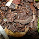
Onixoforlar tipinə bir sinif - İlktraxeyalılar (Prototracheata), bir dəstə, iki fəsilə və təxminən 70 -ə qədər növ daxildir. Müasir onixoforlar sırf quruda yaşayan onurğasızlardır. Onlara Amerika, Afrika , Avstraliya meşələrində , daşların altında , mamırlıqda , nadir hallarda mağaralarda rast gəlinir . Onixorforlar içərisində peripatuslar nisbətən daha yaxşı öyrənilmişdir.
Onixoforların bədəni 4-5 sm , daha iri növlərdə isə 15 sm -ə qədərdir. Onların qurdvarı bədənlərində baş gövdədən aydın seçilmişdir. Başda olan buğumsuz bığcıqlar ilbizin hiss çıxıntılarına oxşardır. Gövdə şaqren kutikula ilə örtülmüşdür , buğumlanmamışdır. Cüt ətraflar metamer təkrarlanır və kəpənək tırtıllarının yalançı ayaqlarını xatırladır. Baş - akrondan və 3 buğumdan ibarətdir. Başın dorsal hissəsində bir cüt sadə gözcüklər yerləşir. Hiss çıxıntıları akronda yerləşir. Birinci baş buğumu interkalyar olub , çıxıntısı yoxdur. İkinci baş buğumunda ağızın yanlarında skleritləşmiş çənələr, üçüncü buğumda isə müdafiəyə xidmət edən , selikli maye ifraz edən vəzili törəmələr yerləşmişdir. Bu maye vasitəsilə peripatuslar öz şikarını yapışdırır və onu hərəkətsizləşdirir. İlktraxeyalılar gövdəsi 14 - 40 cüt ayaqlara malikdir. Ayaqlar bədənin qarın divarından əmələ gəlmişdir və cüt skleritləşmiş caynağa malikdir. Onixoforlar çox yavaş hərəkət edirlər . Dəri - əzələ kisəsi peripatusların torpağı qazmasında böyük rol oynayır. İlktraxeyalıların bədəni birqatlı epiteli ilə örtülüdür . Epiteli nazik kitukulanı əmələ gətirir . Kutikulanın tərkibində xitin də vardır. Həlqəvi qurdlarda olduğu kimi epitelinin altında dəri -əzələ kisəsini əmələ gətirən həlqəvi və uzununa əzələlər yerləşmişdir. Əzələlər hamardır.
3 şöbədən ibarətdir . Ağız boşluğuna tüpürcək vəzilərinin cüt axarları açılır. Bu vəzilər selomoduktların şəkildəyişməsindən əmələ gəlmişdir.
Metamer yerləşən şəklidəyişmiş cüt selomoduktlardan ibarətdir.
Gövdədə olan ayaqların əsasında metamer yerləşir . Hər bir ifrazat orqanının bir ucu ilgəkşəkilli kanalla selom kisəsinə açılan qıfdan , digər ucu sidik kisəsinə açılan kanaldan təşkil olunmuşdur . Sidik kisəsi ifrazat dəliyi ilə nəhayətlənir .
Traxeyalardır. Bədənin üstündə çoxlu sayda nəfəsgahlar – stiqmalar vardır. Bu stiqmalar traxeyalara açılır . Traxeyalar dəri vəzilərindən əmələ gələn buğumlanmamış orqandır.
Pilləkənvari tipdir. Baş beyin 3 şöbədən ibarətdir və gözləri, bığcıqları sinirləndirir . Beyindən çıxan udlaqətrafı konnektivlər iki yan sinir sütunlarını əmələ gətirir . Bu sinir sütunları bədənin dal ucunda birləşirlər. Sinir sütunları arasında çoxlu kommisurlar vardır. Onixoforların sinir sitemi yastı qurdların sinir sisteminə oxşardır. Belə ki , onixoforlarda sinir düyünləri formalaşmamışdır, sinir hüceyrələri sütun üzərində bərabər paylanmışdır.
Zəif inkişaf etmişdir. Gözcük və bığcıqlardan əlavə , dərinin üzərində çoxlu lamisə sensillaları vardır.
Onixoforlar ayrıcinslidirlər. Qonadaları cütdür. Erkək cinsi orqanlar cüt toxumluqdan , toxum borusundan və toxum kisəsindən ibarətdir .Toxum boruları birləşərək tək toxumxaricedici kanalı əmələ gətirir . Dişi cinsi orqanları yumurtalıqlardan , cüt yumurta borularından və balalıqdan ibarətdir. Balalıq - balalıq yoluna açılır . Onixoforlarda cinsi dəlik sonuncu iki cüt ayaqların arasında yerləşmişdir . Spermatafor vasitəsilə çoxalırlar, mayalanma daxilidir . Bir neçə onixofor müstəsna olmaqla , demək olar ki, bütün onixoforlar diri bala doğurlar , az hallarda yumurta qoyurlar . İnkişafları düzənidir . Doğulmuş balalar nisbətən iri olur.[1]
B.İ.Ağayev, Z.A.Zeynalova. Onurğasızlar zoologiyası. Bakı , «Təhsil» 2008 , 568 səh
Onixoforlar; Caynaqdaşıyıcılar; İlktraxeylər (lat. Onychophora < yun.. onychos — caynaq, phoros — daşıyıcı) Quruda yaşayan rütubətsevən onurğasız heyvan tipi.
Els onicòfors (Onycophora) constitueixen un fílum d'animals terrestres d'aspecte envellutat, semblants a les erugues, l'existència dels quals està registrada des del Cambrià. Estan estretament emparentats amb els artròpodes però els seus nombrosos parells d'apèndixs, els lobòpodes, no són realment articulats.
Presenten un cos allargat, cilíndric i sense segmentar, tot i ser metamèrics. El cap té un parell de tentacles força llargs i anellats a la base dels quals es troben els ulls. La boca s'obre anteriorment en posició ventral i al voltant es troben tot un seguit de lòbuls cutanis. Tenen un parell de papil·les bucals contràctils, una a cada costat de la boca, que segreguen un líquid enganxifós que utilitzen per a capturar els petits artròpodes dels que s'alimenten. Les potes acaben en dues urpes.
El fílum compta en l'actualitat exclusivament amb formes terrestres dividides en dues famílies:[1]
Els onicòfors es troben ja a fòssils del Cambrià, per exemple als sediments de Burgess Shale. La distribució actual és un reflex de la seva història evolutiva, ja que es troben a zones discontínues que corresponen a fragments del primitiu continent de Gondwana.
Habiten indrets foscs i humits, a la fusta en putrefacció, l'humus i sota les pedres.
Els onicòfors presenten diverses estratègies reproductives i poden ser:[2][3][1]
Les cries s'assemblen en a les formes adultes si bé sovint són de color blanc. No reben atenció parental i a vegades poden ser devorades per la mare.
Els onicòfors (Onycophora) constitueixen un fílum d'animals terrestres d'aspecte envellutat, semblants a les erugues, l'existència dels quals està registrada des del Cambrià. Estan estretament emparentats amb els artròpodes però els seus nombrosos parells d'apèndixs, els lobòpodes, no són realment articulats.
Drápkovci (Onychophora) jsou kmen živočichů. Jedná se o velmi starobylou skupinu, která se v podstatě za posledních 500 milionů let nezměnila.[1] Zatím bylo popsáno asi 200 druhů, ale předpokládá se, že celkový počet bude výrazně vyšší. Jde o článkovce s homonomní metamerií.
Jedinci mohou dorůstat až 20 cm délky. Jméno dostali podle dvojice drápků, které zakončují nečlánkované končetiny, nesoucí tělo nad podkladem. Stejnoměrně článkované tělo pokrývá kutikula s chloupky, kterou jednou za čas svlékají. Při každém svlékání přibývá počet článků i panožek. Na hlavě mají pár kroužkovaných tykadel a na jejich bázi jsou poměrně vyspělé oči s rohovkou i čočkou. První pár panožek (onychopodií) je přeměněn v kusadla, v ústech je navíc několik masitých pedipalp, vylučujících lepivý sekret. Vylučovací soustavu tvoří metanefridie. Nervová soustava: shodná jako u členovců, složena ze tří oddílů. Pár ventrálních nervových provazců se nachází v každém článku, nad řitním otvorem se spojují. Cévní soustava je otevřená, silně redukovaná díky vzdušnicovému typu dýchání. Přítomna jedna hřbetní céva s párem ostií na každém článku. Dýchací soustava: dýchají vzdušnicemi. Protože nemohou vzdušnice uzavírat (jak to například dělá hmyz), hrozí jim vyschnutí a musí se stále zdržovat ve vlhku. Na povrchu těla se nachází velké množství pórů se svazky tracheálních kapilár. Díky nim dochází k transportu kyslíku k jednotlivým buňkám.
Jsou to gonochoristé s přímým vývojem. Některé druhy kladou vajíčka, jiné rodí živá mláďata. U amerických živorodých druhů dokonce nacházíme jednoduchou placentu vyživující zárodek a vývoj embrya může trvat i 15 měsíců. V okamžiku vylíhnutí, či narození jsou mláďata podobná dospělcům.
Žijí převážně na jižní polokouli ve vlhkých tropech. Je to jediný kmen živočichů, který je zcela suchozemský. Všichni drápkovci jsou masožraví. Z ústních žláz vystřikují lepkavou tekutinu, která na vzduchu tuhne. Užívají ji hlavně k lovu, k znehybnění kořisti, ale i pro obranu. Na jejich vlastním těle se tato vlákna rozpadají.
Převážně jde o noční dravce, kteří napadají různé členovce a další malé živočichy (například slimáky) až do velikosti útočníka. Kořist je paralyzována a poté do ní drápkovec napustí trávicí enzymy, které obsah těla rozpustí. Díky svalnatému hltanu je pak obsah těla vysáván (na principu pumpy).
Celý kmen obsahuje asi 200 druhů. Příkladem je drápkonoš kapský (Peripatopsis capensis), modře zbarvený, 5 cm velký a žijící v jižní Africe.
Aktuální klasifikace (2013):[2]
(† označuje vyhynulé taxony; české názvy podle BioLib[3])
Kmen: Onychophora Grube, 1853 - drápkovci
Drápkovci (Onychophora) jsou kmen živočichů. Jedná se o velmi starobylou skupinu, která se v podstatě za posledních 500 milionů let nezměnila. Zatím bylo popsáno asi 200 druhů, ale předpokládá se, že celkový počet bude výrazně vyšší. Jde o článkovce s homonomní metamerií.
Die Stummelfüßer (Onychophora – wörtlich Krallenträger) bilden einen eigenen Tierstamm aus der Gruppe der Häutungstiere (Ecdysozoa) und lassen sich vereinfacht als Würmer mit Beinen beschreiben. Hauptsächlich auf der Südhalbkugel verbreitet, leben sie räuberisch von kleineren Tieren wie zum Beispiel Insekten, die sie oft durch Verspritzen eines klebrigen Schleims erbeuten. In der modernen Zoologie sind sie nicht zuletzt durch die lebendgebärende Fortpflanzungsweise und die ausgefallenen Begattungsformen vieler Arten bekannt. Mögliche Vorfahren der Tiere, die Lobopoden aus der erdgeschichtlichen Epoche des Kambriums, sind in der Paläontologie von großem Interesse.
Stummelfüßer sind sehr wahrscheinlich nahe mit Gliederfüßern (Arthropoda) und Bärtierchen (Tardigrada) verwandt und bilden mit diesen beiden das Taxon Panarthropoda. Die erste Gattung wurde 1825 von Lansdown Guilding (1797–1831) wissenschaftlich beschrieben, der die Tiere noch als abgewandelte Schnecken (Gastropoda) ansah; der Name Onychophora wurde erst 1853 durch Adolf Eduard Grube geprägt.
Stummelfüßer sind wurmförmige, segmentierte Tiere mit zylindrischem, bauchseitig abgeflachten Körperquerschnitt und reihenförmig angeordneten ungegliederten Körperanhängen, den Stummelfüßen. Die Tiere werden zwischen 0,5 und 20 Zentimeter lang, wobei der Durchschnitt bei etwa 5 Zentimetern liegt, und weisen zwischen 13 und 43 Beinpaare auf. Die Farbe ihrer von zahlreichen feinen Querringen bedeckten Haut ist oft unauffällig orangefarben, rot oder braun, manchmal aber auch leuchtend grün, blauschwarz, goldfarben oder weiß, gelegentlich auch farbig gemustert.
Die äußerlich unauffällige und nur am regelmäßigen Abstand der Beinpaare erkennbare Segmentierung zeigt sich in der regelmäßigen Aneinanderreihung von Hautöffnungen (Ostien), Ausscheidungsorganen (Nephridien) und Nervenzellkonzentrationen (Ganglien). Die einzelnen Körperabschnitte sind kaum spezialisiert; selbst der Kopf entwickelt sich nur wenig anders als ein beliebiges Hinterleibssegment. Die Segmentierung wird anscheinend durch das auch bei anderen Tiergruppen nachweisbare Gen engrailed festgelegt, das während der Embryonalentwicklung jeweils an der hinteren Segmentgrenze und in der Wachstumszone der Stummelfüße aktiviert wird.
Das namensgebende Merkmal der Stummelfüßer sind die fachlich als Oncopodien bezeichneten kegelförmigen, sackartigen Körperanhänge, die innen hohl sind, keine Gelenke aufweisen und terminal mit einem Paar Krallen versehen sind. Obwohl ihre Zahl zwischen den Arten beträchtlich variieren kann, sind sie sich im Aufbau grundsätzlich sehr ähnlich. Ihre Festigkeit beruht auf dem hydrostatischen Druck der enthaltenen Körperflüssigkeit.
Bewegt werden die Beine meist passiv durch Kontraktion und Dehnung des Gesamtkörpers. In jedem Bein verlaufen allerdings auch interne Muskeln, die es verkürzen und biegen können; aufgrund der fehlenden Gelenke kann diese Verbiegung an jeder Stelle entlang der Längsseite des Beines erfolgen.
Die Ausmündungen zweier verschiedener Organbildungen einzelner Arten befinden sich in der Nähe des Beinansatzes:
An den Füßen sitzt je ein Paar einziehbarer verhärteter (sklerotisierter) Krallen aus Chitin, von denen der wissenschaftliche Name des Taxons herrührt, der sich von dem Griechischen onyches, „Krallen“ und pherein, „tragen“, ableitet. Am Klauenansatz befinden sich drei bis sechs dornige „Kissen“, auf denen das Bein in Ruhestellung aufsetzt und auf welchen die Tiere auf glattem Untergrund laufen; die Krallen werden hauptsächlich auf unebenem Terrain eingesetzt, um festen Halt zu erlangen.
Neben den Beinpaaren gibt es drei weitere Körperanhänge, die sich an dem aus drei Segmenten bestehenden Kopf befinden:
Alle drei Strukturen entsprechen ihrer evolutionären Herkunft nach den Beinpaaren der anderen Segmente.
Anders als die Gliederfüßer besitzen Stummelfüßer kein festes Außenskelett (Exoskelett). Stattdessen wird ihre Körperform wie bei einem wassergefüllten Ballon durch den leicht erhöhten Druck der Innenflüssigkeit auf die Körperwand aufrechterhalten: Weil diese Flüssigkeit bei den durch Muskelkraft auftretenden Drücken inkompressibel ist, also ihr Volumen nicht ändert, können Muskeln dagegen Arbeit verrichten. Wegen des Skelettmaterials Wasser spricht man auch von einem Hydroskelett.
Die Körperwandung selbst besteht aus einer nicht-zelligen Außenhaut, der Cuticula, einer einlagigen Zellschicht, der Epidermis, als Innenhaut und in der Regel drei darunter gelegenen Muskelschichten, die in Bindegewebe eingebettet sind.
Die Cuticula ist etwa einen Mikrometer dünn, mit feinen Härchen besetzt und ähnelt in Zusammensetzung und Aufbau der Cuticula der Gliederfüßer. So besteht sie aus α-Chitin und diversen Proteinen, nicht jedoch aus Kollagen und lässt sich in eine außen liegende Epicuticula und eine darunter liegende Procuticula unterteilen, die sich ihrerseits aus Exo- und Endocuticula zusammensetzt. Dieser mehrschichtige Aufbau ist für die hohe Flexibilität der Außenhaut verantwortlich, die es den Tieren ermöglicht, sich noch in die engsten Spalten zu quetschen. Obwohl sie nach außen wasserabstoßend wirkt, ist sie nicht in der Lage, Feuchtigkeitsverluste zu verhindern, was zur Folge hat, dass Stummelfüßer nur in einem Mikroklima mit hoher Luftfeuchte leben können.
Die Oberfläche der Cuticula ist mit zahlreichen feinen Papillen übersät; die größeren von ihnen tragen die als Härchen sichtbaren Sinnesborsten. Die Papillen sind ihrerseits mit kleinen Schüppchen bedeckt und verleihen so der Haut ein samtartiges Aussehen. Sie fühlt sich bei Berührung auch entsprechend trocken-samtig an, wofür ihre wasserabweisende Natur verantwortlich ist. Im Rahmen der durch das Hormon Ecdyson vermittelten Häutung (Ecdysis) wird sie regelmäßig, manchmal alle vierzehn Tage, abgestoßen.
Erneuert wird sie dabei von der darunter gelegenen einschichtigen Innenhaut, der Epidermis, die im Gegensatz zur Cuticula aus lebenden Zellen besteht. Ihr unterliegt eine dicke Schicht Bindegewebe, das in erster Linie von Kollagenfasern gebildet wird, die entweder parallel oder senkrecht zur Körperlängsachse ausgerichtet sind. Innerhalb des Bindegewebes liegen drei ununterbrochene Schichten unspezialisierter glatter Muskulatur. Die äußere, verhältnismäßig dicke Schicht wird von den Ringmuskeln gebildet, die innerste von den ebenfalls voluminösen Längsmuskeln. Dazwischen liegen die dünnen Diagonalmuskeln, die sich schraubenförmig entlang der Körperachse nach vorne und hinten winden. Zwischen Ring- und Diagonalmuskulatur existieren feine Blutkanäle, die unterhalb der oberflächlich erkennbaren Querringe der Haut liegen und für diese Pseudosegmentierung genannte Musterung verantwortlich sind.
Unterhalb der innersten Muskelschicht liegt die Körperhöhle. Sie wird durch so genannte Dorsoventralmuskeln, die von der Mitte der Bauchseite quer durch sie hindurch zu den Rändern der Rückenseite verlaufen, im Querschnitt in drei Regionen geteilt: einen zentralen Mittelteil und zwei jeweils bauchseitig links und rechts gelegene Seitenteile, die auch die Beine einschließen.
Die Körperhöhle selbst ist ein so genanntes Pseudocoelom, das im speziellen Fall auch als Hämocoelom bezeichnet wird. Ein Pseudocoel ist im Gegensatz zu einem echten Coelom nicht von einer echten, von embryonalem Mesoderm abgeleiteten Zellschicht umgeben. Ein Coelom ist jedoch um die Keimdrüsen und die der Ausscheidung dienenden Nephridien herum ausgebildet.
Wie der Name Hämocoelom bereits andeutet, ist die Körperhöhle mit einer blutartigen Flüssigkeit gefüllt, in die alle Organe eingebettet sind; auf diese Weise können sie leicht mit im Blut zirkulierenden Nährstoffen versorgt werden. Diese Flüssigkeit ist farblos, da sie keine Pigmente enthält; aus diesem Grund dient sie auch nur in geringem Maße dem Sauerstofftransport. In der Blutflüssigkeit bewegen sich zwei verschiedene Typen von Blutzellen oder Hämozyten, die Amöbozyten und Nephrozyten. Erstere haben vermutlich eine Funktion bei der Abwehr von Bakterien und anderen Fremdkörpern; bei einigen Arten spielen sie auch bei der Fortpflanzung eine Rolle. Nephrozyten speichern dagegen Giftstoffe oder wandeln sie in eine für die als Nephridien bezeichneten Ausscheidungsorgane verwertbare Form um.
Das Hämocoelom wird durch eine horizontale Scheidewand, das Diaphragma, in zwei Teile geteilt, den rückseitig gelegenen pericardialen Sinus und den bauchseitig gelegenen perivisceralen Sinus. Ersterer umgibt das schlauchförmige Herz, letzterer die anderen Organe. Das Diaphragma ist mehrfach perforiert, so dass ein Flüssigkeitsaustausch zwischen den beiden Teilhöhlungen möglich ist.
Das Herz selbst ist ein aus einschichtigem Epithelgewebe bestehender Ringmuskelschlauch mit je zwei seitlichen Öffnungen (Ostien) pro Segment. Während unbekannt ist, ob das Hinterende offen oder geschlossen ist, mündet es zur Vorderseite hin direkt in die Körperhöhlung. Da es abgesehen von den feinen, zwischen den Muskulaturschichten der Körperwandung befindlichen Blutgefäßen und einem Paar Arterien, welche die Antennen versorgen, keine Adern gibt, spricht man von einem offenen Blutkreislauf.
Der zeitliche Ablauf des Pumpvorgangs lässt sich in zwei Teile zerlegen, Diastole und Systole. Während der Diastole fließt das Blut aus dem pericardialen (herznahen) Sinus durch die Ostien in das Herz. Sobald die Systole beginnt, schließen sich die Ostien, die Herzmuskulatur zieht sich zusammen, verringert so das Herzvolumen und pumpt auf diese Weise Blut aus dem Vorderende des Herzens in das periviscerale (organnahe) Hämocoelom. Die verschiedenen Gewebe dort werden auf diese Weise mit Nährstoffen versorgt, bevor das Blut schließlich wieder durch die Perforationen des Diaphragmas in den pericardialen Sinus zurückgelangt. Neben der Pumptätigkeit des Herzens haben auch Körperbewegungen einen Einfluss auf die Blutzirkulation.
Die Sauerstoffaufnahme der Tiere erfolgt zum einen durch einfache Diffusion über die gesamte Körperoberfläche hinweg, bei einigen Arten möglicherweise auch über ihre Coxalvesikel an den Stummelbeinen. Am wichtigsten ist jedoch der Gasaustausch über feine unverzweigte Röhrchen, die Tracheen, die von der Körperoberfläche tief in den Körper zu den verschiedenen Organen, ganz besonders aber zum Herz hinziehen.
Die Wandung dieser meist weniger als drei Mikrometer dicken Strukturen besteht nur aus einem extrem dünnen Häutchen, durch die der Sauerstoff leicht diffundieren kann. Sie entspringen an winzigen Öffnungen, den Spirakeln, die ihrerseits jeweils zu mehreren in einer dellenförmigen Einbuchtung der Außenhaut, dem Atrium, zusammengefasst sind. Die Zahl der so entstehenden Tracheenbündel lässt sich mit durchschnittlich 75 je Körpersegment angeben; gehäuft treten sie vor allem auf der Rückseite der Tiere auf.
Anders als Gliederfüßer, die ihre Tracheenöffnungen kontrollieren können, sind die Stummelfüßer dazu nicht in der Lage. Ihre Tracheen sind immer geöffnet, was bei Trockenheit große Wasserverluste zur Folge hat. Aus diesem Grund sind Stummelfüßer auf Lebensräume mit hoher Luftfeuchtigkeit angewiesen.
Der Verdauungstrakt beginnt mit dem subterminal, also etwas hinter dem vordersten Punkt des Körpers auf der Bauchseite gelegenen Mund, in dem Beute durch die mit feinen Zähnchen besetzten Kiefer mechanisch zerkleinert werden kann. Zwei Speicheldrüsen münden durch einen gemeinsamen Leiter in den sich anschließenden Schlund, der bereits den ersten Teil des Vorderdarms darstellt. Der von ihnen produzierte Speichel enthält Schleim und hydrolytische Enzyme, die in und außerhalb der Mundhöhle bereits die Verdauung einleiten. Evolutionsgeschichtlich leiten sich die Speicheldrüsen wahrscheinlich von als Nephridien bezeichneten Ausscheidungsorganen ab, die sich als homologe Organe in den anderen Körpersegmenten befinden.
Der Schlund selbst ist sehr muskulös und dient dazu, die vorverdaute teilverflüssigte Nahrung einzusaugen und durch die Speiseröhre, die den hinteren Teil des Vorderdarms bildet, in den Mitteldarm zu pumpen. Dieser ist anders als der Vorderdarm nicht mit Cuticula ausgekleidet und besteht nur aus einer einlagigen Schicht Epithelgewebe, die anders als bei anderen Tieren keine auffälligen Einbuchtungen aufweist. Beim Eintritt in den Mitteldarm werden Nahrungspartikel von einer schleimhaltigen peritrophischen Membran umhüllt, die eine mechanische Beschädigung des Darmgewebes durch scharfkantige Partikel verhindern soll. Das Darmepithel selbst sondert weitere Verdauungsenzyme ab und absorbiert die aufgeschlossenen Nährstoffe; der Großteil der Verdauung findet allerdings bereits extern oder in der Mundhöhle statt. Unverdauliche Reste gelangen in den auch Rektum genannten Hinterdarm, der wiederum mit Cuticula ausgekleidet ist und sich bauchseitig nahe dem Hinterende der Tiere in den After öffnet.
In fast jedem Segment befindet sich ein Paar als Nephridien bezeichneter Ausscheidungsorgane, die sich von Coelomgewebe ableiten. Sie bestehen jeweils aus einem kleinen Säckchen, das über einen geißelbesetzten Gang, Nephridiodukt genannt, mit einer bauchseitig am nächstgelegenen Beinansatz befindlichen Öffnung, der Nephridiopore, verbunden ist. Das Säckchen ist mit speziellen Zellen, den Podozyten besetzt, die eine Ultrafiltration des Blutes durch die Trennwand von Hämocoelom und Nephridium ermöglichen. Der Primärurin wird durch selektive Rückgewinnung von Nährstoffen und Wasser sowie durch Absonderung von Gift- und Abfallstoffen im Nephridiodukt noch in seiner Zusammensetzung verändert, bevor er an der Nephridiopore an die Außenwelt abgegeben wird. Das wichtigste stickstoffhaltige Ausscheidungsprodukt ist die wasserunlösliche Harnsäure; sie kann in festem Zustand mit nur wenig Wasser an die Umgebung abgegeben werden. Dieser als uricotelisch bezeichnete Ausscheidungsmodus stellt eine Anpassung an das Landleben und die Notwendigkeit, sparsam mit Wasser umzugehen, dar.
Ein ehemaliges Nephridienpaar im Kopf wurde sekundär in die Speicheldrüsen umgewandelt, ein weiteres im letzten Segment der Männchen übernimmt heute eine Drüsenfunktion, die anscheinend bei der Fortpflanzung zum Einsatz kommt.
Das Nervensystem besteht in erster Linie aus dem Gehirn und zwei bauchseitigen Nervensträngen, die durch zahlreiche Querverbindungen miteinander verbunden sind. Nach neueren Erkenntnissen handelt es sich aber nicht um ein „Strickleiternervensystem“ wie bei den Gliederfüßern, da weder segmental angeordnete Ganglien existieren noch die Querverbindungen segmental angeordnet sind[1].
Das vorne und rückseitig gelegene Gehirn besteht aus einer linken und einer rechten Hälfte, die zusammen mit Nervenverbindungen einen Ring um die Speiseröhre bilden. Anstelle einer Dreiteilung des Gehirns in Richtung der Körperlängsachse, wie man sie bei den eng verwandten Gliederfüßern (Arthropoda) findet, existieren nur zwei Gehirnabschnitte.[2]
Vom Gehirn gehen etwa fünfzehn Nervenpaare aus, die sowohl sensorische, als auch motorische Nervenzellen enthalten. Die Hauptnerven des Kopfbereichs sind die Nervi optici, die Augen und Gehirn verbinden und zwei Sinnesnerven, die in die Antennen ziehen und für die Vermittlung von Berührungs- und chemischen Reizen verantwortlich sind. Die wichtigsten Körpernerven sind dagegen zwei weit auseinander liegende, bauchseitige Nervenstränge, die sich den gesamten Körper entlang längsseitig nach hinten ziehen. Körperwand und -anhänge werden durch gemischte Nerven versorgt, die sowohl sensorische, der Sinneswahrnehmung dienende als auch motorische, die Muskeln ansprechende Signale weiterleiten.
Der gesamte Körper inklusive der Stummelfüße ist mit zahlreichen Papillen übersät, warzigen Vorsprüngen, die eine auf mechanische Reize ansprechende (mechanorezeptive) Borste an der Spitze tragen, die jeweils mit weiter innen liegenden Sinnesnervenzellen verbunden ist. Auch die Mundpapillen, die Ausgänge der Schleimdrüsen, haben vermutlich eine Funktion bei der Sinneswahrnehmung.
Die auf den „Lippen“, dem Labrum, gelegenen Sinneszellen oder Sensillen dienen dagegen der Wahrnehmung chemischer Reize, weshalb man von Chemorezeptoren spricht. Sie finden sich auch auf den beiden Antennen, die als die wichtigsten Sinnesorgane der Stummelfüßer angesehen werden können.
Mit Ausnahme einiger weniger, meist höhlenlebender Arten befindet sich am Ansatz der Antennen je ein einfach gebautes Auge (Ocellus), das aus einer chitinhaltigen Kugellinse sowie Hornhaut und Netzhaut besteht und durch je einen Nervus opticus mit dem Gehirn verbunden ist. Die Netzhaut setzt sich aus zahlreichen Pigmentzellen und Photorezeptoren zusammen; bei letzteren handelt es sich um leicht modifizierte Geißelzellen, deren Geißelmembran ein lichtempfindliches Pigment auf der Oberfläche trägt.
Das Auge der Onychophora zeigt weder im Bau noch in der Innervation eine Beziehung zum Facettenauge der Arthropoden. Vorgeschlagen worden ist eine Homologie zwischen diesem Auge und den Stirnaugen (Ocellen) z. B. der Insekten[3].
Beide Geschlechter verfügen über paarige Keimdrüsen, die durch als Gonodukte bezeichnete Ausführgänge in eine gemeinsame Geschlechtsöffnung, die Gonopore, münden, welche sich auf der hinteren Bauchseite befindet. Sowohl Keimdrüsen als auch Gonodukte leiten sich von echtem Coelomgewebe ab.
Bei den Weibchen sind die beiden Eierstöcke in der Mitte zusammengewachsen und an die horizontal verlaufende Scheidewand geheftet. Die Gonodukte sind unterschiedlich ausgeprägt, je nachdem, ob es sich um lebendgebärende oder eierlegende Arten handelt. Bei Ersteren teilt sich jeder Ausführgang in einen schlanken Eileiter und eine geräumige „Gebärmutter“, den Uterus, in dem die Embryos heranwachsen. Die nur einfach vorhandene Vagina, mit der beide Uteri verbunden sind, mündet an der Gonopore nach außen. Bei eierlegenden Arten, deren Gonodukte gleichförmig aufgebaut sind, liegt die Geschlechtsöffnung an der Spitze eines langen Eierlegeapparats, des Ovipositors. Die Weibchen vieler Arten verfügen darüber hinaus über einen Receptaculum seminis genannten Samenspeicher, in dem Spermazellen der Männchen vorübergehend oder auch längerfristig aufbewahrt werden können.
Männchen verfügen über zwei getrennte Hoden sowie die dazugehörigen Samenbläschen (vesicula seminalis) und Ausführgänge (vasa efferentia). Letztere vereinigen sich zu einem gemeinsamen Samenleiter, dem vas deferens, der sich wiederum zum Ejakulationsleiter verbreitert an der Gonopore nach außen öffnet. Direkt neben beziehungsweise hinter dieser liegen bei den Männchen zwei Paar spezieller Drüsen, die vermutlich eine Hilfsfunktion bei der Fortpflanzung haben; die Drüsen des hinteren Paars werden auch als Analdrüsen bezeichnet.
Eine penisartige Struktur konnte bisher nur bei Männchen der Gattung Paraperipatus nachgewiesen, aber noch nicht in Aktion beobachtet werden. Männchen zahlreicher australischer Arten weisen dagegen wie bereits erwähnt spezielle Strukturen am Kopf auf, die anscheinend Aufgaben bei der Samenübertragung auf die Weibchen übernehmen.
Stummelfüßer leben in tropischen Lebensräumen und in der gemäßigten Zone der Südhalbkugel; sie weisen sowohl eine zirkumtropische, als auch eine zirkumaustrale Verbreitung auf. Einzelne Arten findet man in Mittel- und Südamerika, auf den Karibischen Inseln, im äquatorialen Westafrika und in Südafrika, des Weiteren in Nordindien, Indonesien und Teilen Malaysias, auf Neu-Guinea, in Australien und in Neuseeland. Die Gesamtheit der heutigen Verbreitungsgebiete weist auf den wahrscheinlichen Ursprung des Taxons auf dem ehemaligen Superkontinent Gondwana hin, weswegen man auch von einer Gondwana-Verteilung spricht.
Alle Stummelfüßer sind landlebend (terrestrisch) und bevorzugen dunkle Lebensräume mit hoher Luftfeuchtigkeit. In Frage kommen dafür insbesondere die Regenwälder der Tropen und gemäßigten Zonen, wo sich die Tiere in Mooskissen, in der Laubstreu des Bodens, unter Baumstämmen und Steinen, in verrottendem Holz oder in Termitengängen finden lassen. Auch in unbewaldetem Grasland kommen sie vor, wenn ausreichend Bodenspalten existieren, in die sie sich während des Tages zurückziehen können.
Zwei Arten leben in Höhlen, einem Lebensraum, an den sie durch ihre Fähigkeit, sich in kleinste Ritzen zu zwängen, hervorragend angepasst sind und der ihnen konstante Lebensbedingungen garantiert. Da die entscheidenden Voraussetzungen für ein Höhlenleben wahrscheinlich bereits vor der Besiedlung dieses Habitats vorlagen, spricht man auch von Exaptation. Die Landwirtschaft hat Stummelfüßern anscheinend neue Lebensräume erschlossen; jedenfalls finden sie sich in Zentralamerika und der Karibik auch in Kakao- und Bananenplantagen.
Weil die Austrocknungsgefahr tagsüber und bei trockenem Wetter am größten ist, verwundert es nicht, dass Stummelfüßer in der Regel nachts und bei Regenwetter aktiv sind. Unter kalten oder trockenen Umweltbedingungen suchen sie Bodenspalten auf, in denen sie ihren Körper in einen Ruhezustand versetzen. Sie wenden sich von starken Lichtquellen ab und sind somit negativ phototaktisch.
Die bisher größte gemessene Populationsdichte liegt mit etwa 10 Individuen pro Quadratmeter Boden sehr niedrig; Stummelfüßer sind auch in ihren Lebensräumen nur sehr schwer zu finden.
Um von der Stelle zu kommen, krabbeln die Tiere mithilfe ihrer Beine vorwärts; sie bewegen dabei, anders als die Gliederfüßer, immer beide Beine eines Paares gleichzeitig. Eine Berührung der Bauchseite mit dem Substrat wird nach Möglichkeit vermieden, der Körper also durch die Stummelfüße oberhalb des Bodens gehalten. Die an den Füßen befindlichen Klauen werden nur auf hartem, rauem Untergrund eingesetzt, wo ein fester Halt erforderlich ist; auf weichen Substraten wie etwa Moos laufen die Tiere auf den am Klauenansatz befindlichen Fußkissen.
Die eigentliche Fortbewegung geschieht weniger durch den Einsatz der Beinmuskulatur, sondern stattdessen durch eine lokale Veränderung der Körperlänge. Diese kann von den Tieren durch Einsatz der Ring- und Längsmuskulatur beeinflusst werden: Wird erstere kontrahiert, verringert sich der Körperquerschnitt und das entsprechende Segment dehnt sich, da sein Volumen aufgrund der enthaltenen inkompressiblen Flüssigkeit des Hämocoeloms konstant bleiben muss – dies ist die übliche Wirkungsweise eines Hydroskeletts wie es auch bei verschiedenen Würmern zum Einsatz kommt. Durch die Dehnung heben sich die Beine des betroffenen Körperabschnitts und schwingen vorwärts. Lokale Kontraktion der Längsmuskeln verkürzt dagegen das entsprechende Segment und bewegt auf diese Weise das zugehörige Beinpaar, dass sich nun im Kontakt mit dem Substrat befindet, nach hinten; dieser Teil des Bewegungszyklus ist der effektive Beinschlag, der für die eigentliche Fortbewegung verantwortlich ist. Durch das Nervensystem werden die einzelnen Muskeldehnungen und -entspannungen der Segmente derart aufeinander abgestimmt, dass Kontraktionswellen durch den Körper laufen, die dazu führen, dass ein Beinpaar nach dem anderen nach vorne schwingt und sich dann auf dem Untergrund nach hinten bewegt. Die auf diese Weise erzielten Geschwindigkeiten schwanken zwischen etwa einem Millimeter und etwas mehr als einem Zentimeter pro Sekunde.
Stummelfüßer sind räuberische Organismen, die in der Lage sind, Tiere zu erbeuten, die erheblich größer sind als sie selbst. Zum Beutespektrum gehören etwa Asseln (Isopoda), Termiten (Isoptera), Echte Grillen (Gryllidae), Staubläuse (Psocoptera), Schaben (Blattodea), Tausendfüßer (Myriapoda), Spinnen (Araneae), diverse Würmer und selbst größere Schnecken (Gastropoda). Sie gelten als ökologisches Äquivalent der Hundertfüßer (Chilopoda).
Potentielle Opfer spüren sie zumeist mit Hilfe der Antennen auf und verfolgen sie dann bis in kleinste Ritzen. Während kleinere Beute direkt getötet wird, nutzen sie bei größeren Tieren eine weiße leimartige und stark proteinhaltige Flüssigkeit, die von den beiden Schleimdrüsen produziert wird, um ihre Opfer unbeweglich zu machen. Sie wird durch die Poren der Mundpapillen über eine Entfernung von bis zu 30 Zentimetern verspritzt und härtet an Luft sehr schnell aus, so dass sich die Beute in der stark klebrigen Substanz verfängt und schließlich unbeweglich wird. Auf der wasserabweisenden Haut der Stummelfüßer selbst klebt die Substanz dagegen nicht, so dass die Tiere sich ihrem Opfer ungefährdet auf kürzeste Distanz nähern können. Durch Injektion toxischen Speichels wird es nun abgetötet und vorverdaut; die scharfen Kiefer zerschneiden die Beute dann in feine Stücke, die über den Mund in den Verdauungstrakt gelangen.
Wichtige Fressfeinde der Tiere sind in erster Linie diverse Spinnen (Araneae) und Hundertfüßer (Chilopoda), daneben aber auch Nagetiere (Rodentia) und Vögel, in Zentralamerika etwa die Gilbdrossel (Turdus grayi). Die zu den Korallenottern (Micrurus) gezählte Schlangenart Micrurus hemprichii ernährt sich sogar beinahe ausschließlich von Stummelfüßern. Zur Abwehr rollen sich manche Arten reflexartig zu einer Spirale zusammen; daneben können sich die Tiere auch durch Schleimauswurf gegen kleinere Gegner zur Wehr setzen.
Diverse Milben (Acari) sind als Ectoparasiten bekannt, die sich auf der Haut der Stummelfüßer festsetzen. Bei Hautverletzungen kommt es zudem meist zu bakteriellen Infektionen, die fast immer tödlich enden.
Stummelfüßer vermehren sich durchwegs auf sexuellem Wege. Einzige bekannte Ausnahme ist die Art Epiperipatus inthurni, bei der bislang keine Männchen nachgewiesen werden konnten und die sich parthenogenetisch vermehrt.
Die Spezies weisen in vielen Fällen einen merklichen Sexualdimorphismus auf – die Weibchen sind meist größer als die Männchen und verfügen, falls die Beinzahl innerhalb einer Art variabel ist, auch über mehr Beine. Bei vielen Arten werden sie nur einmal im Leben besamt. Dies kann dazu führen, dass die Begattung schon stattfindet, wenn die Fortpflanzungsorgane der Weibchen noch nicht ausgereift sind, bei Macroperipatus torquatus beispielsweise schon im Alter von drei Monaten. Die übertragenen Spermien werden in diesem Fall in einem speziellen Speicherorgan aufbewahrt, wo sie für längere Zeiträume befruchtungsfähig bleiben können.
Die Befruchtung findet bei Stummelfüßern grundsätzlich intern statt, die Art der Samenübertragung variiert allerdings recht stark. Bei den meisten Arten, etwa denen der Gattung Peripatus, wird ein Spermienpaket, die Spermatophore, in der Geschlechtsöffnung des Weibchens platziert. Wie im Einzelnen dies geschieht, ist in den meisten Fällen allerdings noch unbekannt, ein regelrechter Penis ist nur bei Arten der Gattung Paraperipatus nachgewiesen. Bei zahlreichen australischen Arten existieren Grübchen oder spezielle stilett- oder hakenförmige Strukturen am Kopf; bei Florelliceps stutchburyae etwa presst das Männchen einen langen Dorn gegen die Geschlechtsöffnung der Weibchen und positioniert sehr wahrscheinlich auf diese Weise seine Spermatophore dort. Es wird dabei durch das Weibchen unterstützt, das mit den Klauen des letzten Beinpaares das Männchen umklammert hält. Eine außergewöhnliche Art der Spermienübertragung erfolgt bei zwei Arten der Gattung Peripatopsis: Hier platziert das Männchen zwei millimetergroße Spermatophoren auf dem Rücken oder den Flanken des Weibchens. Amöbozyten aus dem Blut des Weibchens sammeln sich daraufhin auf der Innenseite der Ablagerungsstelle und bauen durch Sekretion von Enzymen die unter dem Spermienpaket befindliche Körperwandung sowie die Hülle der Spermatophore ab. Die dadurch freigesetzten Spermien bewegen sich dann frei durch das Hämocoel zu den Eierstöcken, durchdringen deren Außenwand und befruchten schließlich die Eizellen. Warum es durch die selbstverursachte Hautverletzung nicht zu bakteriellen Infektionen kommt, ist noch unverstanden.
Bei Stummelfüßern findet man sowohl eierlegende (ovipare), eierlebendgebärende (ovovivipare) und lebendgebärende (vivipare) Formen.
Die Zahl der Nachkommen eines Weibchens schwankt zwischen 1 und 23 pro Jahr; die zwischen 6 und 17 Monate andauernde Entwicklung von der befruchteten Eizelle zum Erwachsenenstadium verläuft grundsätzlich direkt ohne Larvenstadium; vermutlich ist dies auch der ursprüngliche Entwicklungsmodus. Maximal können Stummelfüßer bis zu 6 Jahre alt werden.
Die globale Gefährdung der Stummelfüßer-Vielfalt lässt sich nur schwer einschätzen; viele Arten sind nur von der Typ-Lokalität, also dem Ort, von dem das erste beschriebene Exemplar stammte, bekannt. Niedrige Populationsdichten, die meist nächtliche Lebensweise der Tiere, möglicherweise noch unerfasste jahreszeitliche Einflüsse und geschlechtsspezifische Unterschiede erschweren die Erhebung vertrauenswürdiger Angaben zusätzlich.
Bisher wurden nur elf Arten hinreichend genau untersucht, um Angaben über ihre Häufigkeit machen zu können. Die International Union for the Conservation of Nature and Natural Resources listet mit der Ausgabe 2009 auf der Roten Liste wie folgt:
Ursache der Bedrohung ist in erster Linie der Verlust und die Fragmentierung des Lebensraums durch zunehmende Industrialisierung, Trockenlegung von Feuchtgebieten und Brandrodungen zu Landwirtschaftszwecken. Viele Arten haben dazu eine von Natur aus niedrige Populationsdichte und ein geografisch eng beschränktes Verbreitungsgebiet, was dazu führt, dass schon kleinere lokale Störungen der betroffenen Ökosysteme zum Aussterben ganzer Populationen und gegebenenfalls Arten führen können. Regional spielt daneben auch das Sammeln von Tieren für Universitäten oder Forschungsinstitute eine Rolle.
Der Schutz der Stummelfüßer ist regional sehr unterschiedlich ausgeprägt; in manchen Staaten wie Südafrika existieren Sammelbeschränkungen und Exportkontrollen, in anderen wie Australien nur letztere; aus vielen Staaten sind überhaupt keine spezifischen Schutzmaßnahmen bekannt. Weltweit einzigartig ist ein Schutzprogramm in Tasmanien: In einem dortigen Forstbezirk kommt ein eigener „velvet worm conservation plan“ zum Einsatz, der speziell auf eine Stummelfüßer-Art zugeschnitten ist.
Gezüchtet werden können Stummelfüßer bisher noch nicht. Entsprechende Überlegungen bestehen aber, nicht nur mit dem Hintergedanken, Populationen für spätere Auswilderungen bereitstellen zu können, sondern auch, um die betroffene Öffentlichkeit besser über die Tiere und die Notwendigkeit ihres Schutzes aufzuklären. Aufgrund der oft bunten Farben und der großen Bedeutung für die Evolutionsgeschichte käme zum Beispiel eine Haltung in „Insektenzoos“ in Frage.
Unter den heutigen Formen sind die Stummelfüßer wahrscheinlich sehr eng mit den Gliederfüßern (Arthropoda) verwandt, einem sehr umfangreichen Taxon, zu dem man etwa die Krebstiere (Crustacea), Insekten (Insecta) und Spinnentiere (Arachnida) zählt. Mit ihnen teilen sie unter anderem das gehäutete und aus α-Chitin sowie nicht-kollagenen Proteinen zusammengesetzte Außenskelett, die von echtem Coelomgewebe umgebenen Keimdrüsen und Ausscheidungsorgane, das offene Blutsystem mit rückseitig gelegenem Schlauchherzen, eine in pericardiale und periviscerale Höhlen aufgeteilte Leibeshöhle, Atmung durch Tracheen und eine ähnliche Embryonalentwicklung. Dazu kommt die Segmentierung mit je zwei Körperanhängen pro Segment. Antennen, Mandibeln und Oralpapillen sind dagegen vermutlich nicht zu den entsprechenden Körperteilen der Gliederfüßer homolog, das heißt, sie haben sich wohl unabhängig von diesen entwickelt.
Eine weitere eng verwandte Gruppe sind die relativ obskuren Bärtierchen (Tardigrada), denen allerdings aufgrund ihrer sehr geringen Größe eine Reihe von Merkmalen der Stummel- und Gliederfüßer wie Blutkreislauf und separate Atmungsstrukturen fehlen. Gemeinsam bilden Stummelfüßer, Gliederfüßer und Bärtierchen ein monophyletisches Taxon, die Panarthropoda, das heißt, alle drei Gruppen zusammengenommen umfassen alle Nachkommen ihres letzten gemeinsamen Vorfahren.
Innerhalb der Panarthropoda wurden die Stummelfüßer meist aufgrund einer gewissen Formverwandtschaft mit den Bärtierchen zu einem Taxon Protoarthropoda zusammengefasst. Mit dieser Bezeichnung sollte zum Ausdruck gebracht werden, dass beide, Stummelfüßer und Bärtierchen, noch nicht die volle Entwicklungshöhe der Gliederfüßer erreicht haben. Moderne systematische Theorien verwerfen derartige Vorstellungen von „primitiven“ und „hoch entwickelten“ Lebensformen und orientieren sich stattdessen ausschließlich an den stammesgeschichtlichen Verwandtschaftsverhältnissen der beteiligten Taxa. Diese sind noch nicht zur Gänze aufgeklärt; es gilt jedoch als relativ wahrscheinlich, dass die Schwestergruppe der Stummelfüßer von einem Taxon gebildet wird, das sowohl Bärtierchen und Gliederfüßer umfasst und als Tactopoda (manchmal aber auch Tritocerebra) bezeichnet wird:
Panarthropoda TactopodaBärtierchen (Tardigrada)
Gliederfüßer (Arthropoda)
Stummelfüßer (Onychophora)
In die weitere Verwandtschaft der Stummelfüßer wurden lange Zeit die Ringelwürmer (Annelida) gestellt. Mit ihnen verbindet sie unter anderem der wurmartige Körper, die dünne und flexible Außenhaut, eine schichtige Muskulatur, paarig angelegte Ausscheidungsorgane sowie ein einfach gebautes Gehirn und einfache Augen. Ausschlaggebend war jedoch die bei beiden Gruppen vorhandene Segmentierung, die nur wenig Spezialisierung der Einzelsegmente erkennen lässt. Die Parapodien genannten Körperanhänge der Ringelwürmer entsprechen demnach den Gliedmaßen der Stummelfüßer.
In dem von Georges Cuvier geprägten Articulata-Konzept gelten die Stummelfüßer daher als evolutionäres Bindeglied zwischen Ringelwürmern und Gliederfüßern: Wurmartige Vorläufer entwickelten demnach zuerst Parapodien, die sich dann über Stummelbeine als Zwischenglied letztlich zu den Körperanhängen der Gliederfüßer weiterentwickelten. Die Stummelfüßer selbst galten aufgrund ihres strukturellen Konservatismus folglich als lebende Fossilien. Diese Ansicht kommt paradigmatisch in der Aussage des französischen Zoologen A. Vandel zum Ausdruck:
Die moderne Systematik bemüht sich, Kriterien wie eine „höhere“ oder „niedrigere“ Entwicklungsstufe der betrachteten Lebewesen ebenso außen vor zu lassen wie Unterscheidungen in Haupt- und Seitenäste – ihr gelten nur die aufgrund kladistischer Methodik erschlossenen Verwandtschaftsbeziehungen als real. Für das Articulata-Konzept spricht aus dieser Sicht immer noch der bei allen betroffenen Tiergruppen segmentierte Körper, das Vorhandensein gepaarter Segmentanhänge, die paarweise Anlage von Ausscheidungsorganen in jedem Segment und vor allem ein strickleiterartig aufgebautes Nervensystem auf der Basis eines bauchseitig gelegenen Doppelnervenstrangs.
Die Vertreter eines alternativen und heute mehrheitlich bevorzugten Konzepts, der so genannten Ecdysozoa-Hypothese, stellen dagegen Ringelwürmer und Panarthropoda in zwei sehr unterschiedliche Großgruppen, die Lophotrochozoen (Lophotrochozoa) einerseits und die Häutungstiere (Ecdysozoa) andererseits. Seit der erstmaligen Aufstellung des Konzepts sind zahlreiche weitere Daten zu seiner Bekräftigung hinzugekommen. Aufgrund z. B. der gleichartigen Ausprägung der Hox-Gene[5], der Gene für die Organisation der Extremitäten[6] und bekräftigt durch einen molekularen Stammbaum auf Basis der mitochondrialen DNA[7] besteht an der Position der Stummelfüßer innerhalb der Ecdysozoa kaum noch ein Zweifel.
Bei den vorgenannten Gemeinsamkeiten von Ringelwürmern und Stummelfüßern handelt es sich also entweder um konvergent entstandene oder um „primitive“ Merkmale, so genannte Symplesiomorphien, die beide betroffene Gruppen unverändert von den gemeinsamen Vorfahren von Lophotrochozoen und Häutungstieren übernommen haben. Im ersteren Falle hätte sich etwa das Strickleitersystem im Laufe der Evolutionsgeschichte zweimal unabhängig entwickelt, im zweiteren handelte es sich um ein sehr altes Merkmal, das keine besonders enge Verwandtschaft zwischen Ringelwürmern und Panarthropoda begründen kann.
Das Ecdysozoa-Konzept sieht die weitere Verwandtschaft der Stummelfüßer in einem Cycloneuralia genannten Taxon, zu dem man neben Faden- (Nematoda) und Saitenwürmern (Nematomorpha) auch drei eher obskure Gruppen, Hakenrüssler (Kinorhyncha), Priapswürmer (Priapulida) und Korsetttierchen (Loricifera) zählt.
Urmünder (Protostomia) Häutungstiere (Ecdysozoa)Panarthropoda (Gliederfüßer, Stummelfüßer, Bärtierchen)
Cycloneuralia (Fadenwürmer, Saitenwürmer und weitere)
Lophotrochozoen (Lophotrochozoa) (Ringelwürmer, Weichtiere und weitere)
Weitere
Besonders charakteristisch für die Cycloneuralia ist ein um die Mundöffnung herumgelegener, zirkumoraler, Nervenring, den die Befürworter des Ecdysozoa-Konzepts in modifizierter Form auch im Innervierungsmuster, also den Details der Nervenanordnung, der Panarthropoda wiedererkennen. Hinzu kommen ein allen Gruppen gemeinsamer Häutungsmechanismus, die Ecdysis, und molekularbiologisch aufgefundene Gemeinsamkeiten. Ein Problem der Ecdysozoa-Hypothese ist die subterminale Mundstellung der Stummelfüßer: Anders als bei den Cycloneuralia befindet sich der Mund nicht am Vorderende des Körpers, sondern liegt bauchseitig dahinter. Es gibt allerdings durch entwicklungsbiologische Untersuchungen, insbesondere hinsichtlich der Entstehung der Kopfnerven, Hinweise darauf, dass dies nicht immer der Fall war und der Mund ursprünglich terminal, also endständig angelegt war. Dies wird durch den Fossilbefund gestützt.
Die fossile Überlieferung der Stummelfüßer schließt Fossilien aus drei verschiedenen Zeitabschnitten ein, namentlich den frühen Epochen des Erdaltertums Kambrium und Ordovizium, dem späteren Karbon sowie dem Tertiär in der Erdneuzeit. Die kambrischen und ordovizischen Formen, deren Zugehörigkeit zu den Stummelfüßern umstritten ist, lebten noch allesamt im Meer, während die späteren Arten bereits als terrestrisch gelten.
Traditionell werden alle fossilen Formen in ein separates Taxon Xenusia gestellt, während die modernen als Euonychophora bezeichnet werden. Diese Klassifikation sagt allerdings nichts über die tatsächlichen stammesgeschichtlichen Zusammenhänge aus, weshalb sie bei kladistisch arbeitenden Taxonomen nicht akzeptiert ist.
Die ältesten annähernd vollständig erhaltenen Fossilien stammen aus dem unteren Kambrium. Die auffälligsten Merkmale, die diese als Lobopoden bezeichneten Formen mit den modernen Stummelfüßern teilen, sind die wurmartige Körperform und die zahlreichen Stummelbeine. Eine Besonderheit vieler Lobopoden sind dagegen zahlreiche Panzerplatten, die Scleriten, die oft rückseitig den ganzen Körper mitsamt dem Kopf bedecken; da sie in langen, spitz zulaufenden Rückenstacheln auslaufen, hatten sie aller Wahrscheinlichkeit nach eine defensive Funktion zum Schutz vor Fressfeinden. Individuelle Scleriten lassen sich als Bestandteil der so genannten „Small-Shelly-Fauna“ (SSF) schon vor Anbruch des Kambriums vor 543 Millionen Jahren nachweisen – demnach haben sich die Lobopoden beziehungsweise ihre unmittelbaren Vorfahren bereits in der Epoche des Ediacarium des Proterozoikums herausgebildet und waren somit die ersten auf Beinen laufenden Tiere.
Zu den bekannteren Gattungen zählt man etwa Aysheaia, die aus dem kanadischen Burgess-Schiefer bekannt ist und die von allen Lobopoden den modernen Stummelfüßern am ähnlichsten sieht – ein Paar Kopfanhänge lassen sich als Vorläufer der Antennen deuten. Xenusion war anscheinend in der Lage, sich mit den Stacheln nach außen zusammenzurollen, was Einblicke in die Verteidigungsstrategie der Lobopoden erlaubt. Die bei weitem berühmteste Lobopoden-Gattung ist hingegen Hallucigenia. Sie erhielt von dem Paläontologen Simon Conway Morris aufgrund ihres bizarren Aussehens ihren wissenschaftlichen Namen. Von ihm mit langen, stelzenförmigen Beinen und rätselhaften fleischigen Rückenauswüchsen rekonstruiert, galt sie lange als Paradebeispiel dafür, dass die Natur im Kambrium mit den verschiedensten und bizarrsten Körperbauplänen experimentierte. Es zeigte sich jedoch durch weitere Funde, dass die Rekonstruktion oben und unten vertauscht hatte; durch Interpretation der "Stelzen" als Rückenstacheln wurde klar, dass die fleischigen Auswüchse auf dem Rücken in Wirklichkeit Beine waren. Die zweite Rekonstruktion vertauschte jedoch ihrerseits Vorder- und Hinterseite des Tieres; erst nach weiteren Untersuchungen stellte sich heraus, dass Conway-Morris die Fossilien in dieser Hinsicht richtig orientiert hatte.
Lobopoden-Fossilien finden sich heute in Nordamerika, China und Nord- beziehungsweise Nordost-Europa. Die Fundorte lagen im Kambrium aufgrund der Kontinentaldrift jedoch wesentlich weiter südlich im tropischen und gemäßigten Gürtel der Erde und entsprachen damals flachen Lagunengewässern.
Die Lebensweise der Lobopoden liegt weitgehend im Dunkeln, einige Arten ernährten sich anscheinend carnivor, fraßen also andere Tiere wie etwa Schwämme (Porifera) oder seewalzenähnliche Stachelhäuter (Holothuroidea) der Gattung Eldonia. Dass sie sich wie moderne Stummelfüßer häuteten, gilt als nachgewiesen.
Stellten sie noch im Kambrium einen beträchtlichen Anteil an der Artenvielfalt, reduzierte sich diese drastisch im Ordovizium; spätestens mit dem Anbruch des Silurs gelten die Lobopoden im engeren Sinne als ausgestorben. Der US-amerikanische Paläontologe Stephen Jay Gould sieht dies als Symptom eines großflächigen Zusammenbruchs des Artenreichtums des Kambriums, die ein Ende der „Experimentierphase“ ankündigte, seine Thesen gelten jedoch als umstritten.
Lobopoden werden traditionell aufgrund der sehr ähnlichen Morphologie mit den Stummelfüßern in Zusammenhang gebracht; nach dieser Ansicht waren sie Stammlinienvertreter dieses Taxons:
Stummelfüßerdiverse Lobopoden-Gattungen (meereslebend, paraphyletisch)
Udeonychophora (alternativ: Onychophora) (landlebend, umfassen moderne und fossile Arten)
Gelegentlich bezeichnet der wissenschaftliche Name Onychophora nur die landlebenden Formen, während die größere Gruppe, die auch die verschiedenen Lobopoden einschließt dann als Lobopodia bezeichnet wird.
Eine wichtige Alternativsicht geht davon aus, dass die Lobopoden stattdessen Stammlinienvertreter der Panarthropoda und Tactopoda waren:
Panarthropoda N.N.Stummelfüßer (Onychophora) inklusive ausgestorbener landlebender Arten
diverse Lobopoden-Gattungen (paraphyletisch)
Tactopoda (Bärtierchen + Gliederfüßer)
Aysheaia
Die Gattung Aysheaia, die in besonders vielen Merkmalen mit den modernen Stummelfüßern übereinstimmt, bildet demnach die Schwestergruppe aller anderen Panarthropoda, die sich sodann in die Stummelfüßer und alle restlichen Formen aufteilen. Die verbliebenen Lobopoden-Gattungen gelten nun als Vertreter derjenigen Entwicklungslinie, die zu den modernen Bärtierchen und Gliederfüßern hinführt. Aus der kladistischen Analyse lässt sich dann ein Trend hin zu verstärkter äußerer Segmentierung und zu einer Abstimmung der feinen Körper- und Beinringung auf die Lage der Segmentgrenzen feststellen.
Das Resultat lässt sich allerdings nur schwer mit der Articulata-Hypothese vereinbaren, weil es bedeutet, dass die ausgeprägte Segmentierung von Ringelwürmern und Gliederfüßern unabhängig voneinander entstanden sein muss. Für die Stummelfüßer hieße dies, dass das Fehlen einer deutlich erkennbaren äußerlichen Segmentierung ein ursprüngliches, „primitives“ und kein sekundäres Merkmal darstellt. Die zweite Hypothese zur systematischen Stellung der Lobopoden verträgt sich daher weitaus besser mit dem oben angesprochenen Ecdysozoa-Konzept.
Vier terrestrische Formen der Stummelfüßer sind bekannt, bei denen es im Gegensatz zu den Lobopoden über die systematische Stellung keinen großen Zweifel gibt.
Wann sich der Übergang an Land vollzog, ist unbekannt; als plausibel gilt, dass er zwischen Ordovizium und spätem Silur, also vor etwa 490 bis 420 Millionen Jahren, in der Gezeitenzone stattfand. Die typische Panzerung der Lobopoden könnte, falls sie bei den Vorfahren der modernen Arten jemals vorhanden war, zu diesem Zeitpunkt verloren gegangen sein, vielleicht um sich durch die gewonnene Flexibilität besser in engen Lebensräumen bewegen zu können. In der Gezeitenzone könnten sich auch die verschiedenen Anpassungen an das Landleben herausgebildet haben, etwa die Tracheen, die demnach unabhängig von denen der Insekten entstanden wären oder der interne Befruchtungsmodus, der auch bei Wassermangel eine Übertragung der Samenzellen in eine lebensfreundliche Umgebung möglich macht. Über den Zeitpunkt der Umfunktionierung der Nephridien in Speichel- und genitale Hilfsdrüsen lässt sich nur spekulieren; bei den zum Beutefang eingesetzten Schleimdrüsen ist immerhin bekannt, dass sie unter Wasser funktionsunfähig sind, woraus vorsichtig geschlossen werden kann, dass sie sich wohl erst an Land ausbildeten, vielleicht ursprünglich zur Abwehr von Fressfeinden.
Wo immer der Übergang letztlich stattfand, es war anscheinend keine die Fossilienbildung begünstigende Umgebung; jedenfalls existiert aus dieser Frühzeit kein einziges Fossil. Die aus dem tropischen Gürtel des späten Karbon bekannte Art Helenodora inopinata war sehr wahrscheinlich bereits landlebend und unterscheidet sich nur wenig von den modernen Formen. Aus diesem Grund und der immer noch großen Ähnlichkeit zu Lobopoden wie Aysheaia gelten Stummelfüßer als Paradebeispiel für evolutionäre Stasis und eine bradytelische Evolutionsrate, bei der sich die biologische Organisation des Gesamtorganismus nur sehr langsam wandelt, weil starke stabilisierende Selektion die gesamte Entwicklungslinie in einem engen Korridor des morphologisch-anatomischen „Raums“ eingrenzt und größere Abweichungen von der „typischen“ Stummelfüßerform nicht toleriert.
Aus dem Erdmittelalter ist nur ein Fossil bekannt, die kreidezeitliche Art Cretoperipatus burmiticus, die in Bernstein aus dem südostasiatischen Myanmar gefunden wurde und auf ein Alter von 100 Millionen Jahren datiert wird. Sie kann sogar bereits einer der modernen Familien, den Peripatidae, zugeordnet werden.
Da heutige Stummelfüßer eine so genannte Gondwana-Verteilung aufweisen, spricht vieles dafür, dass ihr letzter gemeinsamer Vorfahre auf ebendiesem Urkontinent lebte. Dass auch außerhalb Gondwanas oder der heutigen Überreste dieses Kontinents landlebende Stummelfüßer existierten, beweist die Existenz von Succinipatopsis balticus, einer 44 Millionen Jahre alten Art, die sich in baltischem Bernstein aus der Periode des unteren Eozäns erhalten hat. Das Verbreitungsgebiet der Stummelfüßer muss also einstmals wesentlich größer gewesen sein als heute – wann und aus welchem Grund alle nicht-Gondwana-Arten ausstarben, ist unbekannt.
Die vierte fossile Art ist Tertiapatus dominicanus, die aus Bernstein von der Karibik-Insel Hispaniola bekannt ist. Es ist möglich, aber nicht beweisbar, dass sie schon einer der beiden modernen Familien, den Peripatidae, angehörte. Die Art lässt sich auf ein Alter von 17 bis 20 Millionen Jahren datieren.
An beiden Fossilien des Tertiärs lassen sich deutlich die Schleimdrüsen erkennen, die sich somit spätestens zu diesem Zeitpunkt, wahrscheinlich aber schon wesentlich früher herausgebildet haben müssen. Formell werden die beiden tertiären Formen in einem Taxon Tertiapatoidea zusammengefasst, das aber vermutlich nur ein Formtaxon darstellt.
Die modernen Stummelfüßer bilden eine so genannte monophyletische Gruppe, umfassen also alle Nachkommen ihres gemeinsamen Vorfahren. Wichtige gemeinsame abgeleitete Merkmale (Synapomorphien) sind zum Beispiel die Kieferwerkzeuge (Mandibeln) des zweiten und die Mundpapillen mit zugehörigen Schleimdrüsen des dritten Körpersegments, bauchseitig verlaufende Nervenstränge mit zahlreichen Querverbindungen pro Segment und die spezielle Form der Tracheen.
Bis 2004 wurden etwa 155 moderne Arten beschrieben, die 47 Gattungen zugeordnet werden; die tatsächliche Artenzahl ist aber wahrscheinlich etwa doppelt so groß. Am bekanntesten ist die Typgattung Peripatus, die bereits 1825 beschrieben wurde und im englischsprachigen Raum auch stellvertretend für alle Stummelfüßer steht.
Alle Gattungen werden einer von zwei Familien zugeordnet, deren Verbreitungsgebiete nicht überlappen, sondern durch aride Gebiete oder Meere voneinander getrennt sind.
Die Stummelfüßer (Onychophora – wörtlich Krallenträger) bilden einen eigenen Tierstamm aus der Gruppe der Häutungstiere (Ecdysozoa) und lassen sich vereinfacht als Würmer mit Beinen beschreiben. Hauptsächlich auf der Südhalbkugel verbreitet, leben sie räuberisch von kleineren Tieren wie zum Beispiel Insekten, die sie oft durch Verspritzen eines klebrigen Schleims erbeuten. In der modernen Zoologie sind sie nicht zuletzt durch die lebendgebärende Fortpflanzungsweise und die ausgefallenen Begattungsformen vieler Arten bekannt. Mögliche Vorfahren der Tiere, die Lobopoden aus der erdgeschichtlichen Epoche des Kambriums, sind in der Paläontologie von großem Interesse.
Stummelfüßer sind sehr wahrscheinlich nahe mit Gliederfüßern (Arthropoda) und Bärtierchen (Tardigrada) verwandt und bilden mit diesen beiden das Taxon Panarthropoda. Die erste Gattung wurde 1825 von Lansdown Guilding (1797–1831) wissenschaftlich beschrieben, der die Tiere noch als abgewandelte Schnecken (Gastropoda) ansah; der Name Onychophora wurde erst 1853 durch Adolf Eduard Grube geprägt.
Birlamchi traxeyalilar (Рrotrаcheatа) — onixoforalar (onychophora) tipining , asosan, tropik va moʻʼtadil mintaqalarda tarqalgan sinfi. Tuzilishi halqali chuvalchanglarga oʻxshash. Qurtsimon tanasi boʻgʻimlarga (13 dan 43 gacha) boʻlingan, boʻgʻimlarining ikki yonida bir juftdan boʻgʻimlarga boʻlinmagan oddiy oyoqlari va oyoqlarining uchida bir juftdan xitinlashgan tirnoqchalari bor. Tana uz. 4—15 sm, yupka xitinli kutikula bilan qoplangan. Boshida bir juft uzun moʻylovi, ular asosida bir juft oddiy koʻzi bor. Boshining pastki tomonida soʻrgʻichlar bilan oʻralgan ogʻzi va ikkita ogʻiz oldi soʻrgʻichlari joylashgan. Ogʻzida bir juft xitin plastinkadan iborat jagʻi boʻladi. Tana boʻshligi aralash (miksotsel). Bosh miyasi 3 ta boʻlimdan iborat. Nafas organi vazifasini sodda tuzilgan, oʻzaro tutashmagan bir tutam traxeya naychalari bajaradi. Qon aylanish sistemasi ochiq. Ichagi naysimon, ayirish organi har bir oyogʻining ostki tomonida joylashgan metanefriydan iborat. Ayrim jinsli, asosan tirik tugʻadi. 70 ta turi bor. Jan. va Gʻarbiy Afrikada, Markaziy va Jan. Amerikada, Indoneziyada, Avstraliyaning jan. sohillarida, Yangi Zelandiya va Shim. Hindistonda tarqalgan. Toshlar, chirigan yogochlarning ostida yashaydi. Yirtqich, oʻljasini ogʻizoldi soʻrgʻichlaridan chiqaradigan yopishqoq suyuq modda yordamida tutadi.
Birlamchi traxeyalilar (Рrotrаcheatа) — onixoforalar (onychophora) tipining , asosan, tropik va moʻʼtadil mintaqalarda tarqalgan sinfi. Tuzilishi halqali chuvalchanglarga oʻxshash. Qurtsimon tanasi boʻgʻimlarga (13 dan 43 gacha) boʻlingan, boʻgʻimlarining ikki yonida bir juftdan boʻgʻimlarga boʻlinmagan oddiy oyoqlari va oyoqlarining uchida bir juftdan xitinlashgan tirnoqchalari bor. Tana uz. 4—15 sm, yupka xitinli kutikula bilan qoplangan. Boshida bir juft uzun moʻylovi, ular asosida bir juft oddiy koʻzi bor. Boshining pastki tomonida soʻrgʻichlar bilan oʻralgan ogʻzi va ikkita ogʻiz oldi soʻrgʻichlari joylashgan. Ogʻzida bir juft xitin plastinkadan iborat jagʻi boʻladi. Tana boʻshligi aralash (miksotsel). Bosh miyasi 3 ta boʻlimdan iborat. Nafas organi vazifasini sodda tuzilgan, oʻzaro tutashmagan bir tutam traxeya naychalari bajaradi. Qon aylanish sistemasi ochiq. Ichagi naysimon, ayirish organi har bir oyogʻining ostki tomonida joylashgan metanefriydan iborat. Ayrim jinsli, asosan tirik tugʻadi. 70 ta turi bor. Jan. va Gʻarbiy Afrikada, Markaziy va Jan. Amerikada, Indoneziyada, Avstraliyaning jan. sohillarida, Yangi Zelandiya va Shim. Hindistonda tarqalgan. Toshlar, chirigan yogochlarning ostida yashaydi. Yirtqich, oʻljasini ogʻizoldi soʻrgʻichlaridan chiqaradigan yopishqoq suyuq modda yordamida tutadi.
Wadudu-ute ni viumbehai vinavyofanana na nyungunyungu wafupi na wanene. Wana mnasaba na vidudu-dubu na arithropodi. Jina lao linarejea uwezo wao wa kurushia ute katika vitundu viwili vya matezi nyuma ya mdomo ili kujitetea au kukamata mawindo.
Mwili wa wadudu-ute umerefuka na una mapingili mengi. Kila pingili linabeba jozi la miguu isipokuwa mapingili matatu ya kichwa. Tofauti na arithropodi miguu hii haina viungo lakini ni kama vifuko vilivyojaa na hemolimfi (“damu”). Inaweza kusogea kwa msaada wa musuli chini ya ngozi. Kichwa kinabeba vipapasio viwili kwenye pingili la kwanza, lakini hivi havina homolojia na vile vya arithropodi. Pingili la pili lina kipenyo kinachofanya kazi ya mdomo lakini, tena, hiki hakina homolojia na mdomo wa arithropodi. Kuna mataya (mandibili) mawili yenye ncha kali katika mdomo. Vitundu vya matezi ya ute vipo kwenye pingili la tatu.
Ngozi ya wadudu-ute ina tabaka la nje la kitini na tabaka la ndani la seli. Chini ya ngozi kuna matabaka matatu ya musuli. Nje ya ngozi inabeba nywele fupi zinazokupa hisi ya mahameli (asili ya jina la kiingereza velvet worm).
Wasomaji wanaombwa kuchangia mawazo yao kwenye ukurasa wa majadiliano ya makala.
Kamusi za Kiswahili hazina jina kwa mnyama huyu au wanyama hawa.
Un onicoforo es un membro de un filo minor de ecdisozones, sesionida, con oios peti, antenas, multe duples de gamas, e glandes cual produi melma.
└─o Panartropodo └─o Lobopodo ├─o Arconicoforo o Luolixanido (estinguida) └─o ├─o Onicoforo │ ├─o Paronicoforo o Onicodictido (estinguida) │ └─o Euonicoforo │ ├─o Elenodorido (estinguida) │ ├─o Peripatido │ └─o Peripatopsido └─o Xenusio (estinguida) ├─o Protonicoforo (estinguida) │ ├─o Aixeaido (estinguida) │ └─o Xenusido (estinguida) └─o Scleronicoforo (estinguida) ├─o Alusijenido (estinguida) ├─o Eoconcarido (estinguida) └─o Cardiodictido (estinguida)
Un onicoforo es un membro de un filo minor de ecdisozones, sesionida, con oios peti, antenas, multe duples de gamas, e glandes cual produi melma.
Los onicofòrs (Onychophora) son d'animals terrèstres de 1 a 15 cm de long, segmentats, portant un parelh d'apendices — apelats lobopòdis — pas articulats sus cada segment. Pòdon comptar de 15 a mai de 40 segments.
Lo nom d'onicofòr proven del grèc "oνυχοϛ" (ongle) e "Φορειν" (portar) vòl dire literalament "que pòrta d'onglas" en referéncia a las àrpias que possedisson los lobopòdis.
Onychophora (frae Auncient Greek, onyches, "claws"; an pherein, "tae cairy"), commonly kent as velvet worms (due tae thair velvety textur an somewhit wirmlik appearance) or mair ambiguously as peripatus (efter the first descrived genus, Peripatus), is a phylum o elongate, saft-bodied, mony-leggit panarthropods.[1][2]
Onychophora, najbliži bosanski naziv crvonošci i baršunaste gliste, su posebno koljeno natkoljena Ecdysozoa. Obično se opisuju kao crvi s nogama, na što i ukazuje latinski korijen naziva. Naseljavaju uglavnom Južnu hemisferu, hraneći se malenim životinjama kao što su insekti koje love štrcanjem ljepljive sluzi (zbog čega ih zovu i "pljujući crvi"). Suvremenoj zoologiji zanimljivi su zbog načina razmnožavanja, odnosno oplodnje većine poznatih vrsta.[1][2] Prema izgledu, različito su ih upoređivali sa crvima s nogama, gusjenicama i blatnjacima.[3]
Opisano je oko 200 vrsta baršunastih crva, iako je pravi broj vrsta vjerovatno veći. Dvije postojeće porodice baršunastih crva su Peripatidae i Peripatopsidae. Imaju osebujnu rasprostranjenost, pri čemu su peripatidi pretežno ekvatorijalni i tropski, dok se peripatipsidi nalaze južno od ekvatora. Jedini je tip u Animalia koji je u potpunosti endemski za kopnena okruženja.[4]
Smatra se da je ovo koljeno su vrlo vjerojatno blisko srodno koljenima Arthropoda i Tardigrada, pa zajedno s njima čine takson Panarthropoda. Zato su ih ranije često opisivali "pola kolutićavci, pola člankonošci". Prvi rod ovog koljena opisao Landsdown Guilding 1825, smatrajući ih tada još neobičnim Gastropodama. Naziv Onychophora dao im je Grube tek 1853.
To su jedine vrste koje su preostale iz perioda kambrija kada je ovo koljeno bilo vrlo brojno, a mnogi smatraju da je danas u procesu izumiranja.
Onychophora su crvolike, segmentirane životinje, cilindričnog, a sa trbušne strane malo spljoštenog tijela. Imaju niz parova kožnih izraslina kojima se koriste kao nogama. Dugee su između 0,5 i 20 cm, a imaju između 13 i 43 para lažnih nožica. Vrlo su raznolikih boja s tankim poprečnim prugama, no pojedine vrste mogu imati i drugačije šare.
Spolja je sasvim neupadljiva segmentiranost i prepoznatljiva samo po ravnomjernom razmaku između nožica; ispoljava se i ravnomjernim, segmentnim otvorima u koži (ostije)Nemaju organa za izlučivanje (nefridije) i koncentriranog nervnog tkiva. Pojedini segmenti gotovo uopće nisu specijalizirani, čak se i glava razvija neznatno drugačije od bilo kojeg drugog narednog segmenta.
Jedno od karagterističnih obilježja ovog koljena su "nožice" (lobopode). To su čunjasto-kesaste izrasline koje su iznutra šuplje i nemaju članke. Iako je njihov broj jako varira od vrste do vrste, građa im je kod svih vrlo slična. Čvrstoću im daje hidrostatskki pritisak tjelesne tekućine.
Ovakve životinje kreću se uglavnom pasivno, skupljanjem i opuštanjem kompletnog cijelog tijela. U svakoj nožici pružaju se mišići koji je mogu skratiti i savijati. Budući da nemaju članke, ovo savijanje može biti na bilo kojem mjestu dužinskog profila.
Svaka nožica ima dvije uvlačive kandže od stvrdnutog hitina. Kandžice se uvlače u tri do šest trnovitih "jastučića" na koje se žibotinja oslanja pri mirovanju, ili prilikom kretanja po glatkoj podlozi. Kandžice se uglavnom upotrebljavaju za kretanja po neravnoj podlozi, omogućavajući tijelu čvrst oslonac.
Pored parnih nožica, Onychophora imaju još tri tjelesne izrasline na glavi, koja jr sastavljena od tri segmenta. Na prvom je par prstenastih antena koje funkcioniraju kao čulni organi, a u njihovoj bazi nalaze se vrlo jednostavne oči, izuzev nekoliko vrsta koje su slijepe. Na drugom i trećem glavenom segmentu nalaze se dijelovi usnog aparata.
Različito od Arthropoda, Onychophora nemaju egzoskelet. Umjesto njega, oblik tijela im održava hidroskelet. Tjelesni zid sastoji se od vanjskog kožnog sloja koji nije građen od ćelija, te jednoslojne epiderme, kao unutrašnje strane kože, a obično ispod nje je sloj mišićnog tkiva koje je uronjeno u vezivno tkivo.
Ove životinje Imaju otvorenu cirkulaciju, a dišu jednostavnom difuzijom. Nervni sistem je ljestvičastog tiapa. Čula su im na vrhovima čekinjastih dlačica na bradavičastim izraslinama, koje su povezane sa unutrašnjim nervnim ćelijama. Slijepe su samo neke pećinske vrste.
Sve su odvojenih spolova, uz veoma čestu pojavu imaju spolnog dimorfizma. Pritom su ženke obično krupnije od mužjaka, a mnoge vrste se pare prije nego ženka dostigne spolnu zrelost; to se dešava samo jednom u životu. Razmnožavanje može biti oviparno, ovoviviparno ili viviparno, zavisno od vrste.
Onychophora su grabljivice i mogu savladati i životinje koje su značajno veće od njih. Manje životinje ubijaju direktno, dok veće demobiliziraju ljepljivom tečnošću bogatom proteinima. Štrcaljke su u antenama na glavi, a efikasne su naudaljenosti i do 30 cm. Na taj način onesposobljavaju čak i veće puževe. Inače ih se smatra ekološkim ekvivalentom stonoga (Chilopoda).
Baršunasti crvi su u svojim sadašnjim oblicima vjerovatno usko povezani s člankonožcima, vrlo opsežnim taksonom koji uključuje, naprimjer, rakove, insekte i paukaše. Oni između ostalog svi imaju:
Segmentacija, s dva tjelesna dodatka po segmentu, također je slična. No, antene, mandibule i usne papile vjerovatno nisu homologne odgovarajućim strukturama kod artropoda, tj. vjerojatno su evolurale neovisno. Druga blisko povezana skupina su relativno nejasne vodeni medvjedi (Tardigrada); međutim, zbog vrlo male veličine, ovima nedostaju neke karakteristike baršunastih crva i artropoda, kao što su sistem cirkulacije i odvojene respiratorne strukture. Baršunasti crvi, člankonožaci i vodeni medvjedi zajedno formiraju monofiletskii takson, Panarthropoda, tj. tri grupe zajedno pokrivaju sve potomke svog posljednjeg zajedničkog pretka. Zbog određenih sličnosti oblika, baršunasti crvi su obično bili grupirani s vodenim medvjedima, kako bi tvorili takson Protoarthropoda. Ovo bi podrazumijevalo da i baršunasti crvi i vodeni medvjedi još nisu toliko razvijeni kao člankonožaci. Moderne sistematske teorije odbacuju takve koncepcije "primitivnih" i "visoko razvijenih" organizama i umjesto toga razmatraju isključivo historijske odnose među njima. Ti odnosi još nisu u potpunosti shvaćeni, ali smatra se vjerovatnim da sestrinske grupe baršunastih crva formiraju takson označen kao Tactopoda
Baršunasti crvi (Onychophora)
Vodeni medvjedi (Tardigrada)
Dugo se smatralo da su baršunasti crvi povezani sa [[annelida] ]ma. Između ostalog imaju tijelo nalik crvu;
Moderna taksonomija ne proučava kriterije poput „viših“ i „nižih“ razvojnih stanja ili razlike između „glavnih“ i „sporednih“ grana – smatraju se relevantnim samo porodični odnosi označeni kladističkim metodima. S ovog stajališta, nekoliko zajedničkih karakteristika i dalje podržava hipotezu Articulata - segmentirano tijelo; upareni dodaci na svakom segmentu; parni raspored organa za uklanjanje otpada u svakom segmentu, a prije svega, nervni sistem sličan vrpcama temeljen na dvostrukom nervnom lancu, koji leži duž trbuha. Alternativni koncept, koji je danas najčešće prihvaćen, je takozvana hipoteza Ecdysozoa. Ovo svrstava annelide i Panarthropoda u dvije vrlo različite skupine: prve u Lophotrochozoa i druge u Ecdysozoa. Sekvence mitohondrijskih gena također pružaju potporu ovoj hipotezi.[5] Zagovornici ove hipoteze pretpostavljaju da su se spomenute sličnosti između anelida i baršunastih crva razvijale konvergentno ili su primitivne karakteristike prošle nepromijenjene od zajedničkog pretka i na Lophotrochozou i na Ecdysozoa. Naprimjer, u prvom bi se slučaju živčani sistem s vrpcama razvijao u dvije skupine neovisno, dok je u drugom slučaju to vrlo stara karakteristika, koja ne podrazumijeva osobito prisnu vezu između Annelida i Panarthropoda. Koncept Ecdysozoa dijeli takson na dvoje:Panarthropoda u koju su smješteni baršunasti crvi, i sestrinska skupina Cycloneuralia, koja sadrži nitaste crve (Nematoda), crve konjske dlake (Nematomorpha) i tri prilično nejasne skupine: zmajevi blata (Kinorhyncha); penisoliki crvi (Priapulida i crvi čekinjaste glave(Loricifera).
Protostomia EcdysozoaPanarthropoda (Arthropoda, Onychophora, Tardigrada)
Cycloneuralia (Nematoda konjskodlaki crvi i ostali)
Lophotrochozoa (Annelida, Mollusca i ostali)
Ostali
Posebno karakteristično za Cycloneuralia je prsten "cirkularnih" živaca oko otvora za usta, koji zagovornici hipoteze Ecdysozoa također u modificiranom obliku prepoznaju u pojedinostima živčanih obrazaca Panarthropoda. Obje skupine dijele i zajednički mehanizam za odbacivanje kože (ekdiza) i molekularno biološke sličnosti. Jedan od problema hipoteze o Ecdysozoa je supterminalni položaj usta baršunastih crva: za razliku od Cycloneuralia, usta nisu na prednjem kraju tijela, već leže dalje ispod trbuha. Međutim, istraživanja o njihovoj razvojnoj biologiji, posebno u vezi s razvojem živčanih glava, sugeriraju da to nije uvijek bilo tako i da su usta izvorno bila terminalna (smještena na vrhu tijela). To podržavaju i fosilni zapisi. Hipoteza o člankonožcima iz skupine matičnih skupina vrlo je široko prihvaćena, ali neka stabla sugeriraju da onikofora mogu zauzeti drugačiji položaj; njihova anatomija mozga bliža je onoj Chelicerata nego bilo kojem drugom člankonošcu.[6] Moderni baršunasti crvi formiraju monofiletnu skupinu, u kojoj su svi potomci njihovog zajedničkog pretka. Važne uobičajene karakteristike derivata (sinapomorfija) uključuju, naprimjer, mandibule drugog segmenta tijela i oralne papile i pridružene sluzne žlijezde trećeg, živčane trake koje se protežu duž donje strane s brojnim poprečnim vezama po segmentu; te poseban oblik traheja. Do 2011. godine opisano je oko 180 modernih vrsta, uključujući 49 roda;[7] stvarni broj vrsta je vjerovatno otprilike dvostruko veći. Prema novijoj studiji, do sada su opisane 82 vrste porodice Peripatidae i 115 vrsta Peripatopsidae. Međutim, među ovih 197 vrsta, 20 su nomina dubia zbog velikih taksonomskih nedosljednosti.[8] Najpoznatiji je tip je rod Peripatus , opisan još 1825. godine i koji se u zemljama engleskog jezika smatra reprezentativnim za sve baršunaste crve. Svi rodovi dodijeljeni su jednoj od dviju porodica, čiji se rasponi distribucije ne preklapaju, već su razdvojeni sušnim područjima ili oceanima:
Onychophora, najbliži bosanski naziv crvonošci i baršunaste gliste, su posebno koljeno natkoljena Ecdysozoa. Obično se opisuju kao crvi s nogama, na što i ukazuje latinski korijen naziva. Naseljavaju uglavnom Južnu hemisferu, hraneći se malenim životinjama kao što su insekti koje love štrcanjem ljepljive sluzi (zbog čega ih zovu i "pljujući crvi"). Suvremenoj zoologiji zanimljivi su zbog načina razmnožavanja, odnosno oplodnje većine poznatih vrsta. Prema izgledu, različito su ih upoređivali sa crvima s nogama, gusjenicama i blatnjacima.
Opisano je oko 200 vrsta baršunastih crva, iako je pravi broj vrsta vjerovatno veći. Dvije postojeće porodice baršunastih crva su Peripatidae i Peripatopsidae. Imaju osebujnu rasprostranjenost, pri čemu su peripatidi pretežno ekvatorijalni i tropski, dok se peripatipsidi nalaze južno od ekvatora. Jedini je tip u Animalia koji je u potpunosti endemski za kopnena okruženja.
Smatra se da je ovo koljeno su vrlo vjerojatno blisko srodno koljenima Arthropoda i Tardigrada, pa zajedno s njima čine takson Panarthropoda. Zato su ih ranije često opisivali "pola kolutićavci, pola člankonošci". Prvi rod ovog koljena opisao Landsdown Guilding 1825, smatrajući ih tada još neobičnim Gastropodama. Naziv Onychophora dao im je Grube tek 1853.
To su jedine vrste koje su preostale iz perioda kambrija kada je ovo koljeno bilo vrlo brojno, a mnogi smatraju da je danas u procesu izumiranja.
Onychophora (frae Auncient Greek, onyches, "claws"; an pherein, "tae cairy"), commonly kent as velvet worms (due tae thair velvety textur an somewhit wirmlik appearance) or mair ambiguously as peripatus (efter the first descrived genus, Peripatus), is a phylum o elongate, saft-bodied, mony-leggit panarthropods.
Los onicofòrs (Onychophora) son d'animals terrèstres de 1 a 15 cm de long, segmentats, portant un parelh d'apendices — apelats lobopòdis — pas articulats sus cada segment. Pòdon comptar de 15 a mai de 40 segments.
Lo nom d'onicofòr proven del grèc "oνυχοϛ" (ongle) e "Φορειν" (portar) vòl dire literalament "que pòrta d'onglas" en referéncia a las àrpias que possedisson los lobopòdis.
Sametwirmer (Onychophora) san en stam faan diarten. Jo like wirmer mä bian, an kem fööraal üüb a süüdelk eerdheleft föör. Sametwirmer san nai mä laspuateten (Arthropoda) an beerdiarten (Tardigrada).
Sametwirmer (Onychophora) san en stam faan diarten. Jo like wirmer mä bian, an kem fööraal üüb a süüdelk eerdheleft föör. Sametwirmer san nai mä laspuateten (Arthropoda) an beerdiarten (Tardigrada).
Онихофоралар (Onychophora), алгачкы трахеялуулар – омурткасыз жандыктар тиби. Денеси сегменттелген (13төн 43кө чейин). Формасы эжеке бээ саа сымал, Узундугу 15 смге жетет, денеси жука кутикула менен капталган. Ар сегментинде хитин тырмакчалуу экиден жөнөкөй буттары болот. Башында оозу, 2 узун антеннасы, 2 көзү, хитин жаагы жана 2 ооз соргучу бар. Дем алуу органы начар өнүккөн кекиртектен турат. Кан айлануусу ачык. Тамак сиңирүү системасы түтүк сымал. Айрым жыныстуу, дээрлик бардыгы тирүү тууйт. 1 классы, 70 түрү бар. Түштүк жарым шардын тропик жана мелүүн алкактарындагы токойлордо чиринди арасында жашайт. Жырткычтары тамагын ооз соргучтарынан чыгарган илешкек суюктук менен кармайт.
नखी या 'ओनिकोफोरा' (Onychophora) एक्डीसोजोवा संघ है जिसमें लगभग १८० जातियाँ हैं।
'ओनिकोफोरा' शब्द की व्युत्पत्ति ग्रीक भाषा से है। इसका अर्थ है 'नख रखनेवाले' 'नखी' (Gr. - Onychus claw+phorus - bearing)। इस समूह में चलकृमि (walking worms) आते हैं। ये छोटे नखर रखनेवाले प्राणी हैं, जिनके पैर में मुड़े हुए नखर रहते हैं। ये आर्थ्रोपोडा संघ (phylum) के प्राचीनतम, आकर्षक एवं विशिष्ट प्राणी हैं। इनकी प्रत्यक्ष प्राचीन आकृति ही आर्थ्रोपोडा का लक्षण है। साथ ही साथ ये आर्थ्रोपोडा संघ के शेष प्राणियों से इतने भिन्न हैं कि कुछ विशेषज्ञ इन्हें स्तवंत्र समूह में रखना पसंद करते हैं। इनमें वृक्कक (nephridium) ऐसी रचनाएँ भी होती हैं, जो खंडित कृमियों (ऐनिलिडा संघ) में पाई जाती हैं। इस तरह अनेक प्रकार से यह जंतु दो संघों (ऐनिलिडा और आर्थ्रोपोडा) को संबद्ध करने का कार्य करता है।
ये स्थलचर प्राणी हैं। इनका बाह्यत्वक् कोमल और पतला होता है। सिर तीन खंडों का बना होता है, जो शरीर से भिन्न नहीं किया जा सकता। इन खंडों में से एक मुखपूर्व (pre-oral) और अन्य दो मुखपश्च (post oral) होते हैं। मुखपूर्ववाले खंड में एक जोड़ा शृंगिका और मुखपश्चवाले खंडों के क्रमानुसार एक जोड़ा हनु और एक जोड़ा मुखांकुरक रहता है।
इनके नेत्र सरल वेसिकिल (vesicle) होते हैं। शरीर के सारे खंड समान और प्रत्येक में पार्श्वपाद की भाँति एक जोड़ा अखंडित अवयव होता है, जिसके अंत में दो हँसियाकार नखर होते हैं। हर खंड में एक जोड़ा उत्सर्गीं नलिका रहती है। श्वसन क्रिया श्वासनली द्वारा होती है, जिसके श्वासछिद्र सारे शरीर पर अव्यवस्थित रूप में बिखरे रहते हैं। देहगुहा रुधिरगुहा (haemo-coele) है और हृदयावरक गुहा में युग्मित, पार्श्विक, अपवाही रध्रं खुलते हैं। जननेंद्रियों में रोमिका (cilia) होती है और परिधान सीधे होता है।
नखी समूह दो कुलों, (१) पेरिपैटिडी (Peripatidae) और (२) पेरिपटॉप्सिडी (Peripatopsidae), में विभाजित है।
पेरिपैटिडी कुल में भूमध्यरेखीय प्राणी अंतर्भूत हैं, जिनमें २२ से लेकर ४३ जोड़े तक पाद होते हैं और इसमें चार वंश, पेरिपेटस (Peripatus), ओवोपेरिपेटस (Ovoperipatus), टिफ्लोपेरिपेटस (Typhloperipatus) तथा मेसोपेरिपेटस (mesoperipatus) सम्मिलित हैं।
पेरिपटॉप्सिडी (Peripatopsidae) कुल में ऑस्ट्रलेशियन (Australasian) प्राणी हैं, जिनमें १४ से लेकर २५ जोड़े तक पाद होते हैं। इस कुल में पाँच वंश, पेरिपैटॉप्सिस (Peripatopsis), पेरिपैटॉयड्स (Peripatoids), ओओपेरिपेटस (Ooperipatus), ऑपिस्थोपेटस (Opisthopatus) और पैरापेरिपैटस (Paraperipatus) हैं।
इस समूह में प्राणियों की संख्या अति अल्प हैं, किंतु सबकी रचना एक समान होती है। इसके लगभग सत्तर स्पीशीज़ (species) पाए जाते हैं, जिनका वितरण अविच्छिन्न रूप से विश्व के समस्त उष्ण प्रदेशों में है। ये अयनवृत्त संबंधी देशों, जैसे अफ्रीका, ऑस्ट्रेलिया, उत्तरी और दक्षिणी अमरीका, एशिया, वेस्ट इंडीज़, मलाया, सुमात्रा, भारत और कुछ अन्य प्रदेशों के जंगलों में पाए जाते है।
इस समूह का स्थानीय प्रदेशों और संसार के विस्तीर्ण और पृथक् पृथक् भागों में पाया जाना इस तथ्य का द्योतक है कि संभवत: पुरातन काल में ये अधिक सफल और विस्तृत रूप में थे, किंतु अब ये क्रमश: लुप्त होते जा रहे हैं।
उपर्युक्त सभी नौ वंशों में इतना अधिक सादृश्य है कि इन सभी के लिए पेरिपेटस (Peripatus) शब्द प्रयोग में आता है। केवल पैरों की संख्या के अंतर को छोड़कर इनके विभिन्न स्पीशीज़ बाह्य आकार में एक से दिखाई देते हैं।
इस लेख में सुगमता और सुविधा के लिए पेरिपेटस शब्द का प्रयोग इस समूह के अंतर्गत आनेवाले सभी स्पीशीज़ के लिए किया गया है। इस वंश का नामकरण गिल्डिंग (Guilding) ने सन् १८२६ में किया और आर्थ्रोपोडा संघ के प्राचीनतम सदस्य के रूप में इसकी वास्तविक सत्ता मोज़ली (Moseley) ने, इसके शरीर में श्वासनली के अन्वेषण के पश्चात् पहली बार सन् १८७४ में, स्वीकार की थी।
पेरिपेटस विशेष प्रकार का प्राणी है, जो स्थायी रूप से सीलन भरे और एकांत स्थानों में रहता है। यह साधारणतया चट्टानों, पत्थरों, लट्ठों तथा पेड़ों की छाल के नीचे पाया जाता है। सूखे स्थानों में यह नहीं रह पाता और प्रकाश से विरक्ति होने के कारण दिन में कदाचित् ही दिखाई देता है। इस प्रकार ये लज्जालु, समाज से दूर ही रहनेवाले, अंधकार के प्रेमी और अधिक कार्यशील जंतु हैं। संभवत: यह जंतु मांसाहारी है और छोटे कीटाणुओं तथा अन्य जंतुओं का भक्षण करता है।
पेरिपेटस कैटरपिलर की भाँति ५० या ७५ मिलीमीटर लंबा, बेलनाकार जंतु है। स्पीशीज़ कैपेन्सिस (Capensis) के जंतु लगभग ५० मिलीमीटर लंबे होते हैं। त्वचा कोमल और मखमली होती है, जिस पर उभड़ी हुई चक्रकार रेखाएँ होती हैं। इसके शरीर में बाह्य खंडीभवन दृष्टिगोचार नहीं होता। इस खंडीभवन का अनुमान केवल उसके कई जोड़े ठूँठे अवयवों से होता है, जिनमें से एक युग्मित उपांग प्रत्येक आंतरिक खंड के लिए होता है। इस प्रकार इन खंडों की संख्या तथा इनका रंग भिन्न-भिन्न स्पीशीज़ में भिन्न होता है। साधारणत: इस जीव का पृष्ठीय भाग गहरा धूसर, जैतूनहरित या भूरे से लेकर ईटं के रंग तक का और उदरदेशीय भाग हल्के रंग का तथा अग्र और पश्च भाग गावदुम (Tapering) होता है। शिरोभाग में इसका मुख नीचे की ओर होता है और इसमें कड़ी हुन होती है। मुख के दोनों ओर एक धारहीन मुखांकुरक होता है। इन मुखांकुरकों पर एक एक श्लेष्मक ग्रंथि खुलती है। जब इस जंतु पर किसी प्रकार का आघात अथवा आक्रमण होता है तब इन ग्रंथों से श्लेष्मिक स्राव निकलता है। शरीर के पश्चभाग में गुदा स्थित होती है और प्रजनन छिद्र केवल एक होता है, जो गुदा से थोड़ा आगे नीचे की ओर रहता है। प्रत्येक पाद की जड़ में अंदर की ओर वृक्क रध्रं होता है। नर मादा की अपेक्षा छोटे और संख्या में कम होते हैं और कुछ स्पीशीज़ के नरों में, पादों के निचले भाग में उत्सर्जन रध्रं के ठीक बाहर की ओर श्रोणी या क्रुरा (coxal or crural) ग्रंथियों के छिद्र होते हैं।
स्पीशीज़ के नरों में, पादों के निचले भाग में उत्सर्जन रध्रं के ठीक बाहर की ओर श्रोणी या क्रुरा (coxal or crural) ग्रंथियों के छिद्र होते हैं।
पाचक नलिका छोटी, सरल और सीधी होती है तथा इस जंतु में एक जोड़ा लारग्रंथि भी होती है। परिवहनीय तंत्र एक पृष्ठीय, नलिकाकार हृदय है, जो शरीर की संपूर्ण लंबाई में फैला होता है।
श्वासनलिका के झुंड या गुच्छे सारे शरीर में विस्तृत होते हैं। खंडों में व्यवस्थित वृक्क ऐनिलिडा (Annelida) की भाँति होता है। मस्तिष्क सिर में होता है और लंबाई में फैली हुई दो समांतर अधरतंत्रिका रज्जु होती हैं, जो एक दूसरे से बहुत सी परियोजियों (commissures) द्वारा जुड़ी हुई होती हैं। इनमें लिंगभेद और जनपिंड नलिकाकार होता है।
नखी या 'ओनिकोफोरा' (Onychophora) एक्डीसोजोवा संघ है जिसमें लगभग १८० जातियाँ हैं।
ಒನಿಕೊಫೊರ: ಸಂಧಿಪದಿಗಳ ವಂಶದ ಒಂದು ವರ್ಗ. ಇದರಲ್ಲಿ ಸು.7 ಜಾತಿಗಳಿದ್ದರೂ ಅವೆಲ್ಲವೂ ಒಂದನ್ನೊಂದು ಬಹಳಮಟ್ಟಿಗೆ ಹೋಲುವುದರಿಂದ ಎಲ್ಲವನ್ನೂ ಒಂದೇ ಜಾತಿಯೆಂದು ಭಾವಿಸುವುದು ಉಂಟು. ಅದೇ ಪೆರಿಪೇಟಸ್ ಜಾತಿ. ಇದರಲ್ಲಿ ಹಲವಾರು ಪ್ರಭೇದಗಳಿವೆ. ಈ ಜಾತಿಗೆ ಸಕ್ರಮ (ಸಿಸ್ಟಮ್ಯಾಟಿಕ್) ಪ್ರಾಣಿವಿಜ್ಞಾನದಲ್ಲಿ ಒಂದು ಮುಖ್ಯಸ್ಥಾನ ಇದೆ.
೧೮೨೬ರಲ್ಲಿ ಇವನ್ನು ಗಿಲ್ಡಿಂಗ್ ಎಂಬಾತ ಮೊದಲಬಾರಿಗೆ ಸಂಗ್ರಹಿಸಿದ. ಇತರ ಸಂಧಿಪದಿಗಳಿಗಿಂತ ಹೆಚ್ಚಾಗಿ ಇವು ಶಂಖವಿಲ್ಲದ ಬಸವನ ಹುಳುವನ್ನು (ಸ್ಲಗ್) ಹೋಲುವುದರಿಂದ ಇವನ್ನು ಮೃದ್ವಂಗಿಗಳಿರಬಹುದೆಂದು ಊಹಿಸಿದ. ಇವುಗಳಲ್ಲಿ ಸಂಧಿಪದಿ ವಂಶದ ಗುಣಲಕ್ಷಣಗಳನ್ನು ಕಂಡುಹಿಡಿದ ಕೀರ್ತಿ ಮೋಸ್ಲೆಗೆ ಸಲ್ಲುತ್ತದೆ.
ಸೀಮಿತ ಪ್ರದೇಶಗಳಲ್ಲಿ ಮಾತ್ರ ಕಂಡುಬರುತ್ತವೆ. 1 ನಿಯೊ ಪೆರಿಪೇಟಿಸ್-ವೆಸ್ಟ್ ಇಂಡೀಸ್ ಮತ್ತು ಅಮೆರಿಕ, ಮೆಕ್ಸಿಕೋದಿಂದ ಹಿಡಿದು ರಯೋ ಡಿ ಜನಿರೋ ದವರೆಗೆ ಹರಡಿರುವ 29 ಪ್ರಭೇದಗಳು. 2 ಕಾಂಗೊ ಪೆರಿಪೇಟಸ್-ಪಶ್ಚಿಮ ಆಫ್ರಿಕದ ಕಾಂಗೋದ ಒಂದು ಪ್ರಭೇದ. 3 ಇಯೋ ಪೆರಿಪೇಟಸ್-ಮಲಯದಲ್ಲಿರುವ ಪ್ರಭೇದಗಳು. 4 ಕೇಪೋಪೆರಿಪೇಟಸ್-ನೇಟಾಲಿನಿಂದ ಕೇಪ್ಟೌನಿನವರೆಗಿನ 7 ಪ್ರಭೇದಗಳು. 5 ಮೆಲಾನೋ ಪೆರಿಪೇಟಸ್-ಮೆಲನೇಷ್ಯದಲ್ಲಿನ ಒಂದು ಪ್ರಭೇದ. 6 ಆಸ್ಟ್ರೋ ಪೆರಿಪೇಟಸ್-ಆಸ್ಟ್ರೇಲಿಯ, ನ್ಯೂಜಿ಼ಲೆಂಡ್ ಮತ್ತು ಟಾಸ್ಮೇನಿಯಗಳಲ್ಲಿರುವಂಥದು. 7 ಚಿಲಿಯೋ ಪೆರಿಪೇಟಸ್-ಚಿಲಿಯಲ್ಲಿನ ಒಂದು ಪ್ರಭೇದ. 8 ಟಿಫ್ಲೋಪೆರಿಪೇಟಸ್-ಭಾರತದ ಹಿಮಾಲಯದ ತಪ್ಪಲಲ್ಲಿರುವ ಒಂದು ಪ್ರಭೇದ.
ಇವು ಬಂಡೆ, ಎಲೆ ಅಥವಾ ತೊಗಟೆಗಳ ಕೆಳಗೆ ವಾಸಿಸುತ್ತವೆ. ಇವು ಬೆಳಕನ್ನು ಇಚ್ಚಿಸುವುದಿಲ್ಲವೆಂದು ತೋರುತ್ತದೆ. ಇವು ನಿಶಾಚರಿಗಳು. ಆದ್ದರಿಂದ ಇವು ಆಹಾರಕ್ಕೂ ರಾತ್ರಿಯಲ್ಲೇ ಹೊರಬರುತ್ತವೆ.
ಒನಿಕೊಫೊರ ಒಂದು ವಿಶಿಷ್ಟವರ್ಗ. ಕಾರಣ, ಇದು ವಲಯವಂತಗಳಿಗೂ ಸಂಧಿಪದಿಗಳಿಗೂ ನಡುವೆ ಬೆಸೆಯುವ ಕೊಂಡಿಯಂತಿದೆ. ಸರಳವಾದ ಒಂದು ಜೊತೆ ದವಡೆಗಳು, ಪಸರಿಸಿದ ಶ್ವಾಸದ್ವಾರಗಳು, ಜನನೇಂದ್ರಿಯಗಳ ಸ್ಥಾನ, ಮೃದುವಾದ ಚರ್ಮದ ರಚನೆ ಮತ್ತು ಖಂಡಗಳ ಸಾಮರಸ್ಯ-ಇವು ಒನಿಕೊಫೊರ ವರ್ಗದ ಹಲವು ವಿಶಿಷ್ಟ ಲಕ್ಷಣಗಳು. ಮೇಲ್ಭಾಗದಲ್ಲಿರುವ ಕೊಳವೆಯಂಥ, ಪೆರಿಕಾರ್ಡಿಯಮಿನೊಂದಿಗೆ ಸಂಪರ್ಕವುಳ್ಳ ಹೃದಯವೂ ಜೀರ್ಣಾಂಗಗಳ ಸುತ್ತಲಿರುವ ಹಿಮೋಸೀಲಿಕ್ ದೇಹಾವಕಾಶವೂ ಶ್ವಾಸಾಂಗಗಳಾಗಿ ವರ್ತಿಸುವ ಟ್ರೇಕಿಯಲ್ ನಾಳಗಳೂ ಉಪಾಂಗಗಳಾಗಿ ಮಾರ್ಪಟ್ಟಿರುವ ದವಡೆಗಳೂ ಸಂಧಿಪದಿಗಳ ಗುಣಲಕ್ಷಣಗಳನ್ನು ಪ್ರತಿನಿಧಿಸುತ್ತವೆ. ಸ್ನಾಯುಭರಿತ ಮೈಚರ್ಮ, ಟೊಳ್ಳಾದ ಕಾಲುಗಳು, ಎರಡನೆಯ ಖಂಡದ ಹಿಂದೆ ಇರುವ ಜೊತೆ ಮೂತ್ರಜನಕಾಂಗಗಳು ಮತ್ತು ಜನನೇಂದ್ರಿಯದಲ್ಲಿ ಕಾಣುವ ಶಿಲಕೆಗಳು ಇವು ಒನಿಕೊಫೊರದ ವಲಯವಂತಗಳ ಗುಣವಿಶೇಷಗಳನ್ನು ಸೂಚಿಸುತ್ತವೆ.
ಪೆರಿಪೇಟಸ್ ಸುಂದರವಾದ ಬಣ್ಣಗಳುಳ್ಳ ಪ್ರಾಣಿ. ನೋಡುವುದಕ್ಕೆ ಕಂಬಳಿ ಹುಳುವಿನಂತೆ ಕಾಣುತ್ತದೆ. ಆದರೆ ಇದಕ್ಕೆ ಅನೇಕ ಪಾದ ಹಾಗೂ ಉಪಾಂಗಗಳಿವೆ. ಚರ್ಮದಲ್ಲಿ ಅನೇಕ ಮಡಿಕೆಗಳಿರುವುದರಿಂದ ಇದರ ಖಂಡಗಳು ಎಣಿಕೆಗೆ ಸಿಗುವುದಿಲ್ಲ. ಚರ್ಮ ಮೃದುವಾಗಿ ಮೇಲ್ಭಾಗದಲ್ಲಿ ಸುಂದರವಾದ ಬಣ್ಣಗಳಿವೆ. ಪ್ರಾಣಿಯ ಉದ್ದಕ್ಕೂ ಬೆನ್ನಿನ ಮಧ್ಯದಲ್ಲಿ ಒಂದು ಹೊಳಪಾದ ರೇಖೆ ಇದೆ. ತಲೆಯೇ ದೇಹದ ಮೊದಲನೆಯ ಖಂಡ. ಇದು ದೇಹದ ಉಳಿದ ಖಂಡಗಳಂತೆಯೇ ಮೇಲ್ನೋಟಕ್ಕೆ ಕಂಡರೂ ಮೂರು ಜೊತೆ ಉಪಾಂಗಗಳಿರುವುದರಿಂದ ಇದನ್ನು ಇತರ ಖಂಡಗಳಿಂದ ಸುಲಭವಾಗಿ ಗುರುತಿಸಬಹುದು. ಇದರಲ್ಲಿ ಒಂದು ಜೊತೆ ಸ್ಪರ್ಶಾಂಗಗಳೂ ಸ್ಲೈಂ ಗ್ರಂಥಿಗಳೊಡನೆ ಸಂಪರ್ಕವಿರುವ ಒಂದು ಜೊತೆ ಓರಲ್ ಪ್ಯಾಪಿಲೆಗಳೂ ಎರಡು ದವಡೆಗಳಿರುವ ಅಧೋಮುಖವಾಗಿ ತಿರುಗಿರುವ ಬಾಯೂ ಇವೆ. ಪಕ್ಕಗಳಲ್ಲಿ ಹೊಳೆಯುವ ಹರಳಿನಂಥ ಎರಡು ಕಣ್ಣುಗಳಿವೆ. ದವಡೆಗಳು ಬೇಟೆಯನ್ನು ಅರಿಯುವುದರಲ್ಲಿ ಸಹಾಯ ಮಾಡುತ್ತವೆ. ಗ್ರಹಣಾ ಶಕ್ತಿಯುಳ್ಳ ಸಣ್ಣ ಕೋಡುಗಳಂಥ ಸ್ಪರ್ಶಾಂಗಗಳು ಬೇಟೆಯನ್ನು ಗುರುತಿಸುವ ಅಂಗಗಳು. ಅಲ್ಲದೆ ಈ ಜೀವಿಯ ಚಲನೆಯನ್ನು ಅವು ನಿಯಂತ್ರಿಸುವಂತೆ ತೋರುತ್ತದೆ. ಈ ಅಂಗಗಳು ವಸ್ತುವನ್ನು ಸ್ಪರ್ಶಿಸದೆಯೇ ಅದನ್ನು ಗುರುತಿಸಬಲ್ಲಷ್ಟು ಚುರುಕಾಗಿರುತ್ತವೆ. ಪೆರಿಪೇಟಸ್ಸನ್ನು ಸ್ವಲ್ಪ ಕೆಣಕಿದರೆ ಸಾಕು, ತನ್ನ ಓರಲ್ ಪ್ಯಾಪಿಲೆಗಳ ಮುಖಾಂತರ ಸ್ಲೈಂ ಗ್ರಂಥಿಗಳಲ್ಲಿ ತಯಾರಾದ ಒಂದು ಬಗೆಯ ಅಂಟುದ್ರವವನ್ನು ಸ್ರವಿಸುತ್ತದೆ. ದೇಹದುದ್ದಕ್ಕೂ ಹಲವಾರು ಜೊತೆ ಕಾಲುಗಳಿವೆ. ಕಾಲುಗಳ ಸಂಖ್ಯೆಯಲ್ಲಿ ಒಂದು ಪ್ರಭೇದದಿಂದ ಇನ್ನೊಂದು ಪ್ರಭೇದಕ್ಕೆ ವ್ಯತ್ಯಾಸವಿದೆ. ಪೆರಿಪೇಟಸ್ ಕೆಪೆನ್ಸಿಸಿನಲ್ಲಿ 17 ಜೊತೆ ಕಾಲುಗಳೂ ಇವೆ. ಇನ್ನು ಕೆಲವು ಪ್ರಭೇದಗಳಲ್ಲಿ 29-43 ಜೊತೆಗಳವರೆಗೂ ಇರುವುದುಂಟು. ಕಾಲುಗಳು ದೇಹಕ್ಕೆ ಅಂಟಿರುವ ಭಾಗದಲ್ಲೆ, ಪ್ರತಿಯೊಂದು ಖಂಡದಲ್ಲೂ ಒಂದೊಂದು ಜೊತೆ ವಿಸರ್ಜನಾಂಗಗಳಾದ ನೆಫ್ರೀಡಿಯಾಗಳೂ ಇವೆ. ಕೆಲವು ಪ್ರಭೇದಗಳಲ್ಲಿ ಗಂಡುಗಳಲ್ಲಿ ಕ್ರೂರಲ್ ಗ್ರಂಥಿಗಳೆಂಬ ವಿಶಿಷ್ಟ ಬಗೆಯ ಗ್ರಂಥಿಗಳಿವೆ. ಇವು ಮಾರ್ಪಾಟಾದ ವಿಸರ್ಜನಾಂಗಗಳು. ದೇಹದ ಕೊನೆಯಲ್ಲಿ ಮಲದ್ವಾರವೂ ಕೊನೆಯ ಜೊತೆ ಕಾಲುಗಳ ಮಧ್ಯೆ ಜನನೇಂದ್ರಿಯವೂ ಇವೆ.
ಪೆರಿಪೇಟಸ್ ಮಾಂಸಾಹಾರಿ. ಕೀಟಗಳು, ಗೆದ್ದಲು ಹುಳುಗಳು ಇದರ ಆಹಾರ. ಇದರ ಬೇಟೆಯ ಕ್ರಮ ವಿಚಿತ್ರವಾಗಿದೆ. ಬಾಯಿಯಿಂದ ಅಂಟುದ್ರವ ಹೊರಬರುತ್ತದಷ್ಟೆ. ಹೀಗೆ ಹೊರಬಂದ ದ್ರವ ಕ್ಷಣಮಾತ್ರದಲ್ಲಿ ದಾರದ ಎಳೆಯಂತಾಗುತ್ತದೆ. ಇಂಥ ಎಳೆಗಳ ಜಾಲವೊಂದು ಏರ್ಪಟ್ಟು ಬೇಟೆ ಈ ಜಾಲದಲ್ಲಿ ಸೆರೆಯಾಗುತ್ತದೆ.
ದೇಹದುದ್ದಕ್ಕೂ ಹೀಮೋಸೀಲ್ ಎಂಬ ದೇಹಾವಕಾಶವಿದೆ. ಅದು ಸ್ನಾಯುಗಳಿಂದ ಮಧ್ಯದಲ್ಲಿ ಒಂದು ಅಕ್ಕಪಕ್ಕದಲ್ಲಿ ಎರಡು ಹೀಗೆ 3 ಕೋಣೆಗಳಾಗಿ ವಿಭಾಗವಾಗಿದೆ. ಮಧ್ಯದ ಕೋಣೆಯಲ್ಲಿ ಜೀರ್ಣಾಂಗವೂ ಜನನಾಂಗಗಳೂ ಸ್ಲೈಂ ಗ್ರಂಥಿಗಳೂ ಇವೆ. ಪಾಶರ್ವ್ದಲ್ಲಿನ
ಕೋಣೆಗಳಲ್ಲಿ ನರವ್ಯೂಹವೂ ನೆಫ್ರೀಡಿಯಾಗಳೂ ಇವೆ. ಜೀರ್ಣಾಂಗವು ಬಾಯಿಯಿಂದ ಮಲದ್ವಾರದವರೆಗೂ ಒಂದು ಸರಳ ಕೊಳವೆಯಂತಿದ್ದರೂ ಬಾಯಂಗಳ, ಫ್ಯಾರಿಂಕ್ಸ್, ಜಠರ ಮತ್ತು ಕರುಳುಗಳೆಂದು ವಿಭಾಗಿಸಬಹುದಾಗಿದೆ. ಬಾಯಂಗಳದಲ್ಲಿ ಒಂದು ಜೊತೆ ಲಾಲಾ ಗ್ರಂಥಗಳಿವೆ. ರಕ್ತಚಲನಾಂಗದ ರಚನೆ ಸಂಧಿಪದಿಗಳಲ್ಲಿರುವಂತೆಯೇ ಇದೆ. ಕೊಳವೆಯಂತಿರುವ ಹೃದಯವನ್ನು ಪೆರಿಕಾರ್ಡಿಯಮ್ ಆವರಿಸಿಕೊಂಡಿದೆ. ಖಚಿತವಾದ ರಕ್ತನಾಳಗಳಿಲ್ಲ. ಒಂದೊಂದು ಖಂಡದಲ್ಲೂ ಎರಡೆರಡು ರಂಧ್ರಗಳಿರುವ ಹೃದಯವೇ ರಕ್ತಪರಿಚಲನೆಯ ಏಕೈಕ ನಿರ್ದಿಷ್ಟ ಅವಕಾಶವಾಗಿದೆ.
ಇತರ ಸಂಧಿಪದಿಗಳಲ್ಲಿರುವಂತೆ ಪೆರಿಪೇಟಸ್ಸಿನಲ್ಲೂ ಶ್ವಾಸನಾಳಗಳು ಉಸಿರಾಡುವ ಅಂಗಗಳಾಗಿದ್ದರೂ ಅವುಗಳ ರಚನೆಯಲ್ಲಿ ಕೆಲವು ವೈಚಿತ್ರ್ಯಗಳು ಕಂಡುಬರುತ್ತವೆ. ಚರ್ಮದ ಮೇಲೆ ಇರುವ ಕೆಲವು ಕುಳಿಗಳಿಂದ ಗುಂಪಾಗಿ ಶ್ವಾಸನಾಳಗಳು ಹೊರಟು ಒಳಕ್ಕೆ ಸಾಗಿ ವಿವಿಧ ಅಂಗಗಳಿಗೆ ಸಂಬಂಧವನ್ನು ಕಲ್ಪಿಸುತ್ತವೆ. ಕುಳಿಗಳ ಮಧ್ಯೆ ಒಂದೊಂದು ಶ್ವಾಸದ್ವಾರವಿದ್ದು ಅದರ ಮುಖಾಂತರ ಉಸಿರಾಟ ಸಾಗುತ್ತದೆ. ಪೆರಿಪೇಟಸ್ಸಿನ ಮಿದುಳು ಬಲು ಸರಳವಾಗಿದೆ. ಕೇವಲ ಅನ್ನನಾಳದ ಮೇಲಿನ ಎರಡು ನರಗ್ರಂಥಿಗಳೇ ಈ ಮಿದುಳು. ಇದರಿಂದ ಒಂದು ಜೊತೆ ಉದರ ಭಾಗದ ನರಹುರಿಗಳು ಹೊರಡುತ್ತವೆ. ಇವು ದೇಹದುದ್ದಕ್ಕೂ ಹರಡಿದ್ದು ಹೀಮೋಸೀಲಿಕ್ ದೇಹಾವಕಾಶದ ಪಾರ್ಶ್ವ ಕೋಣೆಗಳಲ್ಲಿ ಇರುತ್ತವೆ. ಮಿದುಳಿನಿಂದ ಚರ್ಮಕ್ಕೂ ಕಣ್ಣುಗಳಿಗೂ ಮತ್ತು ನರಹುರಿಗಳಿಂದ ದೇಹದ ಇತರ ಭಾಗಗಳಿಗೂ ನರಗಳು ಹೊರಡುತ್ತವೆ. ಪೆರಿಪೇಟಸ್ಸಿನಲ್ಲಿ ಲಿಂಗಭೇದವಿದೆ. ಗಂಡಿನಲ್ಲಿ ಒಂದು ಜೊತೆ ಕೊಳೆವೆಯಂತಿರುವ ವೃಷಣಗಳಿವೆ. ಅವುಗಳಿಂದ ಹೊರಡುವ ಎರಡು ನಾಳಗಳು ಕೊಂಚ ದೂರ ದೇಹಾವಕಾಶದಲ್ಲೇ ಸಾಗಿ ಒಂದುಗೂಡಿ ಕೊನೆಯ ಜೊತೆ ಕಾಲುಗಳ ಮಧ್ಯೆ ಇರುವ ಜನನೇಂದ್ರಿಯದ ಮೂಲಕ ಹೊರಕ್ಕೆ ತೆರೆಯುತ್ತವೆ. ಹೆಣ್ಣುಹುಳುವಿನಲ್ಲಿ ಒಂದು ಜೊತೆ ಅಂಡಾಶಯಗಳಿವೆ. ಅವೂ ಕೂಡ ಕೊಳವೆಯಂತಿದ್ದು ಎರಡು ಗರ್ಭಾಶಯಗಳನ್ನು ಹೊಂದಿವೆ. ಗರ್ಭಾಶಯಗಳೆರಡೂ ಒಂದುಗೂಡಿ ಒಂದೇ ಕೊಳವೆಯಾಗಿ ಕೊನೆಯ ಜೊತೆ ಕಾಲುಗಳ ಮಧ್ಯದಲ್ಲಿನ ಜನನೇಂದ್ರಿಯದ ಮುಖಾಂತರ ಹೊರದೆರೆಯುತ್ತವೆ. ಗರ್ಭಾಂಕುರತೆ ದೇಹದ ಒಳಗೇ ನಡೆಯುವುದೆಂದು ತಿಳಿದಿದ್ದರೂ ವೀರ್ಯಾಣುಗಳು ಅಂಡಾಶಯವನ್ನು ಹೇಗೆ ತಲಪುತ್ತವೆ ಎಂಬುದೇ ತಿಳಿದುಬಂದಿಲ್ಲ. ಆಸ್ಟ್ರೇಲಿಯದಲ್ಲಿನ ಕೆಲವು ಪ್ರಭೇದಗಳು ಮೊಟ್ಟೆಗಳ ಮೂಲಕ ಮರಿ ಮಾಡಿದರೂ ಎಲ್ಲವೂ ನೇರವಾಗಿ ಮರಿಗಳಿಗೇ ಜನ್ಮವೀಯುತ್ತವೆ.
ಹೀಗೆ ಸಂಧಿಪದಿಗಳ ಮತ್ತು ವಲಯವಂತಗಳ ಹಲವಾರು ಗುಣಗಳನ್ನು ಹೊಂದಿರುವ ಪೆರಿಪೇಟಸ್ ತನ್ನ ದೇಹದ ಹೊರರಚನೆ ಹಾಗೂ ಒಳರಚನೆಗಳಲ್ಲಿ ಅನೇಕ ವೈಶಿಷ್ಟ್ಯಗಳನ್ನು ತೋರಿಸುತ್ತದೆ.
ವೆಲ್ವೆಟ್ಟಿನಂತೆ ಮೃದುವಾದ ಚರ್ಮ, ರಂಗುರಂಗಿನ ಮೇಲ್ಮೈ, ಎರಡು ರತ್ನಗಳನ್ನು ತಲೆಯ ಎರಡು ಕಡೆಯಲ್ಲೂ ನಿಸರ್ಗವೇ ಹುದುಗಿಸಿಟ್ಟಂತೆ ಕಾಣುವ ಕಣ್ಣುಗಳು-ಇವು ಈ ಪ್ರಾಣಿಯನ್ನು ಸಾಟಿಯಿಲ್ಲದ ಸೌಂದರ್ಯದ ಖನಿಯಂತೆ ಮಾಡಿವೆ ಎಂದು ಪ್ರೊ, ಸೆಜ್ವಿಕ್ ಇದನ್ನು ಮುಕ್ತಕಂಠದಿಂದ ಹೊಗಳಿದ್ದಾನೆ.(ಎಂ.ಎಸ್.ಎಚ್.)
Onychophora /ɒnɪˈkɒfərə/ (from Ancient Greek: ονυχής, onyches, "claws"; and φέρειν, pherein, "to carry"), commonly known as velvet worms (due to their velvety texture and somewhat wormlike appearance) or more ambiguously as peripatus /pəˈrɪpətəs/ (after the first described genus, Peripatus), is a phylum of elongate, soft-bodied, many-legged panarthropods.[1][2] In appearance they have variously been compared to worms with legs, caterpillars, and slugs.[3] They prey upon other invertebrates, which they catch by ejecting an adhesive slime. Approximately 200 species of velvet worms have been described, although the true number of species is likely greater. The two extant families of velvet worms are Peripatidae and Peripatopsidae. They show a peculiar distribution, with the peripatids being predominantly equatorial and tropical, while the peripatopsids are all found south of the equator. It is the only phylum within Animalia that is wholly endemic to terrestrial environments, at least among extant members.[4][5] Velvet worms are generally considered close relatives of the Arthropoda and Tardigrada, with which they form the proposed taxon Panarthropoda.[6] This makes them of palaeontological interest, as they can help reconstruct the ancestral arthropod. In modern zoology they are particularly renowned for their curious mating behaviours and the bearing of live young in some species.
Velvet worms are segmented animals with a flattened cylindrical body cross-section and rows of unstructured body appendages known as oncopods or lobopods (informally: stub feet). They grow to between 0.5 and 20 cm (.2 to 8 in), with the average being about 5 cm (2 in). The number of leg pairs ranges from as few as 13 (in Ooperipatellus nanus) to as many as 43 (in Plicatoperipatus jamaicensis).[7] Their skin consists of numerous, fine transverse rings and is often inconspicuously coloured orange, red or brown, but sometimes also bright green, blue, gold or white, and occasionally patterned with other colours. Segmentation is outwardly inconspicuous, and identifiable by the regular spacing of the pairs of legs and in the regular arrangement of skin pores, excretion organs and concentrations of nerve cells. The individual body sections are largely unspecialised; even the head develops only a little differently from the abdominal segments. Segmentation is apparently specified by the same gene as in other groups of animals, and is activated in each case, during embryonic development, at the rear border of each segment and in the growth zone of the stub feet. Although onychophorans fall within the protostome group, their early development has a deuterostome trajectory, (with the mouth and anus forming separately); this trajectory is concealed by the rather sophisticated processes which occur in early development.[8]
The stub feet that characterise the velvet worms are conical, baggy appendages of the body, which are internally hollow and have no joints. Although the number of feet can vary considerably between species, their structure is basically very similar. Rigidity is provided by the hydrostatic pressure of their fluid contents, and movement is usually obtained passively by stretching and contraction of the animal's entire body. However, each leg can also be shortened and bent by internal muscles.[9] Due to the lack of joints, this bending can take place at any point along the sides of the leg. In some species, two different organs are found within the feet:
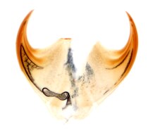
On each foot is a pair of retractable, hardened (sclerotised) chitin claws, which give the taxon its scientific name: Onychophora is derived from the Ancient Greek: ονυχής, onyches, "claws"; and φέρειν, pherein, "to carry". At the base of the claws are three to six spiny "cushions" on which the leg sits in its resting position and on which the animal walks over smooth substrates. The claws are used mainly to gain a firm foothold on uneven terrain. Each claw is composed of three stacked elements, like Russian nesting dolls. The outermost is shed during ecdysis, which exposes the next element in — which is fully formed, so does not need time to harden before it is used.[12] (This distinctive construction identifies many early Cambrian fossils as early offshoots of the onychophoran lineage.[12]) Apart from the pairs of legs, there are three further body appendages, which are at the head and comprise three segments:
The surface of the mandibles is smooth, with no ornamentation.[16] The cuticle in the mandibles (and claws) is distinct from the rest of the body. It has an inner and outer component; the outer component has just two layers (whereas body cuticle has four), and these outer layers (in particular the inner epicuticle) are dehydrated and strongly tanned, affording toughness.[16]

On the third head segment, to the left and right of the mouth, are two openings designated "oral papillae". Within these are a pair of large, heavily internally branched slime glands. These lie roughly in the centre of the body and secrete a sort of milky-white slime, which is used to ensnare prey and for defensive purposes.[17] Sometimes the connecting "slime conductor" is broadened into a reservoir, which can buffer pre-produced slime. The slime glands themselves are probably modified crural glands.[18] All three structures correspond to an evolutionary origin in the leg pairs of the other segments.
Unlike the arthropods, velvet worms do not possess a rigid exoskeleton. Instead, their fluid-filled body cavity acts as a hydrostatic skeleton, similarly to many distantly related soft-bodied animals that are cylindrically shaped, for example sea anemones and various worms. Pressure of their incompressible internal bodily fluid on the body wall provides rigidity, and muscles are able to act against it. The body wall consists of a non-cellular outer skin, the cuticula; a single layer of epidermis cells forming an internal skin; and beneath this, usually three layers of muscle, which are embedded in connective tissues. The cuticula is about a micrometer thick and covered with fine villi. In composition and structure, it resembles the cuticula of the arthropods, consisting of α-chitin and various proteins,[14] although not containing collagen. It can be divided into an external epicuticula and an internal procuticula, which themselves consist of exo- and endo-cuticula. This multi-level structure is responsible for the high flexibility of the outer skin, which enables the velvet worm to squeeze itself into the narrowest crevices. Although outwardly water-repellent, the cuticula is not able to prevent water loss by respiration, and, as a result, velvet worms can live only in microclimates with high humidity to avoid desiccation. The surface of the cuticula is scattered with numerous fine papillae, the larger of which carry visible villi-like sensitive bristles. The papillae themselves are covered with tiny scales, lending the skin a velvety appearance (from which the common name is likely derived). It also feels like dry velvet to the touch, for which its water-repellent nature is responsible. Moulting of the skin (ecdysis) takes place regularly, around every 14 days,[19] induced by the hormone ecdysone. The inner surface of the skin bears a hexagonal pattern.[20] At each moult, the shed skin is replaced by the epidermis, which lies immediately beneath it; unlike the cuticula, this consists of living cells. Beneath this lies a thick layer of connective tissue, which is composed primarily of collagen fibres aligned either parallel or perpendicular to the body's longitudinal axis. The colouration of Onychophora is generated by a range of pigments. The solubility of these pigments is a useful diagnostic character: in all arthropods and tardigrades, the body pigment is soluble in ethanol. This is also true for the Peripatidae, but in the case of the Peripatopsidae, the body pigment is insoluble in ethanol.[21]
Within the connective tissue lie three continuous layers of unspecialised smooth muscular tissue. The relatively thick outer layer is composed of annular muscles, and the similarly voluminous inner layer of longitudinal muscles. Between them lie thin diagonal muscles that wind backward and forward along the body axis in a spiral. Between the annular and diagonal muscles exist fine blood vessels, which lie below the superficially recognisable transverse rings of the skin and are responsible for the pseudo-segmented markings.[9] Beneath the internal muscle layer lies the body cavity. In cross-section, this is divided into three regions by so-called dorso-ventral muscles, which run from the middle of the underbelly through to the edges of the upper side: a central midsection and on the left and right, two side regions that also include the legs.
The body cavity is known as a "pseudocoel", or haemocoel. Unlike a true coelom, a pseudocoel is not fully enclosed by a cell layer derived from the embryonic mesoderm. A coelom is, however, formed around the gonads and the waste-eliminating nephridia.[9] As the name haemocoel suggests, the body cavity is filled with a blood-like liquid in which all the organs are embedded; in this way, they can be easily supplied with nutrients circulating in the blood. This liquid is colourless as it does not contain pigments; for this reason, it serves only a limited role in oxygen transport. Two different types of blood cells (or haemocytes) circulate in the fluid: Amoebocytes and nephrocytes. The amoebocytes probably function in protection from bacteria and other foreign bodies; in some species, they also play a role in reproduction. Nephrocytes absorb toxins or convert them into a form suitable for elimination by the nephridia.
The haemocoel is divided by a horizontal partition, the diaphragm, into two parts: The pericardial sinus along the back and the perivisceral sinus along the belly. The former encloses the tube-like heart, and the latter, the other organs. The diaphragm is perforated in many places, enabling the exchange of fluids between the two cavities. The heart itself is a tube of annular muscles consisting of epithelial tissues, with two lateral openings (ostia) per segment. While it is not known whether the rear end is open or closed, from the front, it opens directly into the body cavity.
Since there are no blood vessels, apart from the fine vessels running between the muscle layers of the body wall and a pair of arteries that supply the antennae, this is referred to as an open circulation. The timing of the pumping procedure can be divided into two parts: Diastole and systole. During diastole, blood flows through the ostia from the pericardial sinus (the cavity containing the heart) into the heart. When the systole begins, the ostia close and the heart muscles contract inwards, reducing the volume of the heart. This pumps the blood from the front end of the heart into the perivisceral sinus containing the organs. In this way, the various organs are supplied with nutrients before the blood finally returns to the pericardial sinus via the perforations in the diaphragm. In addition to the pumping action of the heart, body movements also influence circulation.
Oxygen uptake occurs to an extent via simple diffusion through the entire body surface, with the coxal vesicles on the legs possibly being involved in some species. However, of most importance is gas exchange via fine unbranched tubes, the tracheae, which draw oxygen from the surface deep into the various organs, particularly the heart.
The walls of these structures, which are less than three micrometers thick in their entirety, consist only of an extremely thin membrane through which oxygen can easily diffuse. The tracheae originate at tiny openings, the spiracles, which themselves are clustered together in dent-like recesses of the outer skin, the atria. The number of "tracheae bundles" thus formed is on average around 75 bundles per body segment; they accumulate most densely on the back of the organism.
Unlike the arthropods, the velvet worms are unable to control the openings of their tracheae; the tracheae are always open, entailing considerable water loss in arid conditions. Water is lost twice as fast as in earthworms and forty times faster than in caterpillars.[22] For this reason, velvet worms are dependent upon habitats with high air humidity.

The digestive tract begins slightly behind the head, the mouth lying on the underside a little way from the frontmost point of the body. Here, prey can be mechanically dismembered by the mandibles with their covering of fine toothlets. Two salivary glands discharge via a common conductor into the subsequent "throat", which makes up the first part of the front intestine. The saliva that they produce contains mucus and hydrolytic enzymes, which initiate digestion in and outside the mouth.
The throat itself is very muscular, serving to absorb the partially liquified food and to pump it, via the oesophagus, which forms the rear part of the front intestine, into the central intestine. Unlike the front intestine, this is not lined with a cuticula but instead consists only of a single layer of epithelial tissue, which does not exhibit conspicuous indentation as is found in other animals.
On entering the central intestine, food particles are coated with a mucus-based peritrophic membrane, which serves to protect the lining of the intestine from damage by sharp-edged particles. The intestinal epithelium secretes further digestive enzymes and absorbs the released nutrients, although the majority of digestion has already taken place externally or in the mouth. Indigestible remnants arrive in the rear intestine, or rectum, which is once again lined with a cuticula and which opens at the anus, located on the underside near to the rear end.
In almost every segment is a pair of excretory organs called nephridia, which are derived from coelom tissue. Each consists of a small pouch that is connected, via a flagellated conductor called a nephridioduct, to an opening at the base of the nearest leg known as a nephridiopore. The pouch is occupied by special cells called podocytes, which facilitate ultrafiltration of the blood through the partition between haemocoelom and nephridium.
The composition of the urinary solution is modified in the nephridioduct by selective recovery of nutrients and water and by isolation of poison and waste materials, before it is excreted to the outside world via the nephridiopore. The most important nitrogenous excretion product is the water-insoluble uric acid; this can be excreted in solid state, with very little water. This so-called uricotelic excretory mode represents an adjustment to life on land and the associated necessity of dealing economically with water.
A pair of former nephridia in the head were converted secondarily into the salivary glands, while another pair in the final segment of male specimens now serve as glands that apparently play a role in reproduction.[23]
The entire body, including the stub feet, is littered with numerous papillae: warty protrusions responsive to touch that carry a mechanoreceptive bristle at the tip, each of which is also connected to further sensory nerve cells lying beneath. The mouth papillae, the exits of the slime glands, probably also have some function in sensory perception. Sensory cells known as "sensills" on the "lips" or labrum respond to chemical stimuli and are known as chemoreceptors. These are also found on the two antennae, which seem to be the velvet worm's most important sensory organs.
Except in a few (typically subterranean) species, one simply constructed eye (ocellus) lies behind each antenna, laterally, just underneath the head.[24] This consists of a chitinous ball lens, a cornea and a retina and is connected to the centre of the brain via an optic nerve.[24] The retina comprises numerous pigment cells and photoreceptors; the latter are easily modified flagellated cells, whose flagellum membranes carry a photosensitive pigment on their surface.
The rhabdomeric eyes of the Onychophora are thought to be homologous with the median ocelli of arthropods; this would suggest that the last common ancestor of arthropods may have only had median ocelli.[24] However, the innervation shows that the homology is limited: The eyes of Onychophora form behind the antenna, whereas the opposite is true in arthropods.[25]
Both sexes possess pairs of gonads, opening via a channel called a gonoduct into a common genital opening, the gonopore, which is located on the rear ventral side. Both the gonads and the gonoduct are derived from true coelom tissue.

In females, the two ovaries are joined in the middle and to the horizontal diaphragm. The gonoduct appears differently depending on whether the species is live-bearing or egg-laying. In live-bearing species, each exit channel divides into a slender oviduct and a roomy "womb", the uterus, in which the embryos develop. The single vagina, to which both uteri are connected, runs outward to the gonopore. In egg-laying species, whose gonoduct is uniformly constructed, the genital opening lies at the tip of a long egg-laying apparatus, the ovipositor. The females of many species also possess a sperm repository called the receptacle seminis, in which sperm cells from males can be stored temporarily or for longer periods.
Males possess two separate testes, along with the corresponding sperm vesicle (the vesicula seminalis) and exit channel (the vasa efferentia). The two vasa efferentia unite to a common sperm duct, the vas deferens, which in turn widens through the ejaculatory channel to open at the gonopore. Directly beside or behind this lie two pairs of special glands, which probably serve some auxiliary reproductive function; the rearmost glands are also known as anal glands.[26] A penis-like structure has so far been found only in males of the genus Paraperipatus but has not yet been observed in action.
There are different mating procedures: In some species males deposit their spermatophore directly into the female's genitals opening, while others deposit it on the female's body, where the cuticle will collapse and allowing the sperm cells to migrate into the female. There are also Australian species where the male place their spermatophore on top of their head, which is then pressed against the female's genitals. In these species the head have elaborate structures like spikes, spines, hollow stylets, pits, and depressions, whose purpose is to either hold the sperm and / or assist in the sperm transfer to the female. The males of most species also secrete a pheromone from glands on the underside of the legs to attract females.[27]
Velvet worms live in all tropical habitats and in the temperate zone of the Southern Hemisphere, showing a circumtropical and circumaustral distribution. Individual species are found in Central and South America; the Caribbean islands; equatorial West Africa and Southern Africa; northeastern India;[28][29] Thailand;[30] Indonesia and parts of Malaysia; New Guinea; Australia; and New Zealand.
Fossils have been found in Baltic amber, indicating that they were formerly more widespread in the Northern Hemisphere when conditions were more suitable.[31]
Velvet worms always sparsely occupy the habitats where they are found: They are rare among the fauna which they are a part of.
All extant velvet worms are terrestrial (land-living) and prefer dark environments with high air humidity. They are found particularly in the rainforests of the tropics and temperate zones, where they live among moss cushions and leaf litter, under tree trunks and stones, in rotting wood or in termite tunnels. They also occur in unforested grassland, if there exist sufficient crevices in the soil into which they can withdraw during the day, and in caves.[32] Two species live in caves, a habitat to which their ability to squeeze themselves into the smallest cracks makes them exceptionally well-adapted and in which constant living conditions are guaranteed. Since the essential requirements for cave life were probably already present prior to the settlement of these habitats, this may be described as exaptation.
Some species of velvet worms are able to occupy human-modified land-uses, such as cocoa and banana plantations in South America and the Caribbean, but for others, conversion of rainforests is likely one of the most important threats to their survival (see Conservation).
Velvet worms are photophobic: They are repelled by bright light sources. Because the danger of desiccation is greatest during the day and in dry weather, it is not surprising that velvet worms are usually most active at night and during rainy weather. Under cold or dry conditions, they actively seek out crevices in which they shift their body into a resting state.[33]

The Onychophora forcefully squirt glue-like slime[34] from their oral papillae; they do so either in defense against predators or to capture prey.[35] The openings of the glands that produce the slime are in the papillae, a pair of highly modified limbs on the sides of the head below the antennae. Inside, they have a syringe-like system that, by a geometric amplifier, allows for fast squirt using slow muscular contraction.[36] High speed films show the animal expelling two streams of adhesive liquid through a small opening (50 to 200 microns) at a speed of 3 to 5 m/s (10 to 20 ft/s).[36] The interplay between the elasticity of oral papillae and the fast unsteady flow produces a passive oscillatory motion (30–60 Hz) of the oral papillae.[36] The oscillation causes the streams to cross in mid air, weaving a disordered net; the velvet worms can control only the general direction where the net is thrown.[37]
The slime glands themselves are deep inside the body cavity, each at the end of a tube more than half the length of the body. The tube both conducts the fluid and stores it until it is required. The distance that the animal can propel the slime varies; usually it squirts it about a centimetre,[38] but the maximal range has variously been reported to be ten centimetres,[39] or even nearly a foot,[40] although accuracy drops with range.[41] It is not clear to what extent the range varies with the species and other factors. One squirt usually suffices to snare a prey item, although larger prey may be further immobilised by smaller squirts targeted at the limbs; additionally, the fangs of spiders are sometimes targeted.[41] Upon ejection, it forms a net of threads about twenty microns in diameter, with evenly spaced droplets of viscous adhesive fluid along their length.[38] It subsequently dries, shrinking, losing its stickiness, and becoming brittle.[38] Onychophora eat their dried slime when they can, which is appropriate, because it takes an onychophoran about 24 days to replenish an exhausted slime repository.[41]
The slime can account for up to 11% of the organism's dry weight[41] and is 90% water; its dry residue consists mainly of proteins — primarily a collagen-type protein.[38] 1.3% of the slime's dry weight consists of sugars, mainly galactosamine.[38] The slime also contains lipids and the surfactant nonylphenol. Onychophora are the only organisms known to produce this latter substance.[38] It tastes "slightly bitter and at the same time somewhat astringent".[42] The proteinaceous composition accounts for the slime's high tensile strength and stretchiness.[38] The lipid and nonylphenol constituents may serve one of two purposes. They may line the ejection channel, stopping the slime from sticking to the organism when it is secreted; or they may slow the drying process long enough for the slime to reach its target.[38]

Velvet worms/Onychophora move in a slow and gradual motion that makes them difficult for prey to notice.[41] Their trunk is raised relatively high above the ground, and they walk with non-overlapping steps.[43] To move from place to place, the velvet worm crawls forward using its legs; unlike in arthropods, both legs of a pair are moved simultaneously. The claws of the feet are used only on hard, rough terrain where a firm grip is needed; on soft substrates, such as moss, the velvet worm walks on the foot cushions at the base of the claws.
Actual locomotion is achieved less by the exertion of the leg muscles than by local changes of body length. This can be controlled using the annular and longitudinal muscles. If the annular muscles are contracted, the body cross-section is reduced, and the corresponding segment lengthens; this is the usual mode of operation of the hydrostatic skeleton as also employed by other worms. Due to the stretching, the legs of the segment concerned are lifted and swung forward. Local contraction of the longitudinal muscles then shortens the appropriate segment, and the legs, which are now in contact with the ground, are moved to the rear. This part of the locomotive cycle is the actual leg stroke that is responsible for forward movement. The individual stretches and contractions of the segments are coordinated by the nervous system such that contraction waves run the length of the body, each pair of legs swinging forward and then down and rearward in succession. Macroperipatus can reach speeds of up to four centimetres per second,[41] although speeds of around 6 body-lengths per minute are more typical.[44] The body gets longer and narrower as the animal picks up speed; the length of each leg also varies during each stride.[44]
The brains of Onychophora, though small, are very complex; consequently, the organisms are capable of rather sophisticated social interactions.[45] Behaviour may vary from genus to genus, so this article reflects the most-studied genus, Euperipatoides.[45]
The Euperipatoides form social groups of up to fifteen individuals, usually closely related, which will typically live and hunt together. Groups usually live together; in drier regions an example of a shared home would be the moist interior of a rotting log. Group members are extremely aggressive towards individuals from other logs.[45] Dominance is achieved through aggression and maintained through submissive behaviour.[45] After a kill, the dominant female always feeds first, followed in turn by the other females, then males, then the young.[45]
When assessing other individuals, individuals often measure one another up by running their antennae down the length of the other individual.[45] Once hierarchy has been established, pairs of individuals will often cluster together to form an "aggregate"; this is fastest in male-female pairings, followed by pairs of females, then pairs of males.[45]
Social hierarchy is established by a number of interactions: Higher-ranking individuals will chase and bite their subordinates while the latter are trying to crawl on top of them.[45] Juveniles never engage in aggressive behaviour, but climb on top of adults, which tolerate their presence on their backs.[45] Hierarchy is quickly established among individuals from a single group, but not among organisms from different groups; these are substantially more aggressive and very rarely climb one another or form aggregates.[45] Individuals within an individual log are usually closely related; especially so with males. This may be related to the intense aggression between unrelated females.[45]
Velvet worms are ambush predators, hunting only by night,[41] and are able to capture animals at least their own size, although capturing a large prey item may take almost all of their mucus-secreting capacity.[46] They feed on almost any small invertebrates, including woodlice (Isopoda), termites (Isoptera), crickets (Gryllidae), book/bark lice (Psocoptera), cockroaches (Blattidae), millipedes and centipedes (Myriapoda), spiders (Araneae),[46] various worms, and even large snails (Gastropoda). Depending on their size, they eat on average every one to four weeks.[41] They are considered to be ecologically equivalent to centipedes (Chilopoda). The most energetically favourable prey are two-fifths the size of the hunting onychophoran.[41] Ninety percent of the time involved in eating prey is spent ingesting it; re-ingestion of the slime used to trap the insect is performed while the onychophoran locates a suitable place to puncture the prey, and this phase accounts for around 8% of the feeding time, with the remaining time evenly split between examining, squirting, and injecting the prey.[41] In some cases, chunks of the prey item are bitten off and swallowed; undigestable components take around 18 hours to pass through the digestive tract.[14] Onychophora probably do not primarily use vision to detect their prey; although their tiny eyes do have a good image-forming capacity, their forward vision is obscured by their antennae;[41] their nocturnal habit also limits the utility of eyesight. Air currents, formed by prey motion, are thought to be the primary mode of locating prey; the role of scent, if any, is unclear.[41] Because it takes so long to ingest a prey item, hunting mainly happens around dusk; the onychophorans will abandon their prey at sunrise.[41] This predatory way of life is probably a consequence of the velvet worm's need to remain moist. Due to the continual risk of desiccation, often only a few hours per day are available for finding food. This leads to a strong selection for a low cost-benefit ratio, which cannot be achieved with a herbivorous diet.
Velvet worms literally creep up on their prey, with their smooth, gradual and fluid movement escaping detection.[41] Once they reach their prey, they touch it very softly with their antennae to assess its size and nutritional value. After each poke, the antenna is hastily retracted to avoid alerting the prey.[41] This investigation may last anywhere upwards of ten seconds, until the velvet worm makes a decision as to whether to attack it, or until it disturbs the prey and the prey flees.[41] Hungry Onychophora spend less time investigating their prey and are quicker to apply their slime.[41] Once slime has been squirted, Onychophora are determined to pursue and devour their prey, in order to recoup the energy investment. They have been observed to spend up to ten minutes searching for removed prey, after which they return to their slime to eat it.[41] In the case of smaller prey, they may opt not to use slime at all.[41] Subsequently, a soft part of the prey item (usually a joint membrane in arthropod prey) is identified, punctured with a bite from the jaws, and injected with saliva. This kills the prey very quickly and begins a slower process of digestion.[41] While the onychophoran waits for the prey to digest, it salivates on its slime and begins to eat it (and anything attached to it). It subsequently tugs and slices at the earlier perforation to allow access to the now-liquefied interior of its prey.[41] The jaws operate by moving backwards and forwards along the axis of the body (not in a side-to-side clipping motion as in arthropods), conceivably using a pairing of musculature and hydrostatic pressure.[14] The pharynx is specially adapted for sucking, to extract the liquefied tissue; the arrangement of the jaws about the tongue and lip papillae ensures a tight seal and the establishment of suction.[14] In social groups, the dominant female is the first to feed, not permitting competitors access to the prey item for the first hour of feeding. Subsequently, subordinate individuals begin to feed. The number of males reaches a peak after females start to leave the prey item.[45] After feeding, individuals clean their antennae and mouth parts before re-joining the rest of their group.[45]
Almost all species of velvet worm reproduce sexually. The sole exception is Epiperipatus imthurni, of which no males have been observed; reproduction instead occurs by parthenogenesis.[47] All species are in principle sexually distinct and bear, in many cases, a marked sexual dimorphism: the females are usually larger than the males and have, in species where the number of legs is variable, more legs. The females of many species are fertilized only once during their lives, which leads to copulation sometimes taking place before the reproductive organs of the females are fully developed. In such cases, for example at the age of three months in Macroperipatus torquatus, the transferred sperm cells are kept in a special reservoir, where they can remain viable for longer periods. Fertilization takes place internally, although the mode of sperm transmission varies widely. In most species, for example in the genus Peripatus, a package of sperm cells called the spermatophore is placed into the genital opening of the female. The detailed process by which this is achieved is in most cases still unknown, a true penis having been observed only in species of the genus Paraperipatus. In many Australian species, there exist dimples or special dagger- or axe-shaped structures on the head; the male of Florelliceps stutchburyae presses a long spine against the female's genital opening and probably positions its spermatophore there in this way. During the process, the female supports the male by keeping him clasped with the claws of her last pair of legs. The mating behavior of two species of the genus Peripatopsis is particularly curious. Here, the male places two-millimetre spermatophores on the back or sides of the female. Amoebocytes from the female's blood collect on the inside of the deposition site, and both the spermatophore's casing and the body wall on which it rests are decomposed via the secretion of enzymes. This releases the sperm cells, which then move freely through the haemocoel, penetrate the external wall of the ovaries and finally fertilize the ova. Why this self-inflicted skin injury does not lead to bacterial infections is not yet understood (though likely related to the enzymes used to deteriorate the skin or facilitate the transfer of viable genetic material from male to female). Velvet worms are found in egg-laying (oviparous), egg-live-bearing (ovoviviparous) and live-bearing (viviparous) forms.
A female can have between 1 and 23 offspring per year; development from fertilized ovum to adult takes between 6 and 17 months and does not have a larval stage. This is probably also the original mode of development. Velvet worms have been known to live for up to six years.
The velvet worm's important predators are primarily various spiders and centipedes, along with rodents and birds, such as, in Central America, the clay-coloured thrush (Turdus grayi). In South America, Hemprichi's coral snake (Micrurus hemprichii) feeds almost exclusively on velvet worms.[49] For defence, some species roll themselves reflexively into a spiral, while they can also fight off smaller opponents by ejecting slime. Various mites (Acari) are known to be ectoparasites infesting the skin of the velvet worm. Skin injuries are usually accompanied by bacterial infections, which are almost always fatal.
The South African species Peripatopsis capensis has been inadvertently introduced to Santa Cruz Island in the Galapagos Islands, where it co-occurs with native velvet worms.[50]
The global conservation status of velvet worm species is difficult to estimate; many species are only known to exist at their type locality (the location at which they were first observed and described). The collection of reliable data is also hindered by low population densities, their typically nocturnal behaviour and possibly also as-yet undocumented seasonal influences and sexual dimorphism. To date, the only onychophorans evaluated by the IUCN are:
The primary threat comes from destruction and fragmentation of velvet worm habitat due to industrialisation, draining of wetlands, and slash-and-burn agriculture. Many species also have naturally low population densities and closely restricted geographic ranges; as a result, relatively small localised disturbances of important ecosystems can lead to the extinction of entire populations or species. Collection of specimens for universities or research institutes also plays a role on a local scale.[52] There is a very pronounced difference in the protection afforded to velvet worms between regions: in some countries, such as South Africa, there are restrictions on both collecting and exporting, while in others, such as Australia, only export restrictions exist. Many countries offer no specific safeguards at all. Tasmania has a protection programme that is unique worldwide: one region of forest has its own velvet worm conservation plan, which is tailored to a particular velvet worm species.[52]
In their present forms, the velvet worms are probably very closely related to the arthropods, a very extensive taxon that incorporates, for instance, the crustaceans, insects, and arachnids. They share, among other things, an exoskeleton consisting of α-chitin and non-collagenous proteins; gonads and waste-elimination organs enclosed in true coelom tissue; an open blood system with a tubular heart situated at the rear; an abdominal cavity divided into pericardial and perivisceral cavities; respiration via tracheae; and similar embryonic development. Segmentation, with two body appendages per segment, is also a shared feature.
However, the antennae, mandibles, and oral papillae of velvet worms are probably not homologous to the corresponding features in arthropods; i.e., they probably developed independently.
Another closely related group are the comparatively obscure water bears (Tardigrada); however, due to their very small size, water bears have no need for – and hence lack – blood circulation and separate respiratory structures: shared characteristics that support common ancestry of velvet worms and arthropods.
Together, the velvet worms, arthropods, and water bears form a monophyletic taxon, the Panarthropoda, i.e., the three groups collectively cover all descendants of their last common ancestor. Due to certain similarities of form, the velvet worms were usually grouped with the water bears to form the taxon Protoarthropoda. This designation would imply that both velvet worms and water bears are not yet as highly developed as the arthropods. Modern systematic theories reject such conceptions of "primitive" and "highly developed" organisms and instead consider exclusively the historical relationships among the taxa. These relationships are not as yet fully understood, but it is considered probable that the velvet worms' sister groups form a taxon designated Tactopoda, thus:
Panarthropoda Velvet worms (Onychophora) ![]()
Water bears (Tardigrada) .png)
Arthropods (Arthropoda) 
For a long time, velvet worms were also considered related to the annelids. They share, among other things, a worm-like body; a thin and flexible outer skin; a layered musculature; paired waste-elimination organs; as well as a simply constructed brain and simple eyes. Decisive, however, was the existence of segmentation in both groups, with the segments showing only minor specialisation. The parapodia appendages found in annelids therefore correspond to the stump feet of the velvet worms. Within the Articulata hypothesis developed by Georges Cuvier, the velvet worms therefore formed an evolutionary link between the annelids and the arthropods: worm-like precursors first developed parapodia, which then developed further into stub feet as an intermediate link in the ultimate development of the arthropods' appendages. Due to their structural conservatism, the velvet worms were thus considered "living fossils". This perspective was expressed paradigmatically in the statement by the French zoologist A. Vandel:
Modern taxonomy does not study criteria such as "higher" and "lower" states of development or distinctions between "main" and "side" branches—only family relationships indicated by cladistic methods are considered relevant. From this point of view, several common characteristics still support the Articulata hypothesis — segmented body; paired appendages on each segment; pairwise arrangement of waste-elimination organs in each segment; and above all, a rope-ladder-like nervous system based on a double nerve strand lying along the belly. An alternative concept, most widely accepted today, is the so-called Ecdysozoa hypothesis. This places the annelids and Panarthropoda in two very different groups: the former in the Lophotrochozoa and the latter in the Ecdysozoa. Mitochondrial gene sequences also provide support for this hypothesis.[53] Proponents of this hypothesis assume that the aforementioned similarities between annelids and velvet worms either developed convergently or were primitive characteristics passed unchanged from a common ancestor to both the Lophotrochozoa and Ecdysozoa. For example, in the first case, the rope-ladder nervous system would have developed in the two groups independently, while in the second case, it is a very old characteristic, which does not imply a particularly close relationship between the annelids and Panarthropoda. The Ecdysozoa concept divides the taxon into two, the Panarthropoda into which the velvet worms are placed, and the sister group Cycloneuralia, containing the threadworms (Nematoda), horsehair worms (Nematomorpha) and three rather obscure groups: the mud dragons (Kinorhyncha); penis worms (Priapulida); and brush-heads (Loricifera).
Protostomia Ecdysozoa Panarthropoda
(arthropods, velvet worms, water bears) ![]()
Cycloneuralia
(threadworms, horsehair worms, and others)

![]()
Lophotrochozoa
(annelids, molluscs, and others) ![]()
![]()
![]()
(other Protostomes)
Particularly characteristic of the Cycloneuralia is a ring of "circumoral" nerves around the mouth opening, which the proponents of the Ecdysozoa hypothesis also recognise in modified form in the details of the nerve patterns of the Panarthropoda. Both groups also share a common skin-shedding mechanism (ecdysis) and molecular biological similarities. One problem of the Ecdysozoa hypothesis is the velvet worms' subterminal position of their mouths: Unlike in the Cycloneuralia, the mouth is not at the front end of the body, but lies further back, under the belly. However, investigations into their developmental biology, particularly regarding the development of the head nerves, suggest that this was not always the case, and that the mouth was originally terminal (situated at the tip of the body). This is supported by the fossil record.
The "stem-group arthropod" hypothesis is very widely accepted, but some trees suggest that the onychophorans may occupy a different position; their brain anatomy is more closely related to that of the chelicerates than to any other arthropod.[54] The modern velvet worms form a monophyletic group, incorporating all the descendants of their common ancestor. Important common derivative characteristics (synapomorphies) include, for example, the mandibles of the second body segment and the oral papillae and associated slime glands of the third; nerve strands extending along the underside with numerous cross-linkages per segment; and the special form of the tracheae. By 2011, some 180 modern species, comprising 49 genera, had been described;[55] the actual number of species is probably about twice this. According to more recent study, 82 species of Peripatidae and 115 species of Peripatopsidae have been described thus far. However, among the 197 species, 20 are nomina dubia, due to major taxonomic inconsistencies.[56] The best-known is the type genus Peripatus, which was described as early as 1825 and which, in English-speaking countries, stands representative for all velvet worms. All genera are assigned to one of two families, the distribution ranges of which do not overlap but are separated by arid areas or oceans:

Certain fossils from the early Cambrian bear a striking resemblance to the velvet worms. These fossils, known collectively as the lobopodians, were marine and represent a grade from which arthropods, tardigrades, and Onychophora arose.[62][63] Onychophorans are found in the Cambrian,[64] Ordovician (possibly),[65] Silurian[66] and Pennsylvanian[5][67] periods.
.jpg)
Historically, all fossil Onychophora and lobopods were lumped into the taxon Xenusia, further subdivided by some authors to the Paleozoic Udeonychophora and the Mesozoic/Tertiary Ontonychophora; living Onychophora were termed Euonychophora.[68] Importantly, few of the Cambrian fossils bear features that distinctively unite them with the Onychophora; none can be confidently assigned to the onychophoran crown or even stem group.[69] The exceptions are Hallucigenia and related taxa such as Collinsium ciliosum, which bear distinctly onychophoran-like claws.[62] It is not clear when the transition to a terrestrial existence was made, but it is considered plausible that it took place between the Ordovician and late Silurian—approximately 490 to 430 million years ago—via the intertidal zone.[21]
The low preservation potential of the non-mineralised Onychophora means that they have a sparse fossil record. Stem-group members include Helenodora (Carboniferous), Tertiapatus dominicanus, and Succinipatopsis balticus (Tertiary).[70] A Carboniferous fossil from Montceau-les-Mines, France, Antennipatus possesses clear onychophoran affinities, but its preservation prohibits differentiating between its placement on the stem or crown of the two extant families, or on the onychophoran stem-group.[5] Crown group representatives are known only from amber—there is a single, partial specimen from the Cretaceous,[68] and a more comprehensive record in Eocene deposits from 40 million years ago.[71] However, some of these amber-borne specimens lack slime papillae and separate feet, and thus may belong in the stem group.[69]
The vagaries of the preservation process can make fossils difficult to interpret. Experiments on the decay and compaction of onychophora demonstrate difficulties in interpreting fossils; certain parts of living onychophora are visible only in certain conditions:
More significantly, features seen in fossils may be artefacts of the preservation process: For instance, "shoulder pads" may simply be the second row of legs coaxially compressed onto the body; branching "antennae" may in fact be produced through decay.[72]
{{cite web}}: CS1 maint: url-status (link) Onychophora /ɒnɪˈkɒfərə/ (from Ancient Greek: ονυχής, onyches, "claws"; and φέρειν, pherein, "to carry"), commonly known as velvet worms (due to their velvety texture and somewhat wormlike appearance) or more ambiguously as peripatus /pəˈrɪpətəs/ (after the first described genus, Peripatus), is a phylum of elongate, soft-bodied, many-legged panarthropods. In appearance they have variously been compared to worms with legs, caterpillars, and slugs. They prey upon other invertebrates, which they catch by ejecting an adhesive slime. Approximately 200 species of velvet worms have been described, although the true number of species is likely greater. The two extant families of velvet worms are Peripatidae and Peripatopsidae. They show a peculiar distribution, with the peripatids being predominantly equatorial and tropical, while the peripatopsids are all found south of the equator. It is the only phylum within Animalia that is wholly endemic to terrestrial environments, at least among extant members. Velvet worms are generally considered close relatives of the Arthropoda and Tardigrada, with which they form the proposed taxon Panarthropoda. This makes them of palaeontological interest, as they can help reconstruct the ancestral arthropod. In modern zoology they are particularly renowned for their curious mating behaviours and the bearing of live young in some species.
Onikoforoj aŭ Oniĥoforoj aŭ laŭ la latina scienca nomo Onychophora (el la antikva greka, onyches, "ungoj"; kaj pherein, "porti"), ofte konataj kiel veluraj vermoj (pro ties velureca teksturo kaj iom vormeca aspekto) aŭ pli ambigue kiel peripatus (laŭ la unua priskribita genro, nome Peripatus), estas filumo de longaj, mildkorpaj, multkruraj panartropodoj.[1][2] Laŭ aspekto ili estis diverse komparitaj al vermoj kun kruroj, raŭpoj, kaj limakoj.[3] Ili predas pli malgrandajn animalojn kiel insektoj, kiujn ili kaptas ŝprucinte gluecan ŝlimon. Oni priskribis ĉirkaŭ 200 speciojn, sed la vera nombro de specioj ests tre verŝajne pli granda. La du nunaj familioj de veluraj vermoj estas Peripatedoj kaj Peripatopsedoj. Ili montras pekuliaran distribuon, ĉar peripatedoj estas hegemoniaj ekvatore kaj tropike, dum la peripatopsedoj troviĝas ĉefe sude de ekvatoro. Ĝi estas la nura filumo ene de animaloj kiu estas tute endemia de surteraj medioj.[4] Veluraj vermoj estas konsiderataj proksimaj parencoj de Artropodoj kaj Tardigradoj, kun kiuj ili formas la taksonon Panartropodoj.[5] Tio havigas al ili paleontologian intereson, ĉar ili povas helpi rekonstrui la praŭlaron de artropodoj. En moderna zoologio, ili estas partikulare renomataj pro ilia kurioza kutimaro por pariĝado kaj por idoportado.
Onikoforoj aŭ Oniĥoforoj aŭ laŭ la latina scienca nomo Onychophora (el la antikva greka, onyches, "ungoj"; kaj pherein, "porti"), ofte konataj kiel veluraj vermoj (pro ties velureca teksturo kaj iom vormeca aspekto) aŭ pli ambigue kiel peripatus (laŭ la unua priskribita genro, nome Peripatus), estas filumo de longaj, mildkorpaj, multkruraj panartropodoj. Laŭ aspekto ili estis diverse komparitaj al vermoj kun kruroj, raŭpoj, kaj limakoj. Ili predas pli malgrandajn animalojn kiel insektoj, kiujn ili kaptas ŝprucinte gluecan ŝlimon. Oni priskribis ĉirkaŭ 200 speciojn, sed la vera nombro de specioj ests tre verŝajne pli granda. La du nunaj familioj de veluraj vermoj estas Peripatedoj kaj Peripatopsedoj. Ili montras pekuliaran distribuon, ĉar peripatedoj estas hegemoniaj ekvatore kaj tropike, dum la peripatopsedoj troviĝas ĉefe sude de ekvatoro. Ĝi estas la nura filumo ene de animaloj kiu estas tute endemia de surteraj medioj. Veluraj vermoj estas konsiderataj proksimaj parencoj de Artropodoj kaj Tardigradoj, kun kiuj ili formas la taksonon Panartropodoj. Tio havigas al ili paleontologian intereson, ĉar ili povas helpi rekonstrui la praŭlaron de artropodoj. En moderna zoologio, ili estas partikulare renomataj pro ilia kurioza kutimaro por pariĝado kaj por idoportado.
Los onicóforos (Onychophora, del griego onykhos, "uña" y phorós, "portador") constituyen un filo de ecdisozoos terrestres, con ~180 especies existentes, cuya existencia se conoce desde el periodo Cámbrico, hace más de 515 millones de años. Fueron descritos por primera vez por Lansdown Guilding en 1826, quien pensó que eran moluscos.
El nombre Onychophora, deriva del griego, y significa literalmente "portadores de uñas", ya que cada una de sus muchas extremidades termina en un par de garras. En los libros de texto reciben el nombre de "gusanos aterciopelados" (del inglés velvet worms) pero en el idioma español solo se les llama gusanos o se les confunde con babosas. De hecho, han sido comparados a gusanos con patas, orugas de mariposas y babosas.[1]
Estos organismos segmentados tienen ojos pequeños, antenas, múltiples pares de patas o lobopodios –que no son realmente articulados, y las glándulas de la goma adhesiva (por error llamada baba). Es posible encontrarlos en regiones tropicales y en el Hemisferio Sur. Son cazadores de animales pequeños como los insectos y arácnidos, que atrapan expulsando un moco adhesivo.[2] Se conocen unas 180 especies actuales.[3]
Las dos familias existentes de gusanos aterciopelados son Peripatidae[4] y Peripatopsidae.[5]
Ellos muestran una distribución peculiar, donde los peripátidos son predominantemente ecuatoriales y tropicales, mientras que los peripatópsidos se encuentran los continentes del sur de clima templado.[6] Hoy se sabe que tal distribución a nivel mundial se originó a partir de la ruptura del antiguo supercontinente Pangea, que empezó a fracturarse hace unos 200 millones de años.[7][8]
A pesar de su escasez actual, en la historia de la biología evolutiva se les ha prestado mucha atención debido a su importancia al presentar características que hacían pensar que eran un intermedio evolutivo entre anélidos y artrópodos. Antiguamente se les consideraba parte del taxón Tracheata.[9] pero en la actualidad no se considera que estén emparentados con anélidos sino que forman parte del taxón Panarthropoda.[10] junto a tardígrados y artrópodos. Según los análisis moleculares constituyen el grupo hermano de los artrópodos.[11][12][13]
La locomoción y el sostén corporal se basan en un esqueleto hidrostático, como ocurre en los anélidos, no en un exoesqueleto de piezas articuladas. A diferencia de los artrópodos, los onicóforos tienen un celoma muy desarrollado, que ocupa buena parte del volumen corporal. Músculos y válvulas trabajan trasladando y comprimiendo el líquido intersticial a través de los compartimentos del hemocele, incluidos los lobópodos, y es así como se mueven los órganos, se deforma el cuerpo y se hace posible la locomoción.[4]
El aspecto general de los onicóforos es el de gusanos o babosas con patas. De hecho, su descubridor Guilding los asignó en 1825 dentro del filo Mollusca.
Miden entre 1,4 y 16 cm, aunque algunos llegan a los 25 cm. El cuerpo es alargado, vermiforme, cilíndrico y aparece dotado de entre 14 y 43 pares de apéndices locomotores (varía con la especie y el sexo) llamados lobópodos. Estos no pueden calificarse como patas, sino que son mamelones sin articulación alguna que se adelantan por turno, siguiendo ondas.[1] Cada uno termina en dos pequeñas uñas, que es a lo que alude el nombre del grupo. La arquitectura corporal deriva de una forma segmentada, como los artrópodos, pero se ha perdido en el curso de la evolución. No existe una segmentación externa visible, pero sí pequeñas arrugas transversales[14]
La superficie de su cuerpo está cubierta por bandas transversales de pequeños tubérculos (los mayores presentan setas sensoriales y escamas muy pequeñas). La disposición de los tubérculos parece mostrar una metamerización que, externamente, es en realidad inexistente. Además, presentan una textura aterciopelada de la que procede su nombre común en otros idiomas como el inglés, velvet worms ("gusanos aterciopelados").
Son muy variados en cuanto a color; siendo lo más común un gris o pardo muy oscuros, pero también los hay de colores vivos, amarillos, más o menos anaranjados, rojos, verdes, negros o incluso azules, pudiendo presentar las papilas un aspecto iridiscente. Se conoce al menos una especie blanca, habitante de cuevas, y los individuos recién nacidos pueden ser blanquecinos antes de adquirir los colores adultos.[15]
La cabeza, que no aparece bien diferenciada del tronco, porta tres pares de apéndices.
En primer lugar un par de antenas, en posición más anterior que superior, gruesas y a veces muy largas; detrás de cada una de ellas hay un pequeño ojo simple, dotado de un cristalino esférico.
El segundo par de apéndices son las papilas orales, retráctiles, situadas por debajo de las antenas y apuntando hacia adelante; son de función sensorial y además escupen un líquido pegajoso que emplean en la captura de sus presas y defenderse.
La boca se encuentra en posición ventral en la base de una depresión. Dentro de ellas y escondidas bajo sus labios se encuentra el tercer par de apéndices, las mandíbulas. Como en los artrópodos, y a diferencia de lo que ocurre en vertebrados la dinámica de la boca es lateral.[14] Dispone la boca de un diente dorsal y de un par de glándulas salivares (derivadas de nefridios).
Cada lobópodo es una evaginación del la pared del cuerpo en forma de corta protuberancia cónica terminada en un par de uñas (de donde procede el nombre del phylum, Onychophora). Carece de articulación con el cuerpo por lo que no se trata de verdaderas patas. Aunque el número de pies puede variar considerablemente entre especies, su estructura es básicamente similar. La rigidez es proporcionada por la presión hidrostática del contenido de sus fluidos, y el movimiento generalmente se obtiene al estirar y contraer todo el cuerpo del animal. Sin embargo, cada apéndice también puede acortarse y doblarse debido a los músculos internos.[16]
La pared del cuerpo está organizada de forma semejante a la de los Anélidos, en tres capas: cutícula, epidermis y capas musculares.
La capa externa está formada por una cutícula quitinosa muy delgada (apenas 1 µm de espesor) y flexible en todas sus partes, sin que exista, como en los artrópodos, distinción entre áreas engrosadas (escleritos) y áreas de articulación.[2] Además, no basta para impermeabilizar la superficie corporal ni para constituir exoesqueleto rígido. Esta circunstancia condiciona la ecología y a la mecánica corporal de estos animales. El organismo de los onicóforos no es capaz de una buena regulación fisiológica del equilibrio hídrico, de manera que, como tantos otros filos de origen acuático, restringen su existencia a ambientes muy húmedos. Además son de hábito nocturno, saliendo solo en las horas en las que baja la temperatura y hay menos peligro de desecación.[15] Por otro lado, al carecer de exoesqueleto rígido les faculta para comprimir mucho su cuerpo y poder atravesar por ranuras muy estrechas.
La epidermis consta de células vivas, a diferencia de la cutícula. Debajo de la epidermis hay una gruesa capa de tejido conectivo, que se compone principalmente de fibras de colágeno.
Por debajo del tejido conectivo anteriormente mencionado hay tres capas de tejido muscular liso.
La cavidad general del cuerpo es, en realidad, un hemocele, es decir, sirve de cavidad circulatoria para la hemolinfa. La estructura en senos del hemocele es semejante a la de los Insectos: seno pericárdido, seno periintestinal medio, seno perineural ventral medio y dos senos ventrolaterales,
El verdadero celoma se ha reducido a las cavidades gonadales y a sacos relacionados con los nefridios.
Son glándulas situadas en unas aberturas denominadas "papilas orales". Dichas glándulas secretan una especie de baba blanca y lechosa, usada para la caza de presas y para la defensa. Se sitúan en el tercer segmento de la cabeza.
Ambos sexos presentan glándulas crurales que se cree que desempeñan algún papel en la reproducción. Estas se encuentran en un par o todos los pares de lobópodos. Se abren en el extremo de una papila cerca del nefridioporo [Barnes, 1977].
El tubo digestivo comienza con una boca situada en posición ventral. Dispone en su interior de un par de mandíbulas en forma de pinza de dinámica lateral (como en Artrópodos y al contrario que Vertebrados). Tras ella se encuentra un diente dorsal. y un par de glándulas salivares. Éstas son nefridios modificados y su secreción es digestiva, permitiendo licuar parcialmente a la presa en el interior de su exoesqueleto antes de ser succionados sus jugos.
El siguiente tramo es un intestino anterior revestido de quitina y formado por una faringe y un esófago.
A continuación se encuentra el gran intestino recto, con funciones de digestión y absorción, no revestido por quitina.
El intestino posterior forma asas hacia adelante sobre el intestino y termina en un ano situado en el extremo del cuerpo.[17]
Presentan un sistema circulatorio abierto.
El único vaso es un corazón tubular situado en el interior de un seno pericárdico en posición dorsal. El corazón está abierto por los extremos y presenta un par de aberturas (ostia) en cada segmento. El corazón propulsa la hemolinfa hacia delante, haciéndola circular por los senos del hemocele. Los senos se comunican entre sí gracias a la presencia de orificios.[18]
La sangre es incolora y presenta amebocitos [19] fagocitarios. No obstante, recientemente se ha comprobado que al menos dos especies presentan el pigmento respiratorio hemocianina. Además, esta hemocianina es homóloga de la que presentan los Artrópodos, lo que refuerza la pertenencia de los onicóforos al taxón Panartropoda.
Para respirar los onicóforos han desarrollado un sistema traqueal que se abre al exterior por espiráculos diminutos

distribuidos en gran número a lo largo del cuerpo entre las bandas de tubérculos. El espiráculo abre paso a una fosa traqueal terminada en un atrio del que parte multitud de tráqueas no ramificadas (al contrario que las de Insectos) cada una de las cuales alcanza directamente la parte a la que está destinada.
A diferencia de lo que ocurre en insectos, los espiráculos no se cierran nunca, careciendo del mecanismo para su control.[20] Su estructura indica que este sistema traqueal no es homólogo del de los arácnidos e insectos.[15] Al no poder cerrar los espiráculos, los onicóforos se deshidratan muy rápidamente en ambiente seco, lo que les limita a hábitats húmedos, bajo hojarasca o lugares semejantes y a horario nocturno.
Presentan un par de nefridios por segmento alojados en los senos ventrolaterales del hemocele. Están formados por un saco terminal (vestigio del celoma) que aloja al nefridio propiamente dicho, constituido por el embudo y el nefrostomo ciliados. En la parte distal forma una vejiga contráctil antes del nefridioporo.[21]
El par de nefridioporos se encuentran en la cara interna de la base de cada lobópodo, salvo en los segmentos 4º y 5º, en los que se localizan sobre un tubérculo algo más distal.[22]
Se desconoce la naturaleza de la secreción excretora.
El primer par de nefridios se ha transformado en glándulas salivares. El último par de nefridios de las hembras se han transformado en los gonoductos,
Es un típico sistema nervioso en escalera propio de los Protostomas. Está formado por un cerebro bilobulado bien desarrollado situado sobre el esófago y dos cordones nerviosos ventrales. Hasta el cerebro llega la información sensorial de tentáculos y ojos y envía nervios a boca y mandíbulas. Los cordones nerviosos presentan engrosamiento ganglionares de los que partes comisuras que les unen entre sí y nervios hacia la pared del cuerpo.[23]
Presentan un par de pequeños ojos en la base de cada antena. Cada ojo tiene un gran cristalino esférico y, tras él, una retina bien desarrollada.[24] Este ojo no puede formar imagen por lo que solo les sirve para detectar intensidad lumínica y controlar su ritmo circadiano.
Repartido en la superficie de su cuerpo, especialmente en los tubérculos, hay células sensoriales.
Los ovarios son órganos alargados y fusionados. Se encuentran en la parte posterior en posición dorsal. Cada ovario presentan un conducto procedente de la modificación de un nefridio que presenta un receptáculo seminal y un útero. Los dos úteros se unen y salen al exterior en un gonoporo común que desemboca en la región posteroventral del animal.
Los machos presentan un par de testículos alargados no fusionados y vías genitales complejas. Al igual que en las hembras, los dos conductos se unen en un conducto y salen al exterior por un único gonoporo muy semejante. Los espermatozoides se forman en el conducto común y se encuentran encerrados en espermatóforos de 1 mm de longitud rodeados por una cubierta quitinosa.
Son dioicos (con sexos separados) y con dimorfismo sexual en cuanto al tamaño (siendo las hembras notablemente más grandes) y número de lobópodos (las hembras pueden tener más).
La mayoría de las especies son ovovivíparas; unas pocas son auténticamente vivíparas (la madre nutre al embrión a través de órganos equivalentes a una placenta); solo los géneros Ooperipatus y Symperipatus son ovíparos.[15] Una especie presenta solamente hembras y se reproduce por partenogénesis.
Se cree que las glándulas crurales, presentes en ambos sexos, desempeñan algún papel en la reproducción.
En el género ovovivíparo sudafricano Peripatopsis -que carece de receptáculos seminales- no hay una fecha de reproducción determinada ni se buscan machos y hembras. Cuando se encuentran, el macho trepa por el cuerpo de la hembra y deposita sobre ella un espermatóforo en cualquier lugar, sea en el dorso o en los lados. De esta manera, una hembra puede acumular los de varios machos distintos. Los espermatóforos estimulan a los amebocitos de la hemolinfa para que disuelvan el tegumento y penetren hasta el hemocele (impregnación hipodérmica), por donde circulan hasta llegar hasta los ovarios donde fecundan a los óvulos.[25]
Se desconoce como realizan la transferencia del esperma las especies en las que las hembras presentan receptáculos seminales.
En los onicóforos ovíparos la segmentación es superficial.
En onicóforos ovovivíparos el huevo se desarrolla en el interior del útero.
En onicóforos vivíparos los huevos tiene poco vitelo, son pequeños y la segmentación es superficial (holoblástica). La hembra produce una secreción uterina nutritiva que recoge el embrión mediante una membrana que hace el papel de placenta.
Los onicóforos se encuentran en climas tropicales húmedos,[26][27][28][29][30][31][32] aunque hay algunas especies australasiáticas y sudamericanas de clima templado.[33][34]
Poseen hábitos nocturnos y viven en ambientes oscuros y normalmente húmedos. Se les encuentra en la hojarasca, bajo piedras, entre las hojas de bromelias y también en troncos en descomposición y grietas del suelo, cuevas, orillas de los ríos y vagando en el exterior solo por la noche.[35][36][37]
En las épocas adversas (tiempo seco o frío invernal) permanecen inactivos refugiados en lugares protegidos.
Son animales depredadores, que cazan activamente saltamontes, termitas, y otros pequeños invertebrados, a menudo más grandes que ellos,[38][39] pegándolos al sustrato con la secreción adhesiva que expulsan desde sus papilas orales. Arrojan certeramente dicha secreción de naturaleza proteica en estado líquido a distancias de hasta 30 o 40 cm, la cual en contacto con el aire se coagula, inmovilizando a la presa.[40] El onicóforo se acerca hasta ella y roe su pared corporal, inyectando enzimas digestivas y sorbiendo la papilla semidigerida resultante, un modo de alimentación como el que observamos frecuentemente en arácnidos.[41]
El cuerpo no toca el sustrato, siendo completamente soportados por los lobópodos que se encuentran en posición más ventral que los parápodos de los Anélidos.
Se desplazan mediante la propagación de lentas ondas de contracción/extensión del cuerpo. Durante la extensión los dos lobópodos se elevan y avanzan.
Son animales nocturnos forzados, entre otros motivos, porque se deshidratan en muy pocas horas en ambiente seco.
Cuando se siente molestado, los onicóforos se defienden segregando el limo adhesivo contra el atacante hasta una distancia de 50 cm.
Los onicóforos son uno de los grupos actuales con fósiles conocidos de mayor antigüedad. Están representados en la fauna del Cámbrico inferior de Burgess Shale (datada en 505 millones de años) por al menos dos especies, Aysheaia pedunculata y Hallucigenia sparsa, y es posible que Xenusion, del Precámbrico terminal (datada en 540 millones de años), también sea un onicóforo.
Los fósiles del Paleozoico inferior son todos marinos y dotados de escleritos (piezas esqueléticas de cutícula endurecida) sobre los lobópodos. En Hallucigenia los escleritos son largas espinas rígidas que dieron origen a una interpretación errónea, popularizada luego por Stephen Jay Gould, que las veía como zancos, confundiendo también los extremos anterior y posterior del cuerpo.
Fuera de ese rasgo la anatomía de los onicóforos es sorprendentemente conservadora, lo que los convierte en ejemplos perfectos de lo que la literatura popular llama fósiles vivientes.
El fósil más antiguo de un onicóforo terrestre, Helenodora, es de Mazon Creek, un importante yacimiento cerca de Chicago. Está datado en el Pensilvaniense y no se distingue morfológicamente de los actuales. Se han recuperado fósiles en ámbar del Cretácico de Birmania (Birmania), así como del Terciario Inferior de La Española y de la Región báltica. En los dominicanos se observa la secreción adhesiva de las papilas orales, lo que demuestra que el carácter es muy antiguo.
Los onicóforos son interpretados actualmente como parientes próximos de los artrópodos, pero externos a este grupo.
Todos los onicóforos habitan regiones tropicales o templadas meridionales. La familia Peripatidae vive en América tropical y Antillas, Gabón, Himalaya y sudeste de Asia. La familia Peripatopsidae habita en Chile, Sudáfrica, Australia-Nueva Guinea y Nueva Zelanda. En todos los casos, solo se han encontrado en determinadas áreas, no en regiones extensas. Esta distribución se corresponde con la del paleocontinente de Gondwana, lo que hace pensar que ahí debe situarse el centro de su radiación evolutiva.
La existencia de fósiles en el ámbar báltico extienden la distribución del grupo a Eurasia, fuera de Gondwana, mostrando que, cualquiera que fuera su centro evolutivo, el grupo ha tenido en el pasado una distribución más amplia.
Con Anélidos:
Con Artrópodos:
Existen unas 165 especies[3] repartidas en 10 géneros y dos familias.
El grupo ha recibido distintas categorías taxonómicas y ubicaciones en la clasificación zoológica. En el pasado fueron tratados a menudo como una clase dentro del filo Arthropoda. Actualmente se les incluye con la categoría de filo dentro del superfilo Panarthropoda, donde además de los artrópodos se incluyen también los tardígrados.
Hay dos familias, Peripatidae y Peripatopsidae. Los Peripatidae se encuentran en torno al Caribe, en el Norte de Sudamérica, en África Ecuatorial, en Assam y en el Sudeste Asiático, siempre con áreas limitadas y dispersas. Los Peripatopsidae presentan un área igualmente dispersa, pero más meridional, con localidades en Chile, Sudáfrica, Australia y Tasmania, Nueva Guinea y también Nueva Zelanda.
|isbn= incorrecto (ayuda). |número-autores= (ayuda) Los onicóforos (Onychophora, del griego onykhos, "uña" y phorós, "portador") constituyen un filo de ecdisozoos terrestres, con ~180 especies existentes, cuya existencia se conoce desde el periodo Cámbrico, hace más de 515 millones de años. Fueron descritos por primera vez por Lansdown Guilding en 1826, quien pensó que eran moluscos.
El nombre Onychophora, deriva del griego, y significa literalmente "portadores de uñas", ya que cada una de sus muchas extremidades termina en un par de garras. En los libros de texto reciben el nombre de "gusanos aterciopelados" (del inglés velvet worms) pero en el idioma español solo se les llama gusanos o se les confunde con babosas. De hecho, han sido comparados a gusanos con patas, orugas de mariposas y babosas.
Estos organismos segmentados tienen ojos pequeños, antenas, múltiples pares de patas o lobopodios –que no son realmente articulados, y las glándulas de la goma adhesiva (por error llamada baba). Es posible encontrarlos en regiones tropicales y en el Hemisferio Sur. Son cazadores de animales pequeños como los insectos y arácnidos, que atrapan expulsando un moco adhesivo. Se conocen unas 180 especies actuales.
Las dos familias existentes de gusanos aterciopelados son Peripatidae y Peripatopsidae.
Ellos muestran una distribución peculiar, donde los peripátidos son predominantemente ecuatoriales y tropicales, mientras que los peripatópsidos se encuentran los continentes del sur de clima templado. Hoy se sabe que tal distribución a nivel mundial se originó a partir de la ruptura del antiguo supercontinente Pangea, que empezó a fracturarse hace unos 200 millones de años.
A pesar de su escasez actual, en la historia de la biología evolutiva se les ha prestado mucha atención debido a su importancia al presentar características que hacían pensar que eran un intermedio evolutivo entre anélidos y artrópodos. Antiguamente se les consideraba parte del taxón Tracheata. pero en la actualidad no se considera que estén emparentados con anélidos sino que forman parte del taxón Panarthropoda. junto a tardígrados y artrópodos. Según los análisis moleculares constituyen el grupo hermano de los artrópodos.
Küüsikloomad ehk onühhofoorid (Onychophora) on loomade hõimkond.
Küüsikloomad on segmenteerunud, üksnes maismaal elavad ussid, kes sarnanevad nii lülijalgsete kui ka rõngussidega. Küüsikloomi on teada 2 sugukonda umbes 200 liigiga.
Veel hiljuti vaadeldi küüsikloomi vaherühmana rõngusside ja trahheedega hingavate lülijalgsete vahel. Tänapäeval loetakse neid üldjuhul mitte lülijalgsete klassiks, vaid iseseisvaks loomariigi haruks, mis eraldus rõngusside ja lülijalgsete ühistest esivanematest. Paljud küüsikloomade tunnused esinesid ka maismaalülijalgsete eellastel.[1]
See on ainus loomade hõimkond, kuhu kuuluvad ainult maismaalised liigid. Siiski vajavad nad eluks kõrget õhuniiskust ja väldivad vähimatki tuult, mis suurendaks aurumist. Nende lemmikelupaik on niiskete troopiliste ja lähistroopiliste metsade metsakõdu, õõnsused puude ja kivide all või samblapadjandid kividel. Kuivemates kohtades kaevavad nad end pinnasesse.[1]
Inglise uurijad Manton ja Ramsey tegid katse Aafrika küüsikloomaga Peripatopsis moseley. Nad jätsid need loomad kambrisse, kust lasti läbi kuiva õhku kiirusega 7 m/s. Veerand tunniga kaotasid nood oma kaalust 39%. Näiteks sama kaalu ja umbes sama kujuga vihmaussid, kes on samuti tuntud niiskusearmastajad, kaotasid üksnes 18% kaalust.[1]
Nende liikumiskiirus on kümmekond kehapikkust ehk 40–70 cm minutis. Nad liiguvad oma arvukate jalgade abil, mida on 14–40 paari. [1]
Paljud küüsikloomad võivad liikuda pinnases. Seda teevad nad oma keha eri osade paksuse ja pikkuse muutmise teel nagu vihmaussidki. [1]
Nad söövad teisi loomi, näiteks väikesi putukaid ja ämblikke. Söökloomadele heidavad nad kleepuva lima joa, mis selle oma kohale kleebib. Niimoodi täissülitatud loomi on juba kerge tabada. Vaatamata rahumeelsele välimusele ja suurele aeglusele on küüsikloomad tegelikult aktiivsed ja aplad röövloomad.[1]
Kuigi küüsikloomad on tüüpilised maismaaloomad, kes hukkuvad meres ega talu niiskuse puudust õhus, näitab nende levik küüsikloomade rühma suurt vanust ja laialdast levikut minevikus. Ainult endise laia levikuga saab seletada lähedaste vormide esinemist üksteisest väga kaugel. Nii on Lõuna-Aafrikas elav Opisthopatus lähedane Tšiilis elavale Metaperipatusele, aga Lõuna-Aafrika perekond Peripatopsis on lähedane Uus-Meremaal elavale Paraperipatusele.[1]
Kuid küüsikloomad põlvnevad kindlasti mereloomadest. Juba 1911 avastas Walcott Põhja-Ameerikas Kesk-kambriumi lademeist küüsiklooma sarnase looma, mis sai nimeks Aysheaia pedunculata. 1930 tõendas Hutchinson, et see on küüsikloomadele väga lähedane, kuid ilmselt mereloom.[1]
Küüsikloomadel puudub vähimgi majanduslik tähtsus, kuid nende uurimine lubab mõista muutusi, mis toimusid lülijalgsetega nende üleminekul vee-elult maismaa-elule. Sellepärast pakuvad küüsikloomad bioloogidele suurt huvi.[1]
Küüsikloomad ehk onühhofoorid (Onychophora) on loomade hõimkond.
Küüsikloomad on segmenteerunud, üksnes maismaal elavad ussid, kes sarnanevad nii lülijalgsete kui ka rõngussidega. Küüsikloomi on teada 2 sugukonda umbes 200 liigiga.
Veel hiljuti vaadeldi küüsikloomi vaherühmana rõngusside ja trahheedega hingavate lülijalgsete vahel. Tänapäeval loetakse neid üldjuhul mitte lülijalgsete klassiks, vaid iseseisvaks loomariigi haruks, mis eraldus rõngusside ja lülijalgsete ühistest esivanematest. Paljud küüsikloomade tunnused esinesid ka maismaalülijalgsete eellastel.
See on ainus loomade hõimkond, kuhu kuuluvad ainult maismaalised liigid. Siiski vajavad nad eluks kõrget õhuniiskust ja väldivad vähimatki tuult, mis suurendaks aurumist. Nende lemmikelupaik on niiskete troopiliste ja lähistroopiliste metsade metsakõdu, õõnsused puude ja kivide all või samblapadjandid kividel. Kuivemates kohtades kaevavad nad end pinnasesse.
Inglise uurijad Manton ja Ramsey tegid katse Aafrika küüsikloomaga Peripatopsis moseley. Nad jätsid need loomad kambrisse, kust lasti läbi kuiva õhku kiirusega 7 m/s. Veerand tunniga kaotasid nood oma kaalust 39%. Näiteks sama kaalu ja umbes sama kujuga vihmaussid, kes on samuti tuntud niiskusearmastajad, kaotasid üksnes 18% kaalust.
Nende liikumiskiirus on kümmekond kehapikkust ehk 40–70 cm minutis. Nad liiguvad oma arvukate jalgade abil, mida on 14–40 paari.
Paljud küüsikloomad võivad liikuda pinnases. Seda teevad nad oma keha eri osade paksuse ja pikkuse muutmise teel nagu vihmaussidki.
Nad söövad teisi loomi, näiteks väikesi putukaid ja ämblikke. Söökloomadele heidavad nad kleepuva lima joa, mis selle oma kohale kleebib. Niimoodi täissülitatud loomi on juba kerge tabada. Vaatamata rahumeelsele välimusele ja suurele aeglusele on küüsikloomad tegelikult aktiivsed ja aplad röövloomad.
Kuigi küüsikloomad on tüüpilised maismaaloomad, kes hukkuvad meres ega talu niiskuse puudust õhus, näitab nende levik küüsikloomade rühma suurt vanust ja laialdast levikut minevikus. Ainult endise laia levikuga saab seletada lähedaste vormide esinemist üksteisest väga kaugel. Nii on Lõuna-Aafrikas elav Opisthopatus lähedane Tšiilis elavale Metaperipatusele, aga Lõuna-Aafrika perekond Peripatopsis on lähedane Uus-Meremaal elavale Paraperipatusele.
Kuid küüsikloomad põlvnevad kindlasti mereloomadest. Juba 1911 avastas Walcott Põhja-Ameerikas Kesk-kambriumi lademeist küüsiklooma sarnase looma, mis sai nimeks Aysheaia pedunculata. 1930 tõendas Hutchinson, et see on küüsikloomadele väga lähedane, kuid ilmselt mereloom.
Küüsikloomadel puudub vähimgi majanduslik tähtsus, kuid nende uurimine lubab mõista muutusi, mis toimusid lülijalgsetega nende üleminekul vee-elult maismaa-elule. Sellepärast pakuvad küüsikloomad bioloogidele suurt huvi.
Onychophora (grezieratik, onykhos, "azkazala" eta phoros, "eraman") animalia ornogabe filum bat da, ileguri itsurarekin, beldarren antzarekin. Kanbriar garaitik existitu direla uste da eta artropodoekin zuzenki loturik daude, baina hauek ez bezala, ez daude artikulaturik. Hala ere, zoologo batzuek artropodoetan sartzen dituzte. 165 espezie ezagutzen dira.
Lehorreko animaliak dira, zizarearen antzekoak, baina neurriz haiek baino askoz txikiagoak. Burua soinetik garbi bereizten ez dena, ahoa azpialdean duena eta intsektuen antzeko antenekikoa dute. Gorputzaren alde banatara hanka asko, giltzatuak eta erpedunak dituzte.
Ekuatorez hegoaldeko lurralde bero eta epeletan bizi dira, leku heze eta ilunetan batez ere. Egur ustela eta artropodo txikiak jaten dituzte.
Onychophora (grezieratik, onykhos, "azkazala" eta phoros, "eraman") animalia ornogabe filum bat da, ileguri itsurarekin, beldarren antzarekin. Kanbriar garaitik existitu direla uste da eta artropodoekin zuzenki loturik daude, baina hauek ez bezala, ez daude artikulaturik. Hala ere, zoologo batzuek artropodoetan sartzen dituzte. 165 espezie ezagutzen dira.
Käsnäjalkaiset (Onychophora) on eläinkunnan pääjakso. Joskus se on luettu myös niveljalkaisten alajaksoksi. Ensimmäinen elävä käsnäjalkainen löydettiin vasta vuonna 1825.[1][2]
Käsnäjalkaiset ovat matomaisia, 5–150 mm pitkiä eläimiä, ja niillä on 13–43 kynnellistä käsnämäistä raajaparia.[3] Lajeja tunnetaan noin 180, ne elävät lähinnä trooppisissa ympäristöissä ja enimmäkseen eteläisellä pallonpuoliskolla.[3] Nykylajisto käsittää kaksi heimoa, Peripatidae ja Peripatopsidae.[3][4]
Vanhimmat fossiilit ovat jo kambrikaudelta.[5] Näitä eläimiä pidetään "elävinä fossiileina", jotka ovat sukua ensimmäisille käveleville eläimille. Lajit ovat alkeellisesti jaokkeisia: vain pää on jaokkeinen. Pään ensimmäisessä jaokkeessa on kaksi tuntosarvea ja yleensä silmäpari, toisessa jaokkeessa suu.[3] Ihossa on hieno samettimainen karvoitus.[3] Ruumiinontelo ja muut sisäelimet kuten verta pumppaava sydän ovat kuin niveljalkaisilla.[3] Erityselimet ovat samanlaiset kuin nivelmadoilla.[3] Jaloissa on kitiinikynnet ("kaviot").[3][2] Aivot muistuttavat hämähäkkien aivoja, mutta molekyylisystematiikka ei tue sukulaisuutta näihin. Käsnäjalkaiset hengittävät ihon kautta ja tarvitsevat kostean ympäristön, jotteivät kuivuisi.[3] Ne ovat siirtyneet vedestä maalle luultavasti ordovikikauden aikana.[3] Väriltään ne ovat yleensä sinisiä, mustia, vihreitä tai oransseja.[3]
Käsnäjalkaiset ovat petoja jotka syövät ravinnokseen pieniä selkärangattomia, kovakuoriaisia, termiittejä ja muita hyönteisiä sekä etanoita ja matoja.[3] Ne voivat käydä jopa useita kertoja itseään suuremman saaliin kimppuun. Jotkin lajit tekevät uhrinsa puolustuskyvyttömiksi ruiskuttamalla näiden päälle tahmeaa limaa.[3] Eläimet lisääntyvät niin, että uros sijoittaa tuntosarvillaan ja eturaajoillaan spermapaketin naaraan peräpäähän.[3]
Käsnäjalkaisia muistuttavia fossiililajeja ovat ehkä myöhäisprekambrinen Xenusion sekä kambrikautiset Aysheaia ja Hallucigenia.
Uudenseelanninkäsnäjalkainen[6] (Peripatoides novaezealandiae)
Uudenseelanninkäsnäjalkaisen tuntosarvet
Uudenseelanninkäsnäjalkaisen tuntosarvet
Uudenseelanninkäsnäjalkainen
Uudenseelanninkäsnäjalkaisen raajat
Uudenseelanninkäsnäjalkaisen raajat lähikuvassa jossa kynnet näkyvät
Käsnäjalkaiset (Onychophora) on eläinkunnan pääjakso. Joskus se on luettu myös niveljalkaisten alajaksoksi. Ensimmäinen elävä käsnäjalkainen löydettiin vasta vuonna 1825.
Käsnäjalkaiset ovat matomaisia, 5–150 mm pitkiä eläimiä, ja niillä on 13–43 kynnellistä käsnämäistä raajaparia. Lajeja tunnetaan noin 180, ne elävät lähinnä trooppisissa ympäristöissä ja enimmäkseen eteläisellä pallonpuoliskolla. Nykylajisto käsittää kaksi heimoa, Peripatidae ja Peripatopsidae.
Vanhimmat fossiilit ovat jo kambrikaudelta. Näitä eläimiä pidetään "elävinä fossiileina", jotka ovat sukua ensimmäisille käveleville eläimille. Lajit ovat alkeellisesti jaokkeisia: vain pää on jaokkeinen. Pään ensimmäisessä jaokkeessa on kaksi tuntosarvea ja yleensä silmäpari, toisessa jaokkeessa suu. Ihossa on hieno samettimainen karvoitus. Ruumiinontelo ja muut sisäelimet kuten verta pumppaava sydän ovat kuin niveljalkaisilla. Erityselimet ovat samanlaiset kuin nivelmadoilla. Jaloissa on kitiinikynnet ("kaviot"). Aivot muistuttavat hämähäkkien aivoja, mutta molekyylisystematiikka ei tue sukulaisuutta näihin. Käsnäjalkaiset hengittävät ihon kautta ja tarvitsevat kostean ympäristön, jotteivät kuivuisi. Ne ovat siirtyneet vedestä maalle luultavasti ordovikikauden aikana. Väriltään ne ovat yleensä sinisiä, mustia, vihreitä tai oransseja.
Käsnäjalkaiset ovat petoja jotka syövät ravinnokseen pieniä selkärangattomia, kovakuoriaisia, termiittejä ja muita hyönteisiä sekä etanoita ja matoja. Ne voivat käydä jopa useita kertoja itseään suuremman saaliin kimppuun. Jotkin lajit tekevät uhrinsa puolustuskyvyttömiksi ruiskuttamalla näiden päälle tahmeaa limaa. Eläimet lisääntyvät niin, että uros sijoittaa tuntosarvillaan ja eturaajoillaan spermapaketin naaraan peräpäähän.
Käsnäjalkaisia muistuttavia fossiililajeja ovat ehkä myöhäisprekambrinen Xenusion sekä kambrikautiset Aysheaia ja Hallucigenia.
Les onychophores (Onychophora) ou vers de velours sont un embranchement d'animaux terrestres d’une taille de 1 à 15 cm de long, segmentés, portant une paire d'appendices (appelés lobopodes) non articulés sur chaque segment. Ils peuvent compter de 15 à plus de 40 segments.
L'embranchement fossile des Lobopodia, rattaché aux onychophores, contient des animaux qui ressemblent à un ver mince au corps mou et possédant des paires de robustes appendices articulés et épineux, formant ainsi un exosquelette, défense originale contre les prédateurs. Ainsi Diania cactiformis, surnommé le « cactus ambulant », est une espèce lobopodienne qui possède 10 paires. Daté de 520 millions d'années, les chercheurs suggèrent que cet organisme serait le plus proche parent connu des arthropodes modernes, même s'il n'est pas leur ancêtre commun[1].
Le nom « onychophore » provient du grec ὀνυχος « ongle » et φορειν « porter ». Il veut littéralement dire « qui porte des ongles » en référence aux griffes de leurs lobopodes.
À la différence des onychophores actuels, ces formes fossiles sont toutes marines et dépourvues d'appareil masticateur.
Les anomalocarides sont considérés par certains auteurs comme intermédiaires entre les onychophores et les arthropodes (cette théorie ne fait pas l'unanimité). Ainsi, à l'heure actuelle, les relations exactes entre les tardigrades, les arthropodes et les onychophores restent à établir.
Les onychophores (Onychophora) ou vers de velours sont un embranchement d'animaux terrestres d’une taille de 1 à 15 cm de long, segmentés, portant une paire d'appendices (appelés lobopodes) non articulés sur chaque segment. Ils peuvent compter de 15 à plus de 40 segments.
L'embranchement fossile des Lobopodia, rattaché aux onychophores, contient des animaux qui ressemblent à un ver mince au corps mou et possédant des paires de robustes appendices articulés et épineux, formant ainsi un exosquelette, défense originale contre les prédateurs. Ainsi Diania cactiformis, surnommé le « cactus ambulant », est une espèce lobopodienne qui possède 10 paires. Daté de 520 millions d'années, les chercheurs suggèrent que cet organisme serait le plus proche parent connu des arthropodes modernes, même s'il n'est pas leur ancêtre commun.
Os onicóforos (Onychophora, do grego ὄνυξ, ὄνυχος ónyx, ónychos, "unlla", e φορός phorós, "portador") constitúen un filo de animais invertebrados terrestres semellantes a eirugas, coñecidos dende o Cámbrico. Teñen ollos pequenos e antenas, e están estreitamente emparentados cos artrópodos pero os seus numerosos pares de apéndices, os lobópodos, non son realmente articulados. Coñécense unhas 165 especies.[3]
Os onicóforos son un grupo de animais que presentan caracteres mesturados dos anélidos e os artrópodos, polo que no seu día se lles chamou o elo perdido destes dous grupos, idea que hoxe resulta algo difícil de manter.
A estrutura do corpo non presenta unha segmentación visible, pero en cambio as patas presentan metámeros con apéndices pares. Na cabeza hai un primeiro par de apéndices que son as antenas que son móbiles e que teñen un ollo na súa base. Ao redor da boca hai unhas papilas orais das que sae un chorro dunha substancia pegañenta non tóxica que utilizan tanto para a caza doutros animais como para a defensa.
Ao longo do corpo hai entre 14 e 43 pares de patas segundo as especies, as cales rematan nun par de garras e nunha almofada que serve de apoio durante a locomoción.
A parede corporal está formada por unha fina e flexible cutícula quitinizada que non está endurecida como no caso dos artrópodos. A epiderme está pigmentada, atopándose diversas cores neste tipo de animais: azuis (ver imaxe superior), laranxas, negras etc.
O aparato dixestivo presenta unha boca con tres mandíbulas, unha medio-dorsal e outras dúas laterais. A parte anterior do tubo dixestivo continúa cunha farinxe quitinizada e desemboca nun esófago. O tubo dixestivo medio presenta un intestino grande e tamén quitinoso que vai dende a parte anterior á posterior do animal. O tubo dixestivo posterior ten un curto recto e remata nun ano polo que saen as feces ao exterior.
O aparato circulatorio é aberto presentando un corazón alongado en forma de tubo contráctil que se abre á cavidade pericárdica por un par de buratos en cada metámero que levan o nome de ostíolos.
O aparato respiratorio é de tipo traqueal. Os metámeros teñen moitos orificios chamados espiráculos que conectan coas traqueas que levan o osíxeno directamente a cada órgano. Este sistema é moi pouco eficiente polo que supón unha grande limitación para este tipo de animais xa que non poden abrir e pechar os espiráculos para limitar as perdas de auga tal e como fan os artrópodos.
A excreción e de tipo uricotélico con un par de metanefridios en cada metámero, excepto nos dous primeiros. Os nefridios desembocan nuns nefridióporos que desembocan na base das patas.
O sistema nervioso amosa unhas antenas con papilas táctiles como principais órganos dos sentidos. Tamén hai un par de ollos na base destas. Outros órganos importantes son receptores higroscópicos que son capaces de detectar a presenza de humidade e orientar ao animal na súa busca dun ambiente húmido imprescindible para a súa vida. O seu cerebro é dorsal, e del saen uns nervios cara ás antenas e dous cordóns nerviosos ventrais dos que parten as comisuras nerviosas e nervios periféricos que van a cada segmento.
A reprodución é sexual, con sexos separados: as femias son máis grandes que os machos e o seu aparato reprodutor presenta un par de ovarios cun útero e un oviduto que se ensancha formando un receptáculo seminal. Os machos teñen un par de testículos con vesículas seminais, espermidutos e glándulas accesorias e un conduto exaculador. Poden almacenar os espermatozoides formando espermatóforos.
Estes animais poden ser ovíparos, ovovivíparos e vivíparos. A súa fecundación é interna e o seu desenvolvemento, directo.
O desenvolvemento embrionario segue unha segmentación superficial, abríndose un blastóporo que se alonga dando lugar á boca na parte anterior e ao ano na posterior (protóstomos).
Os onicóforos (Onychophora, do grego ὄνυξ, ὄνυχος ónyx, ónychos, "unlla", e φορός phorós, "portador") constitúen un filo de animais invertebrados terrestres semellantes a eirugas, coñecidos dende o Cámbrico. Teñen ollos pequenos e antenas, e están estreitamente emparentados cos artrópodos pero os seus numerosos pares de apéndices, os lobópodos, non son realmente articulados. Coñécense unhas 165 especies.
Crvonošci (lat. Onychophora), samostalno koljeno unutar natkoljena Ecdysozoa a može ih se, vrlo pojednostavljeno, opisati kao crve s nogama, što i proizlazi iz njihovog hrvatskog imena. Nastanjuju uglavnom južnu polutku, a žive hraneći se malenim životinjama kao na primjer kukcima koje love štrcajući na njih ljepljivu sluz (zbog čega ih ponekad zovu i "pljujući crvi"). Suvremenoj zoologiji zanimljivi su zbog svog načina razmnožavanja, kao i zbog neobičnih načina oplodnje mnogih poznatih vrsta.
Crvonošci su vrlo vjerojatno bliski srodnici člankonožaca (Arthropoda) i dugoživaca (Tardigrada) i zajedno s njima čine takson Panarthropoda. Zbog toga, ranije su ih često opisivali "pola kolutićavac, pola člankonožac". Prvi rod ovog koljena znanstveno je opisao Landsdown Guilding 1825., smatrajući ih tada još neobičnim puževima (Gastropoda); naziv Onychophora dao im je Grube tek 1853.
To su jedine vrste preostale iz razdoblja kambrija kada je ovo koljeno bilo vrlo brojno, i smatra se da se danas nalaze u procesu izumiranja.
Crvonošci su crvolike, segmentirane životinje s cilindričnim, s trbušne strane malo spljoštenim tijelom. Imaju niz parova tjelesnih izraslina kojima se koriste kao nogama. Veliki su između 0,5 i 20 centimetara, a imaju između 13 i 43 para "nožica". Vrlo su različitih boja s tankim poprečnim prugama, no pojedine vrste mogu imati i drugačije šare.
Izvana sasvim neupadljiva segmentiranost prepoznatljiva samo po ravnomjernom razmaku između nožica, pokazuje se i ravnomjernim, redajući se jedan za drugim, otvorima u koži (ostije): organa za izlučivanje (nefridiji) i koncentracije živčanog tkiva. Pojedini segmenti gotovo uopće nisu specijalizirani, čak se i glava razvija neznatno drugačije od bilo kojeg segmenta iza nje.
Obilježje ovog koljena su "nožice" (stručni naziv im je lobopode. To su čunjaste, vrećaste izrasline na tijelu koje su iznutra šuplje i nemaju članke. Iako je broj ovih izraslina od vrste do vrste vrlo različit, njihova građa je kod svih vrlo slična. Čvrstoću im daje hidrostatički pritisak tjelesne tekućine.
Kreću se uglavnom pasivno, skupljanjem i širenjem cijelog tijela. Svakom nožicom pružaju se mišići koji ju mogu skratiti i savijati. Kako nožice nemaju članke, ovo savijanje može biti na bilo kojem mjestu njene duže.
Svaka nožica ima dvije kandžice građene od stvrdnutog hitina koje mogu uvući. Kandžice se uvlače u tri do šest trnovitih "jastučića" na koje se tijelo oslanja u mirovanju, ili kod kretanja po glatkoj podlozi. Kandžice se koriste uglavnom kod kretanja po neravnoj podlozi, kako bi tijelu osigurale čvrst oslonac.
Pored parova nožica, ove životinje imaju još tri tjelesne izrasline koje se nalaze na glavi sastavljenoj isto od tri segmenta. Na prvom segmentu je par prstenastih antena koje služe kao osjetilni organi, a na njihovom "korijenu" nalaze se, osim kod samo nekoliko vrsta koje su slijepe, vrlo jednostavne oči. Na drugom i trećem segmentu glave nalaze se izrasline usnog aparata.
Različito od člankonožaca, crvonošci nemaju vanjski kostur (egzoskelet). Umjesto toga, oblik tijela im održava hidroskelet. Tjelesna stijenka sastoji se od vanjske kože koja nije građena od stanica, epiderme od jednog sloja stanica kao unutrašnje strane kože i u pravilu ispod toga sloj mišićnog tkiva uronjenog u vezivno tkivo.
Imaju otvoreni krvotok, a dišu jednostavnom difuzijom, a živčani sustav im je građen po principu ljestvi. Osjetila su im smještena na vrhovima četkastih dlačica na bradavičastim izraslinama, koje su povezane sa živčanim stanicama unutar tijela. Slijepe su samo neke vrste koje nastanjuju uglavnom špilje.
Sve vrste su odvojenih spolova, i vrlo često imaju izražen spolni dimorfizam. Pri tome su ženke obično duže od mužjaka, a mnoge vrste se pare prije nego ženka dostigne spolnu zrelost, i to samo jednom u životu. Razmnožavanje može biti oviparno, ovoviviparno ili viviparno, kako kod koje vrste.
Crvonošci su grabežljivci i mogu uloviti životinje značajno veće od njih. Manje životinje ubijaju direktno, dok veće onesposobe za kretanje ljepljivom tekućinom vrlo bogatom proteinima. Štrcaju ju iz antena na glavi s udaljenosti i do 30 centimetara. Na taj način onesposobe čak i veće puževe. Inače ih se smatra ekološkim ekvivalentom stonoga (Chilopoda).
Crvonošci (lat. Onychophora), samostalno koljeno unutar natkoljena Ecdysozoa a može ih se, vrlo pojednostavljeno, opisati kao crve s nogama, što i proizlazi iz njihovog hrvatskog imena. Nastanjuju uglavnom južnu polutku, a žive hraneći se malenim životinjama kao na primjer kukcima koje love štrcajući na njih ljepljivu sluz (zbog čega ih ponekad zovu i "pljujući crvi"). Suvremenoj zoologiji zanimljivi su zbog svog načina razmnožavanja, kao i zbog neobičnih načina oplodnje mnogih poznatih vrsta.
Crvonošci su vrlo vjerojatno bliski srodnici člankonožaca (Arthropoda) i dugoživaca (Tardigrada) i zajedno s njima čine takson Panarthropoda. Zbog toga, ranije su ih često opisivali "pola kolutićavac, pola člankonožac". Prvi rod ovog koljena znanstveno je opisao Landsdown Guilding 1825., smatrajući ih tada još neobičnim puževima (Gastropoda); naziv Onychophora dao im je Grube tek 1853.
To su jedine vrste preostale iz razdoblja kambrija kada je ovo koljeno bilo vrlo brojno, i smatra se da se danas nalaze u procesu izumiranja.
Cacing beludru (Onychophora — secara harfiah "pembawa cakar") adalah filum ecdysozoa kecil dengan ~ 180 spesies. Organisme samar-samar tersegmentasi[2] ini memiliki mata kecil, antena, beberapa pasang kaki, dan kelenjar lendir.[3] Mereka telah berbagai telah dibandingkan dengan cacing dengan kaki, ulat, dan siput.[4] Paling umum di daerah tropis dari Belahan Bumi Selatan, mereka memangsa hewan kecil seperti serangga, yang mereka menangkap dengan menyemprotkan suatu mukus perekat. Dalam zoologi modern, mereka sangat terkenal perilaku kawin penasaran mereka dan untuk melahirkan anak.
Cacing beludru (Onychophora — secara harfiah "pembawa cakar") adalah filum ecdysozoa kecil dengan ~ 180 spesies. Organisme samar-samar tersegmentasi ini memiliki mata kecil, antena, beberapa pasang kaki, dan kelenjar lendir. Mereka telah berbagai telah dibandingkan dengan cacing dengan kaki, ulat, dan siput. Paling umum di daerah tropis dari Belahan Bumi Selatan, mereka memangsa hewan kecil seperti serangga, yang mereka menangkap dengan menyemprotkan suatu mukus perekat. Dalam zoologi modern, mereka sangat terkenal perilaku kawin penasaran mereka dan untuk melahirkan anak.
Gli Onicofori (Onychophora, Grube 1853) sono un phylum di invertebrati protostomi celomati che comprende circa 200 specie viventi raggruppate in un unico ordine di un'unica classe.
L'aspetto degli onicofori è quello di vermi con zampe e antenne. Il corpo, approssimativamente cilindrico, non presenta segmentazione esterna ed ha una lunghezza variabile da uno a pochi centimetri. La cuticola extracellulare che lo riveste è fittamente coperta da papille.
Le zampe, in numero variabile da 14 a 43 paia, sono tozze e non articolate e terminano con due grosse unghie che hanno suggerito il nome del phylum (Onicofori significa infatti letteralmente portatori di unghie).
Il capo è l'unica parte del corpo che conserva tracce della metameria che doveva caratterizzare il progenitore delle forme attuali. È composto infatti da tre segmenti: il primo porta due antenne e usualmente due occhi. Nel secondo segmento vi è la bocca.
Come nei tardigradi il celoma è regredito, mentre è presente un emocele, cioè una vasta cavità nella quale fluisce un sangue incolore, la cui funzione è quella di distribuire i nutrienti e non l'ossigeno. La circolazione è simile a quella degli Artropodi: l'emocele è diviso in seni, parzialmente separati da un diaframma e un vaso dorsale funge da cuore.
Anche il sistema respiratorio è analogo a quello di molti artropodi, essendo formato da una fitta serie di tubi (trachee) che partendo dalle aperture esterne (stigmi) portano direttamente l'ossigeno alle cellule dei tessuti interni. Gli stigmi non hanno alcun meccanismo di chiusura, cosicché non vi è modo di esercitare un controllo sulla perdita d'acqua per evaporazione (il che spiega perché questi animali vivano in ambienti molto umidi).
L'apparato digerente è un tubo rettilineo che inizia con la bocca posizionata antero-ventralmente e dotata di un cercine carnoso e due mandibole dentellate, continua con una faringe muscolosa, un corto esofago e un lungo intestino medio e termina con l'ano che sbocca nella parte posteriore del corpo. Una parte rilevante della digestione avviene già nella cavità orale e nella faringe, grazie alla saliva ricca di enzimi idrolitici prodotta da due ghiandole salivari. La faringe, fortemente muscolosa, ha una funzione aspirante e pompa il cibo, attraverso l'esofago, nell'intestino medio.
La cuticola, formata da una procuticola chitinosa e un'epicuticola formata da 4 strati privi di cere, non presenta ghiandole dermali e pori canali, e non essendo sclerificata, non può svolgere la funzione di sostegno dell'esoscheletro degli artropodi e non fornisce un appoggio ai fasci muscolari. Tuttavia essa viene rinnovata periodicamente dall'animale tramite muta. Essa però non essendo impermeabile (per mancanza degli strati cerosi) non riesce ad evitare l'evaporazione dei liquidi interni e di conseguenza gli Onicofori devono vivere necessariamente in ambienti molto umidi quali quelli tropicali. La forma del corpo è stabilizzata dalla pressione del liquido interno.
Il sistema muscolare, simile a quello degli anellidi, è formato da fasci muscolari circolari, longitudinali e diagonali che si trovano al di sotto della membrana basale dell'epidermide.
Il sistema nervoso è basato su due grandi gangli cerebroidi, un cingolo periesofageo e due cordoni nervosi ventrali, collegati da numerose connessure parallele. Dai gangli cerebroidi partono diverse coppie di nervi e in particolare i nervi ottici e i nervi sensori connessi alle antenne.
Le principali strutture sensoriali sono fornite da chemiorecettori, concentrati sulle antenne e sulla bocca, e da recettori tattili che, oltre ad essere concentrati anch'essi sulle strutture precedenti, sono anche sulle papille distribuite sulla superficie del corpo. In quasi tutte le specie alla base delle antenne vi sono anche due ocelli, forniti di una lente e di uno strato retinico.
I sessi sono separati e molte specie mostrano un notevole dimorfismo sessuale, con le femmine in genere più grandi e spesso anche con più zampe. Vi sono specie ovipare e vivipare, ma la maggioranza degli onicofori sono ovovivipari: le uova si sviluppano in una camera incubatrice detta utero.
Lo sviluppo è continuo, senza uno stadio larvale e, a causa della cuticola, richiede diverse mute. La durata della vita può giungere a 6 anni.
Gli onicofori sono presenti in molte aree tropicali o temperate, quasi tutte appartenenti all'emisfero meridionale: in America centrale e meridionale, nell'Africa equatoriale occidentale, in Sudafrica, nell'India settentrionale, in Malaysia e Indonesia, in Australia, Nuova Zelanda e Nuova Guinea. Si tratta di terre che fino al Mesozoico appartenevano tutte al supercontinente Gondwana, della cui passata esistenza gli onicofori forniscono una conferma.
Gli habitat sono sempre terrestri (è l'unico phylum a non avere alcun rappresentante acquatico o semiacquatico) e caratterizzati da alta umidità e scarsa luce, come è il caso della lettiera delle foreste tropicali. Due specie vivono in grotte.
Gli onicofori si nutrono anche di vegetali ma soprattutto di animali, che vengono catturati mediante il getto di una sostanza mucosa emessa dalle due ghiandole mucipare che sboccano nelle papille orali presenti ai lati della bocca. Il muco, che può essere lanciato fino alla distanza di mezzo metro, indurisce rapidamente all'aria, immobilizzando le prede.
Fossili risalenti al Cambriano mostrano che i progenitori degli attuali onicofori (detti protonicofori) erano forme marine. Analisi del DNA e del RNA hanno dimostrato che il phylum è filogeneticamente collegato agli Artropodi, ai Tardigradi e, meno strettamente, ai Nematodi e ad altri phyla minori. Nel 1997 è stato perciò introdotto il superphylum degli Ecdysozoa, che raggruppa tutti questi taxa.
Recenti analisi cladistiche individuano gli Onicofori come i parenti viventi più vicini degli Artropodi.[1]
Gli Onicofori (Onychophora, Grube 1853) sono un phylum di invertebrati protostomi celomati che comprende circa 200 specie viventi raggruppate in un unico ordine di un'unica classe.
Onychophora[1] sunt animalia vermiformia et segmentata, quae pedes in segmentis ferunt. Onychophora (Lobopedia) ex cambrio nota sunt. Hodie in Nova Zelandia, Africa Australi, et America Australi endemica sunt.
Onychophora sunt animalia vermiformia et segmentata, quae pedes in segmentis ferunt. Onychophora (Lobopedia) ex cambrio nota sunt. Hodie in Nova Zelandia, Africa Australi, et America Australi endemica sunt.
Onihofori (Onychophora) ir mitrummīlošu sauszemes bezmugurkaulnieku tips. Pirmo reizi šos dzīvniekus aprakstīja mācītājs un naturālists Gildings (Lansdown Guilding) 1825 gadā. Pētnieks jauno sugu nosauca — Peripatus — par godu alejai (peripatos) Aristoteļa liceja dārzos, un pieskaitīja to pie moluskiem. 1874. gadā naturālists Tomass Belts pārlika to pie daudzkājiem; citi pētnieki tos uzskatīja gan par tārpiem, gan kukaiņiem. Visbeidzot peripatus iedalīja atsevišķā onihoforu tipā.
Onihoforu ķermenis atgādina tauriņu kāpurus, un ir klāts ar mīkstu kutikulu ar daudzām šķērseniskām krokām. Ķermeņa garums 2 — 20 cm. (Macroperipatus, Trinidada); tēviņi mazāki par mātītēm. Krāsojums ir visdažādākais no spilgta (sarkana, zila) līdz blāvam.
Uz galvas — gaļīgu antenu pāris un acu pāris. Mutes dobumā ir āķveidīgu žokļu pāris. Dažu sugu tēviņiem uz galvas vēl ir izaugumi spermatofora nodošanai. Ķermenis ar 14 — 43 neposmotu kāju pāriem. Kājas veido ķemeņa sieniņu izaugumi, kas bruņoti ar nelieliem nadziņiem; tēviņiem parasti ir mazāks kāju skaits. Vēl tēviņiem uz aizmugurējām kājām ir īpaši dziedzeri, kas izdala feromonu, kas pievilina mātītes. Muskuļu sistēma ir ādas muskuļu maisa veidā, kas ir raksturīgi tārpiem. Ārējā slāņa muskuļi ir gredzenveida, iekšējā — gareniski. Gremošanas sistēma ir cauruļveida. Elpošanas orgāni ir traheju kūlīši, kuru stigmas jeb elpatveres ir izkaisītas pa visu ķermeņa virsmu. Izvadorgāni atveras pie kāju pamatnēm. Asinsrites sistēma ir vaļējā — hemolimfa no ķermeņa dobuma plūst uz sirdi, kas būtībā ir cauruļveida muguras asinsvads ar atverēm. Ķermeņa dobums šeit funkcionē, kā hidrostatisks skelets; arī kājas ir pildītas ar asinīm un tām ir speciāli regulējošie vārsti. Onihoforām ir trepjveida nervu sistēma: no virsrīkles ganglija atiet divi gareniski nervu stobri, kas savā starpā savienojas ar šķērseniskām komisūrām.
Līdzīgo īpašību dēļ onihofori tiek uzskatītas par starpposmu starp posmtārpiem un posmkājiem. Tāpat, kā posmtārpiem, tiem ir segmentēts ķermenis ar mīkstām sieniņām, neposmoti izaugumi, pāru nefrīdijas katrā segmentā un nesazarots barības trakts. Ar posmkājiem tos tuvina elpošana ar trahejām un celoma redukcija: telpu starp iekšējiem orgāniem aizpilda hemocels — ar asinīm pildīts dobums.
Onihofori dzīvo tropu un subtropu mežos — zem kritušajām lapām, pūstošos celmos un zem akmeņiem; sastopami arī jūras krastā saskalotajās aļģu kaudzēs. Dzīvošanas vidi nosaka tas, ka plānā (ap 1 μm) kutikula un traheju elpatveres gandrīz nemaz netraucē ūdens iztvaikošanai, tāpēc onihoforām nākas pastāvīgi uzturēties mitrā vidē.
Onihofori ir aktīvi plēsēji, kas medī termītus, zirnekļus un citus sīkos bezmugurkaulniekus. Uz medībām dodas naktī vai lietus laikā. Onihoforām ir savdabīgs medīšanas veids: lai sagūstītu medījumu, tās no speciāliem dziedzeriem, kas atrodas mutes malās, izšļāc lipīgas gļotas. Dažām sugām šāds „spļāviens” var trāpīt upuri attālumā līdz 30 cm. Onihoforiem, tāpat kā zirnekļiem, ir ārējā gremošana: viņu kodiens iešļāc upura ķermenī siekalas, kas satur gremošanas sulas un fermentus. Pēc tam onihofori izveidojušos „buljonu” iesūc mutē.
Onihofori ir šķirtdzimuma dzīvnieki. Tēviņu dzimumsistēma bez sēkliniekiem un sēklvadiem, kas atveras kopējā kanālā, ietver vēl arī papildus dziedzerus, kas izstrādā spermatoforu apvalku — īpašus maisiņus, kuros ir iepakota sperma. Spermatoforus dažu sugu (Peripatopsis) tēviņi piestiprina tieši pie mātīšu ķermeņa. Pēc tam šajā vietā kutikula izšķīst un spermatofotrs iegremdējas ķermeņa dobumā; spermatozoīdi iziet no tā un migrē pie olnīcām. Bet biežāk spermatoforus atstāj substrātā, un mātīte tos uzņem ar savu dzimumatveri.
Apaugļošanās notiek mātītes ķermenī. Mātītes parasti ir oldzīvdzemdētājas (embrijs attīstās no dzeltenuma, bet izšķiļas mātītes dzimumkanālos) vai arī dzīvdzemdētājas. Dzīvdzemdētāju sugām mātītēs izveidojas placenta, caur kuru pie embrija tiek piegādātas barības vielas. Embriju attīstība var notikt vairāk par gadu.
Vienīgā klase — pirmstraheāti (Protracheata) tiek sadalīta divās dzimtās:
Ziemeļu puslodē izplatīti tikai Meksikā. Zināms ap 90 -110 mūsdienu sugām; mitinās mitrās vietās, galvenokārt tropos. Visas mūsdienu sugas ir sauszemes dzīvnieki.
Fosilā veidā zināmi no kembrija vai pat no vēlā proterozoja (Aysheaia, Xenusion) pie tam, kā jūras dzīvnieki. Vieni jau bija līdzīgi mūsdienu onihoforām, citi bija bruņoti ar dzelkšņiem (Hallucigenia).
(†) - izmirušu organismu grupa. ]
Onihofori (Onychophora) ir mitrummīlošu sauszemes bezmugurkaulnieku tips. Pirmo reizi šos dzīvniekus aprakstīja mācītājs un naturālists Gildings (Lansdown Guilding) 1825 gadā. Pētnieks jauno sugu nosauca — Peripatus — par godu alejai (peripatos) Aristoteļa liceja dārzos, un pieskaitīja to pie moluskiem. 1874. gadā naturālists Tomass Belts pārlika to pie daudzkājiem; citi pētnieki tos uzskatīja gan par tārpiem, gan kukaiņiem. Visbeidzot peripatus iedalīja atsevišķā onihoforu tipā.
Fluweelwormen (Onychophora) zijn een stam van dieren die behoren tot de ongewervelden. Fluweelwormen vormen in vergelijking met andere hogere groepen van ongewervelden een kleine en uniforme groep. Ze worden vertegenwoordigd door ongeveer 200 moderne soorten. Van verschillende uitgestorven soorten zijn goed bewaard gebleven fossielen bekend.[2]
Het lichaam lijkt op een kruising tussen een worm en een insect. Fluweelwormen vormen een opmerkelijke groep van dieren omdat ze het licht schuwen en een verborgen leven leiden. Hierdoor was er lange tijd weinig over deze dieren bekend, maar tegenwoordig worden ze door biologen beter bestudeerd. Niet alleen de lichaamsbouw, maar ook de voortplanting en de manier waarop de fluweelwormen jagen zijn uniek en niet te vergelijken met die van andere dieren.
Fluweelwormen komen al meer dan 500 miljoen jaar voor en werden lange tijd beschouwd als 'missing link' tussen de in zee levende wormachtigen en de op het land levende insecten. Nieuwe inzichten doen echter vermoeden dat ze geen voorouders van een van beide groepen zijn, maar samen met de geleedpotigen en de beerdiertjes tot de Panarthropoda behoren, de groep van de 'geleedpotig-achtigen'. Fluweelwormen wijken met name af van de geleedpotigen doordat ze geen gelede poten hebben, maar beweegbare lichaamsuitstulpingen.
De bekendste vertegenwoordigers zijn de soorten uit het geslacht Peripatus (letterlijk: 'wandelaar'). De grootste soort wordt maximaal 20 centimeter lang. Fluweelwormen komen - uitgezonderd Europa en Antarctica - wereldwijd voor en leven in tropische en subtropische gebieden. Het zijn bewoners van vochtige bossen die op kleine diertjes jagen.
Fluweelwormen danken hun Nederlandse naam aan de combinatie van een langwerpig, wormachtig lichaam en de huid die bedekt is met een laagje zeer kleine uitsteekseltjes die doen denken aan een fluweelachtige beharing. Ook in andere talen wordt wel verwezen naar de huid, zoals in het Noors waar de benaming "fluweeldier" of "zijdedier" (fløyelsdyr, silkedyr) wordt gebruikt. De Engelse naam is velvet worm. In andere talen wordt verwezen naar de poten, zoals in het Zweedse klomaskar ("klauwwormen") of het Duitse Stummelfüßer ("stompvoetigen").
De wetenschappelijke naam Onychophora betekent 'nagel-dragers'. Fluweelwormen worden ook wel onychoforen, nageldragers of klauwdragers genoemd, naar de wetenschappelijke naam.
De fluweelworm leeft in tropische en subtropische gebieden. Het verspreidingsgebied beslaat delen van Amerika, Afrika, zuidelijk Azië en Australië, maar in Europa komen geen soorten voor. Fluweelwormen worden verdeeld in twee families. De soorten die tot de familie Peripatidae behoren komen in het noordelijke deel van het verspreidingsgebied voor, de soorten uit de familie Peripatopsidae in het zuidelijke deel.
Bij de familie Peripatidae is het grootste deel van het verspreidingsgebied gelegen in delen van Centraal-Amerika tot het noordelijke deel van Zuid-Amerika. Het gebied omvat het zuiden van Mexico, een deel van de Caraïben (zoals in de Antillen), Brazilië, noordelijk Bolivia en Peru. In Afrika komen soorten voor aan de equatoriale westkust, Kameroen, Gabon, Congo en Equatoriaal-Guinea. In Azië is een geïsoleerde populatie bekend uit India (Assam), terwijl er ook fluweelwormen voorkomen in Myanmar, Laos, Thailand, Malakka en Singapore. Het oostelijkste deel van het verspreidingsgebied bevindt zich op het eiland Borneo, waar de familie voorkomt op vrijwel het gehele eiland, uitgezonderd de oostelijke kuststrook.
De Peripatopsidae komen in Zuid-Amerika alleen voor in een relatief klein gebied in Argentinië en rond de Chileense stad Santiago. In Afrika wordt de familie vertegenwoordigd in het uiterste zuiden, in Zuid-Afrika en Lesotho. In Azië en Oceanië bevindt zich het grootste deel van het verspreidingsgebied, fluweelwormen komen voor in grote delen van Nieuw-Guinea en Nieuw-Zeeland. In Australië vinden we soorten in het uiterst zuidwestelijke deel van West-Australië en verder in een groot gebied in het zuidoosten van Australië in zuidelijk Zuid-Australië, oostelijk en zuidelijk Queensland en een groot deel van Nieuw-Zuid-Wales.
Als men het verspreidingsgebied nader bekijkt, valt op dat de fluweelwormen in een brede band in de tropen voorkomen. De verschillende verspreidingsgebieden worden echter gescheiden door de oceanen. In een ver verleden -toen de continenten nog tegen elkaar aan lagen en Gondwanaland vormden- lagen de verspreidingsgebieden echter dichter bij elkaar. Met het uiteendrijven van de continenten is ook het verspreidingsgebied van de fluweelwormen uit elkaar getrokken.[3]
Fluweelwormen zijn door hun uiterlijke kenmerken niet met andere dieren te verwarren. Het lichaam lijkt op een worm maar draagt twee duidelijke antennes. Het heeft aan weerszijden tientallen pootjes die in golven worden bewogen. Het lichaam is week en wordt niet beschermd door een verhard exoskelet zoals bij de geleedpotigen voorkomt.
Het lichaam lijkt aan de buitenzijde verdeeld in ringen, deze segmentatie komt echter niet terug in de inwendige anatomie.[3] De meeste soorten worden 3 tot 4 centimeter lang, de kleinste soorten worden niet groter dan 5 millimeter.[2] De grootste soorten kunnen een lichaamslengte van 20 centimeter bereiken. Bij de fluweelwormen blijven mannetjes altijd kleiner dan vrouwtjes.[4] Daarnaast hebben vrouwtjes meer pootjes, wat het belangrijkste geslachtsonderscheid is.[2]
De kop van een fluweelworm bestaat uit drie segmenten. De kop is duidelijk te onderscheiden van de achterzijde van het lichaam door de aanwezigheid van de antennes. Deze zijn relatief lang en dik en zijn ontstaan uit het oorspronkelijke eerste potenpaar.[3] De antennes vervullen de belangrijkste zintuiglijke functie; hiermee onderscheidt een fluweelworm prooien van vijanden. Het dier onderzoekt een mogelijke prooi eerst met de antennes voordat het wordt buitgemaakt. De antennes zijn voorzien van zenuwen die direct leiden naar de 'hersenen' van de fluweelworm. De hersenen zijn net als bij andere ongewervelden eenvoudig van structuur en bestaan uit een zenuwknoop of ganglion.
Het tweede potenpaar van de fluweelworm is net als het eerste potenpaar aangepast en heeft ten opzichte van evolutionaire voorgangers een andere functie gekregen. Het tweede potenpaar ligt onder de kop en is in de loop van de evolutie omgevormd tot monddelen die te vergelijken zijn met insecten en mandibels heten. Dit is bij nadere bestudering te zien doordat de monddelen sterk gespierd zijn, net als alle functionele looppoten. Een ander overblijfsel van de looppoot van voorouders is bij de monddelen te zien in de vorm van twee kleine haakjes die bij alle looppoten voorkomen. Bij de tot monddelen omgevormde poten echter zijn de haakjes gemodificeerd tot sikkelvormige structuren die dienen om de prooi open te rijten.
Het derde oorspronkelijke potenpaar is eveneens omgevormd en naar de zijkanten van de kop verplaatst. Deze zogenaamde orale papillen zijn klein en knopvormig. Vanwege de positie doen ze denken aan ogen maar hun functie is wezenlijk anders. De orale papillen zijn net als de antennen hun klauwtjes verloren en bevatten de uitgangen van klierkanalen die over het gehele lichaam aanwezig zijn. Aan de achterzijde van het lichaam zijn ze vertakt, aan de voorzijde bestaan de klieren uit langwerpige kanalen die in de orale papillen uitmonden. De klieren produceren een slijmerige lijmstof die wordt afgeschoten door de orale papil en dient voor de afweer en om prooien te vangen, zie ook onder voedsel.
De ogen van fluweelwormen verschillen met van die van geleedpotigen doordat ze uit een enkele lens bestaan en niet uit samengestelde ogen zoals bij insecten of meerdere rijen ogen zoals bij spinnen en schorpioenen. Met de ogen kan de fluweelworm geen beelden vormen, ze dienen enkel om grove lichtverschuivingen waar te nemen om te zien of het dag is of nacht. Voor een fluweelworm is dit van levensbelang, omdat er overdag meer gevaren zijn dan 's nachts. Daarnaast moeten de dieren hitte vermijden aangezien een fluweelworm twee keer zo snel uitdroogt als een regenworm.[5]
Het lichaam is rolrond en langwerpig van vorm, naar het einde toe wordt het lichaam smaller. Het heeft een fluweelachtige verschijning: de huid bestaat uit een dun laagje chitine, dat nog geen micrometer dik is en niet te vergelijken valt met het exoskelet van geleedpotigen. Naast bepantsering missen fluweelwormen - althans de moderne vormen - ook andere dermale verdedigingsmechanismen, zoals stekels of knobbels, wat ze kwetsbaar maakt. De fluweelachtige textuur ontstaat door miljoenen microscopisch kleine haartjes, die zo klein zijn dat ze een laagje lucht kunnen vasthouden. Hierdoor stoten de haartjes water af en wordt een fluweelworm nooit nat. Een fijne beharing die lucht vasthoudt en zo waterafstotend wordt, is niet uniek. Dit komt ook voor bij planten zoals de lotus, die waterafstotende bladeren heeft. De haartjes die vrijwel het gehele lichaamsoppervlak bedekken, kennen daarnaast een tastzintuiglijke en chemoreceptorische functie. Ze kunnen voelen en ruiken. De haartjes op de antennes en rond de monddelen worden alleen gebruikt om geuren waar te nemen.
Naast haartjes is de huid voorzien van talrijke papillen, die in ringen zijn gerangschikt, waardoor het lichaam gesegmenteerder aandoet dan het in werkelijkheid is. De segmentatie van het lichaam is af te lezen aan de poten. Ieder segment draagt één paar pootjes. Ook de poten zijn voorzien van ringen papillen, waardoor ze een geribbeld oppervlak krijgen. De ringen van papillen worden anulli genoemd. De papillen hebben een zintuiglijke functie. Ze bevatten chemische receptoren die geuren kunnen waarnemen en mechanoreceptoren waarmee bewegingen worden geregistreerd. De meeste papillen zijn klein, maar ook grotere papillen komen voor die kleine borstelige haartjes dragen. Deze hebben een tastzintuiglijke functie. Bij de kleinere papillen komen deze borsteltjes niet voor.
Het lichaam bevat verschillende organen die ook gevonden worden bij andere ongewervelde dieren. Fluweelwormen ademen net als geleedpotigen door lucht in kleine adembuisjes of tracheeën naar de organen te voeren. De zuurstof wordt hierdoor direct naar de organen geleid en niet opgelost in het bloed, zoals bij zoogdieren het geval is. De buisjes zijn korter dan die van andere geleedpotigen. Het hart is buisvormig en loopt door een deel van het lichaam. Het is aan de bovenzijde gelegen.[2] De uitscheiding vindt plaats middels op nieren gelijkende organen. In ieder lichaamssegment zijn afvoerbuisjes aanwezig.[6] De anus bevindt zich helemaal achteraan het lichaam.
Fluweelwormen hebben enkele tientallen paren poten, het precieze aantal verschilt per soort. De meeste soorten hebben ongeveer 20 paar poten.[5] Het totale aantal potenparen kan uiteenlopen van 13 tot 43.[7] De poten hebben een primitieve bouw en zijn niet verhard of voorzien van beschermende platen. Ze bestaan niet uit segmenten zoals de poten van geleedpotige dieren, waar ze enigszins op lijken. De poten van fluweelwormen zijn vlezig en bestaan uit spierweefsel, waardoor ze aandoen als vormeloze uitstulpingen van het lichaam die alle kanten op kunnen bewegen.[3] De poten worden ook wel pseudopoten genoemd, omdat ze niet zijn ontstaan uit ledematen zoals bij andere dieren het geval is. De poten zijn in plaats daarvan te beschouwen als gespierde uitstulpingen van het lichaam, wat direct te zien is als de fluweelworm loopt. De pootjes zijn daarnaast zeer beweeglijk en kunnen van vorm veranderen, worden uitgestulpt en teruggetrokken. De poten van de fluweelworm worden ook wel lobopoden genoemd, wat zoiets als ballonpoten betekent. Omdat deze poten zo uniek zijn voor de fluweelworm wordt ten slotte ook wel de naam oncopoden gebruikt, een verwijzing naar de wetenschappelijke naam van de groep.[4] De poten vallen alleen te vergelijken met de parapodiën van de borstelwormen, dit zijn lichaamsaanghangsels die ook dienen voor de voortbeweging. Borstelwormen zijn sterker verwant aan fluweelwormen dan andere groepen van wormen zoals de ringwormen.[7]
De uiteinden van de pootjes zijn afgerond en de onderzijde is afgeplat. Aan het uiteinde van iedere poot zijn twee haakjes aanwezig, die ook wel onychia worden genoemd. Hiermee kan de fluweelworm zich vasthouden aan het substraat. Ook bij de verwante beerdiertjes komen dergelijke haakjes voor.[3] De poten van de fluweelworm worden tijdens het lopen in golven bewogen, wat sterk doet denken aan de manier waarop miljoenpoten zich voortbewegen.
Fluweelwormen zijn lichtschuwe dieren die erg gevoelig zijn voor uitdroging en daarom uitsluitend 's nachts actief. Overdag verstoppen ze zich tussen bladeren, in rottend hout en onder objecten als stenen en stukken hout.[2] De nachtelijke levenswijze heeft ertoe bijgedragen dat het gedrag van fluweelwormen biologen lange tijd onbekend was. Alle soorten zijn bodembewoners, fluweelwormen kunnen niet klimmen.
Fluweelwormen zijn in evolutionaire zin ontstaan uit typische zeedieren die zich aan leven op land hebben aangepast. Hoewel het ademhalingsstelsel gebruik kan maken van atmosferische lucht, functioneert het alleen als het vochtig is. De waterhuishouding vormt het zwakste punt van de fluweelworm; in een omgeving met een lage luchtvochtigheid droogt het lichaam snel uit. Een fluweelworm verliest in een droge omgeving binnen vier uur een derde van zijn lichaamsgewicht.[5] Fluweelwormen kunnen hierdoor alleen overleven in permanent natte milieus en alle soorten komen dan ook voor in vochtige bossen. Van de Tasmaanse soort Tasmanipatus anophthalmus, die blind is en geheel wit van kleur, werd lange tijd gedacht dat het een bewoner van grotten was vanwege de witte kleur en gereduceerde ogen, wat vaak voorkomt bij grotbewoners. Ook deze soort blijkt echter een bosbewoner die vaak wat dieper in rottend hout of in steenhopen leeft.[8] Bij verstoring kan een fluweelworm slijm afschieten op een vijand om die te verwarren. Het dient om prooien te immobiliseren, zie onder voeding. De fluweelworm zal zich bij een confrontatie met een vijand proberen te verstoppen en te ontsnappen. Fluweelwormen kunnen niet bijten of steken, zijn niet giftig en dragen geen defensieve stekels of vergelijkbare structuren.
Fluweelwormen zijn voor zover bekend zonder uitzondering carnivoor, ze jagen op de bosbodem tussen de bladeren van de strooisellaag op kleine ongewervelde dieren. Op het eerste gezicht lijken fluweelwormen te sloom om op prooien te jagen, vele soorten kunnen een kleverige lijmstof op een prooi spuiten, waardoor deze wordt vastgehecht aan het substraat. De lijmstof bestaat uit slijm dat wordt aangemaakt door grote en vertakte klieren die door het gehele lichaam lopen en uitmonden in de orale papillen. Deze zijn zichtbaar als kleine uitstulpingen aan weerszijden van de kop. De klieren produceren een vloeistof die verandert in een kleverig slijm zodra de substantie met lucht in aanraking komt.[5]
De draadachtige slijmafscheiding kan zo'n 30 tot 50 centimeter ver weg worden afgeschoten waarbij wordt gemikt op de potentiële prooi.[3] Het slijm waaiert uit en komt als een net op het slachtoffer terecht zodat deze bij een voltreffer wordt geïmmobiliseerd. De fluweelworm heeft zelf geen last van het slijm omdat het net als andere substanties zoals vloeistoffen niet aan de huid blijft plakken.
Als de prooi eenmaal is gevangen maakt de fluweelworm met zijn scherpe monddelen een opening in het lichaam. De opname van voedsel vertoont veel overeenkomsten met de 'tafelmanieren' van andere geleedpotigen. De fluweelworm brengt een verterende en proteïnen bevattende vloeistof in de prooi om de weke delen op te lossen. Vervolgens wordt het papje weer opgezogen en naar het spijsverteringskanaal gebracht. De zachte delen rond de mond spelen hierbij een rol, ze sluiten het gat in de prooi af tijdens het zuigen.[3]
De prooien van fluweelwormen bestaan voornamelijk uit kleine diertjes die op de vochtige bosbodem leven, zoals termieten (Isoptera) en houtluizen (Psocoptera). Ook andere kleine diertjes worden wel gegeten en er is een waarneming bekend van een fluweelworm die de spin Hapalopus butantan verorberde, deze prooi behoort tot de vogelspinnen (Theraposidae).
De vertering van een prooi begint bij de monddelen, die de prooi verkleinen zodat deze kan worden opgezogen door de farynx. Dit is een soort pomp die het voedsel naar de oesofagus of 'slokdarm' brengt. Net als andere geleedpotigen bestaat er geen echte scheiding tussen maag en darm. Het geheel bestaat uit een langwerpige, onregelmatig gevormde buis die het grootste deel van het lichaam beslaat en in het midden van het lichaam is gelegen, zowel dorsaal als ventraal gezien.[2]
Fluweelwormen planten zich op een bijzondere wijze voort, de mannetjes en vrouwtjes zoeken elkaar namelijk niet op. Als een mannetje toevallig een soortgenoot treft, plakt hij een spermatofoor of spermacapsule op het lichaam van de soortgenoot. Het maakt hierbij niet uit of dit een vrouwtje of een juveniel is en zelfs als het een ander mannetje betreft wordt deze voorzien van een spermacapsule. Vrouwtjes hebben echter wel een voorkeur als er een tekort is aan vrouwtjes kunnen de weinige exemplaren helemaal volgeplakt worden met capsules. Er is weleens een vrouwtje aangetroffen dat van maar liefst 180 spermapakketjes was voorzien.[3]
Bij een geslachtsrijp vrouwtje zal de huid onder de spermatofoor oplossen waarna het zaad zijn weg vindt in het lichaam en uiteindelijk in de eileider terechtkomt. Lange tijd was dit proces onbegrepen, tegenwoordig is bekend dat de witte bloedcellen hierbij een rol spelen. Ze bewegen zich zodra een spermatofoor op het lichaam is afgezet naar de onderliggende huid, waarna deze wordt afgebroken. De onderzijde van de spermacapsule scheurt, waarbij het sperma vrijkomt. Vervolgens vinden de mannelijke geslachtscellen zich een weg door de bloedbaan naar de eileider van het vrouwtje. Ze dringen in grote hoeveelheden binnen in de eicel. Bij vrouwtjes waarvan de eicel nog niet rijp is worden de spermacellen als voedsel gebruikt door de eicel, zodat deze zich sneller ontwikkelt. Alleen bij geslachtsrijpe vrouwtjes worden de eicellen bevrucht waarna ze uitgroeien tot embryo's.
Een aantal soorten is eierleggend, bij deze soorten hebben de vrouwtjes een legbuis of ovipositor tussen het achterste potenpaar.[9]
De meeste fluweelwormen zijn echter eierlevendbarend. Van een aantal soorten is zelfs bekend dat de embryo's worden gevoed met speciaal hiertoe gevormd weefsel, dat te vergelijken is met de placenta van zoogdieren. De voedingsstoffen worden door de wand van de eileider naar het embryo gevoerd. De draagtijd is voor een ongewerveld dier ongebruikelijk lang en kan tot 13 maanden bedragen. Ieder jaar wordt doorgaans een enkel jong geboren maar als dit ter wereld komt is het lichaam volledig ontwikkeld, er is dus geen larvestadium. De pasgeboren fluweelwormen zijn nog niet geslachtsrijp en kunnen zich pas na enige tijd voortplanten.
Fluweelwormen vormen een zeer oude groep van dieren; vertegenwoordigers van het geslacht Aysheaia zijn bekend uit het Midden-Cambrium en leefden meer dan 540 miljoen jaar geleden.[3] Die soorten waren echter nog zeebewoners. Hun lichaamsbouw is gedurende 500 miljoen jaar nauwelijks veranderd, waardoor de moderne vertegenwoordigers van de groep wel worden gezien als levende fossielen. Lange tijd was de levenswijze van fluweelwormen onbegrepen en werden ze beschouwd als excentrieke dieren. In de encyclopedie The Outline of Science uit 1922 door J. Arthur Thomson werd de groep aangeduid als 'permanent caterpillar', te vertalen als een permanente rups.[10]
De evolutie van de fluweelwormen is vandaag de dag uitvoerig onderzocht omdat biologen al lange tijd willen weten wat de positie van de fluweelwormen is binnen de groep van de ongewervelde dieren. De toewijzing van de groep binnen het dierenrijk is al verschillende malen gewijzigd door de constante aanvoer van nieuwe inzichten. De reden van de vele studies naar de dieren is de aanname dat het een kruising zou zijn tussen een zeebewoner en een insect. Er is nog steeds niet verklaard hoe de enorme groep van insecten uit eerdere dieren zijn ontstaan en hoewel lange tijd gedacht werd dat de fluweelwormen hier een rol in speelden blijken ze een veel oudere groep van ongewervelden te zijn.
Hoe insecten zijn ontstaan uit hun in zee levende voorouders is altijd al een groot raadsel geweest aangezien de vroegst bekende insecten al ontwikkelde vleugels hadden en dus reeds hoog ontwikkeld waren.
Veel indelingen zijn al de revue gepasseerd om de insecten aan zeedieren te koppelen, maar geen enkele bood een sluitende stamboom van zeebewoner naar insect. Zo zouden alle geleedpotigen waaronder de insecten uit ringwormen zijn geëvolueerd vanwege de sterke segmentatie van het lichaam. Ringwormen kennen echter geen vervelling terwijl alle geleedpotigen deze vorm van lichaamsgroei kennen. De Nematoda of rondwormen kennen wel dergelijke groeistadia en blijken genetisch gezien ook sterker verwant aan geleedpotigen in vergelijking met de ringwormen.
Fluweelwormen werden gezien als een oplossing voor het probleem; hun voorouders leefden in zee en de huidige vormen lijken op een mogelijke voorouder van de insecten. Fluweelwormen lijken anatomisch gezien een perfecte kruising tussen een gelede worm (Annelida) en een geleedpotige (Arthropoda, hiertoe behoren de insecten).[5] Niet alleen de uitwendige kenmerken vertonen overeenkomsten, ook de indeling van de organen en andere inwendige kenmerken vertonen een gelijkenis. Zo hebben fluweelwormen in ieder segment nierbuisjes, die bij insecten ontbreken maar wel voorkomen bij de Annelida.[6]
Sommige onderzoekers beschouwden de fluweelwormen als de lang gezochte 'missing link'; een tussenvorm van de Annelida en de Arthropoda.[11] De theorie bestond uit het idee dat de voorouders van de insecten en duizendpotigen eruitzagen als fluweelwormen en dat de eerstgenoemde twee groepen verder zijn geëvolueerd tot de moderne vormen, terwijl de fluweelwormen uiterlijk hetzelfde zijn gebleven.
Uit recenter onderzoek blijkt dat de fluweelwormen behoren tot de groep van 'geleedpotigachtigen' (Panarthropoda). Er is nog geen Nederlandstalige naam voor deze groep. De Panarthropoda hebben weliswaar een primitieve bouw, maar behoren niet tot enige groep van nog voorkomende ringwormen. Over de fluweelwormen is tegenwoordig veel bekend dankzij onderzoek. Daaruit blijkt dat onder andere de lichaamsbouw, de wijze van voortbeweging en de ontwikkeling van ei tot volwassen exemplaar alleen vergelijkbaar is met die van geleedpotige dieren, niet met die van wormachtige dieren.[5]
Fluweelwormen worden tegenwoordig gezien als een zeer oude aftakking van oergeleedpotigen. De meeste van dergelijke aftakkingen zijn lang geleden uitgestorven, maar de fluweelwormen hebben zich dus weten te handhaven. Hierbij zijn ze wat betreft hun uiterlijk nauwelijks veranderd sinds hun voorouders transformeerden van typische zeebewoners naar landdieren.
Fluweelwormen worden vertegenwoordigd door ongeveer 200 moderne soorten en worden tot de Panarthropoda gerekend, samen met de beerdiertjes (Tardigrada, ongeveer 1000 soorten) en de geleedpotigen (Arthropoda).[2] Deze laatste groep omvat ook alle kreeftachtigen en insecten en telt miljoenen soorten. De beerdiertjes zijn zonder uitzondering microscopisch klein en hebben een sterk afwijkende bouw en een meestal aan het water gerelateerde levenswijze. De Panarthropoda is hierdoor een verzameling van diergroepen die op het eerste gezicht weinig met elkaar gemeen hebben, maar de groep is desondanks monofyletisch; alle drie de groepen stammen af van dezelfde voorvader. De naam Pan(-)arthropoda betekent letterlijk 'alle geleedpotigen'.
Van veel fossiele fluweelwormen die tot de Onychophora worden gerekend is onduidelijk of ze echt tot de groep behoren. Om de vele uitgestorven soorten te scheiden van de fluweelwormen die nu voorkomen, worden deze laatsten wel in de klasse Onychophorida en de orde Euonychophora geplaatst. Het Griekse voorvoegsel eu betekent 'goede' of 'echte', zodat de naam vertaald kan worden als 'echte nageldragers'. De moderne fluweelwormen worden verdeeld in twee families en ongeveer vijftig verschillende geslachten. Sommige uitgestorven soorten leefden ongeveer 100 miljoen jaar geleden, maar worden al tot de moderne families gerekend. Een voorbeeld hiervan is Cretoperipatus burmiticus.
Hieronder is een lijst van de families en geslachten van fluweelwormen weergegeven. De lijst van geslachten is gebaseerd op wikispecies,[12] de families zijn gebaseerd op Zootaxa[13]
Familie Peripatidae Evans, 1901 [12 geslachten (1†), 76 gevalideerde soorten (1†), 9 nomina dubia]
Familie Peripatopsidae Bouvier, 1905 [39 geslachten, 108 gevalideerde soorten, 11 nomina dubia]
Familie † Succinipatopsidae Poinar, 2000 [1† geslacht, 1† gevalideerde soort]
Familie † Tertiapatidae Poinar, 2000 [1† geslacht, 1† gevalideerde soort]
Familie † Helenodoridae Poinar, 2000 [1† geslacht, 1† gevalideerde soort]
Referenties
Bronnen
Fluweelwormen (Onychophora) zijn een stam van dieren die behoren tot de ongewervelden. Fluweelwormen vormen in vergelijking met andere hogere groepen van ongewervelden een kleine en uniforme groep. Ze worden vertegenwoordigd door ongeveer 200 moderne soorten. Van verschillende uitgestorven soorten zijn goed bewaard gebleven fossielen bekend.
Het lichaam lijkt op een kruising tussen een worm en een insect. Fluweelwormen vormen een opmerkelijke groep van dieren omdat ze het licht schuwen en een verborgen leven leiden. Hierdoor was er lange tijd weinig over deze dieren bekend, maar tegenwoordig worden ze door biologen beter bestudeerd. Niet alleen de lichaamsbouw, maar ook de voortplanting en de manier waarop de fluweelwormen jagen zijn uniek en niet te vergelijken met die van andere dieren.
Fluweelwormen komen al meer dan 500 miljoen jaar voor en werden lange tijd beschouwd als 'missing link' tussen de in zee levende wormachtigen en de op het land levende insecten. Nieuwe inzichten doen echter vermoeden dat ze geen voorouders van een van beide groepen zijn, maar samen met de geleedpotigen en de beerdiertjes tot de Panarthropoda behoren, de groep van de 'geleedpotig-achtigen'. Fluweelwormen wijken met name af van de geleedpotigen doordat ze geen gelede poten hebben, maar beweegbare lichaamsuitstulpingen.
De bekendste vertegenwoordigers zijn de soorten uit het geslacht Peripatus (letterlijk: 'wandelaar'). De grootste soort wordt maximaal 20 centimeter lang. Fluweelwormen komen - uitgezonderd Europa en Antarctica - wereldwijd voor en leven in tropische en subtropische gebieden. Het zijn bewoners van vochtige bossen die op kleine diertjes jagen.
Fløyelsdyr eller fløyelsormer er en liten gruppe markeaktige dyr som forekommer i varme og fuktige strøk på den sydlige halvkule. De er rovdyr som lever av andre virvelløse dyr. De er fjerne slektninger av leddyrene og har ikke forandret seg nerveneverdig på en halv milliard år. Fossiler av dem går helt tilbake til kambrium.
Kutikulaen er så tynn at de er dårlig beskyttet mot væsketap. En forskjell fra leddyrenes stamart er at fløyelsdyr er landlevende. Derfor må de unngå væsketap ved et nattaktivt levesett i fuktige og skyggefulle miljøer, f.eks. i huler og under løv og råtnende trær. Kroppen er segmentert, men segmenteringen er ikke synlig fra utsiden slik som hos leddyr eller leddormer.[1]
Fløyelsdyr blir 1–20 cm lange og har 12–40 beinpar. Bena er ikke leddelte som hos andre leddyr, de beveges ved hjelp av hydraulikk og kan forkortes eller forlenges etter samme prinsipp som hos klassiske teleskopkikkerter. De tre første (forhenværende) beinparene brukes ikke lenger som sådanne, men er spesialisert på nye funksjoner:[2]
Følehornene befinner seg bak munnåpningen i fosterstadiet, men beveger seg så fremover og forbi munnen senere i utviklingen.
Ved hjelp av oralpapillene spruter fløyelsdyrene lim på byttedyr (små leddyr). Etter at byttedyret er fiksert på denne måten, suger en rund og myk hudfold rundt fløyelsdyrets munnåpning seg fast til offeret for å la munnkrokene arbeide seg gjennom kutikulaen. Fordøyelsessafter sprøytes så inn og det oppløste vevet suges etterhvert ut.[3]
Noen arter legger egg, mens andre føder levende unger via vivipari. De har indre befruktning, både direkte og indirekte. Hos artene med indirekte befruktning avsettes det en sædpakke på hunnen, og hunnens kutikula løses delvis opp hvor den fester seg og gjør den gjennomtrengelig for spermiene. Så snart sædcellene har kommet seg inn i hunnen, svømmer de gjennom hemocoelet hennes til de når frem til eggene og befrukter dem.[4]
I likhet med bjørnedyrene, som er nære slektninger, regnes de ikke med blant de egentlige leddyrene, men er inkludert i gruppen Panarthropoda. De er spesialiserte skapninger hvis stamart tidlig utviklet seg i sin egen retning, og ligner mest på meitemakk med bein, eller på myke tusenbein. Deres anatomi har likevel utviklet seg lite på hundrevis av millioner av år. Utdødde marine arter utviste derimot mer variasjon, og var kledd i både panserplater og pigger, f.eks. Hallucigenia sparsa. Det er mulig at fløyelsdyrene var de første organismer som utviklet evnen til egentlig gange.
De har fremdeles hudmuskelsekken i behold, et trekk som er gått tapt hos de ekte leddyrene. Kutikulaen er temmelig stiv og lite elastisk, men er tett besatt med tverrfurer, som gjør kroppen smidig og bevegelig. Overflaten inneholder dessuten mengder av små papiller som hver er i besittelse av flere korte sansehår, derav deres fløyelsaktige utseende.
Fløyelsdyr kan deles inn i to grupper. Den ene (Peripatidae) har en pantropisk utbredelse, det vil si langs ekvatoren i både Afrika, Sydøst-Asia og Sør-Amerika. Den andre (Peripatopsidae) er til dels nokså fargesprakende. De forekommer på Sørhalvkulen i subtropisk og temperert klima.
Fløyelsdyr eller fløyelsormer er en liten gruppe markeaktige dyr som forekommer i varme og fuktige strøk på den sydlige halvkule. De er rovdyr som lever av andre virvelløse dyr. De er fjerne slektninger av leddyrene og har ikke forandret seg nerveneverdig på en halv milliard år. Fossiler av dem går helt tilbake til kambrium.
Pazurnice (Onychophora), pratchawce (Protracheata) – typ lądowych zwierząt bezkręgowych o robakowatym kształcie ciała, homonomicznej metamerii, jednej parze czułków i licznych odnóżach. Pazurnice posiadają cechy zarówno pierścienic (Annelida), jak i stawonogów (Arthropoda). Tworzą reliktową grupę o budowie nieznacznie zaledwie zmienionej od paleozoiku. Dotychczas opisano około 200 gatunków[2], z których większość występuje w tropikalnej strefie półkuli południowej, a część w strefach umiarkowanych Australii i Nowej Zelandii. Zasiedlają miejsca wilgotne i kamieniste w pobliżu cieków wodnych. Żywią się materią organiczną i drobnymi bezkręgowcami.
Pazurnice mierzą zwykle około 5 cm długości, największe osiągają 15 cm. Ich ciało, składające się z segmentów-pierścieni, jest delikatnie spłaszczone na stronie brzusznej. Z każdego segmentu wyrasta para odnóży, wyposażonych w pazurki, od których pochodzi nazwa pazurnice. Nazwa pratchawce (pro- + tracheata) nawiązuje do koncepcji wywodzącej pochodzenie tchawkowców (Tracheata) od Protracheata, które dawniej uważane były za przodków lądowych stawonogów (zobacz Uniramia).
Do cech upodabniających pazurnice do pierścienic naukowcy zaliczają homonomiczną metamerię ciała, liczne warstwy mięśni wora powłokowo-mięśniowego, ułożenie metanefrydiów oraz pęcherzykowaty typ oczek[3] (oczy kubkowe).
Z drugiej strony pazurnice oddychają tchawkami, ich ciało pokrywa chityna, mają zdolność linienia, otwarty układ krwionośny i hemocel, a na głowie mają parę członowanych czułków, co nadaje im cechy stawonogów.
Przechodzą rozwój prosty – czyli bez stadium larwalnego; są rozdzielnopłciowe.
Jest dużo skamieniałości, z tego względu przedstawiona systematyka uwzględniała grupy wymarłe. Współcześnie żyją tylko przedstawiciele rzędu Euonychophora obejmującego rodziny Peripatidae i Peripatopsidae, tradycyjnie wydzielane na podstawie położenia gonoporów[4]. Badania genetyczne nie potwierdzają[5] jednak takiego podziału.
Na podstawie:
Halanych et al. Evidence from 18S ribosomal DNA that the lophophorates are protostome animals. „Science”. 267 (5204), s. 1641–1643, 1995. DOI: 10.1126/science.7886451 (ang.).
Edgecombe et al. Higher-level metazoan relationships: recent progress and remaining questions. „Organisms Diversity and Evolution”. 11, s. 151–172, 2011. DOI: 10.1007/s13127-011-0044-4 (ang.).
Pazurnice (Onychophora), pratchawce (Protracheata) – typ lądowych zwierząt bezkręgowych o robakowatym kształcie ciała, homonomicznej metamerii, jednej parze czułków i licznych odnóżach. Pazurnice posiadają cechy zarówno pierścienic (Annelida), jak i stawonogów (Arthropoda). Tworzą reliktową grupę o budowie nieznacznie zaledwie zmienionej od paleozoiku. Dotychczas opisano około 200 gatunków, z których większość występuje w tropikalnej strefie półkuli południowej, a część w strefach umiarkowanych Australii i Nowej Zelandii. Zasiedlają miejsca wilgotne i kamieniste w pobliżu cieków wodnych. Żywią się materią organiczną i drobnymi bezkręgowcami.
Pazurnice mierzą zwykle około 5 cm długości, największe osiągają 15 cm. Ich ciało, składające się z segmentów-pierścieni, jest delikatnie spłaszczone na stronie brzusznej. Z każdego segmentu wyrasta para odnóży, wyposażonych w pazurki, od których pochodzi nazwa pazurnice. Nazwa pratchawce (pro- + tracheata) nawiązuje do koncepcji wywodzącej pochodzenie tchawkowców (Tracheata) od Protracheata, które dawniej uważane były za przodków lądowych stawonogów (zobacz Uniramia).
Do cech upodabniających pazurnice do pierścienic naukowcy zaliczają homonomiczną metamerię ciała, liczne warstwy mięśni wora powłokowo-mięśniowego, ułożenie metanefrydiów oraz pęcherzykowaty typ oczek (oczy kubkowe).
Z drugiej strony pazurnice oddychają tchawkami, ich ciało pokrywa chityna, mają zdolność linienia, otwarty układ krwionośny i hemocel, a na głowie mają parę członowanych czułków, co nadaje im cechy stawonogów.
Przechodzą rozwój prosty – czyli bez stadium larwalnego; są rozdzielnopłciowe.
Jest dużo skamieniałości, z tego względu przedstawiona systematyka uwzględniała grupy wymarłe. Współcześnie żyją tylko przedstawiciele rzędu Euonychophora obejmującego rodziny Peripatidae i Peripatopsidae, tradycyjnie wydzielane na podstawie położenia gonoporów. Badania genetyczne nie potwierdzają jednak takiego podziału.
Gromada: † Xenusia Rząd: † Archonychophora Rodzina Luolishaniidae Rząd: † Protonychophora Rodzina Aysheaiidae Rodzina Xenusiidae Rząd: † Scheronychophora Rodzina Eochochariidae Rodzina Hallucigeniidae Rodzina Cardiodyctyidae Rząd: † Paronychophora Rodzina Onychodyctyidae Gromada: Udeonychophora Rząd: † Ontonychophora Rodzina Helenodoridae Rodzina Succinipatopsidae Rodzina Tertiapatidae Rząd: Euonychophora Rodzina Peripatidae (10 rodzajów, 87 gatunków) Rodzina Peripatopsidae (39 rodzajów, 140 gatunków)Os onicóforos (Onychophora, do grego: onyx, garra + pherein, possuir), comumente conhecidos como vermes aveludados (devido à sua textura parecida com veludo e à sua aparência vermiforme) é um filo de animais segmentados, bilaterais, vermiformes terrestres. Devido às muitas papilas na superfície do corpo, esses animais costumam ser denominados também de minhocas-com-pernas, lagartas ou lesmas, no entanto, todas terminologias falsas, pois pertencem a um filo distinto.[1] Foram descritos, pela primeira vez, pelo reverendo Lansdown Guilding, em 1826, como uma espécie de lesma (molusco). Atualmente, estes organismos fazem parte do grupo proposto Panarthropoda (panartrópodes), do clado Ecdysozoa (ecdizoários), devido principalmente a características mitogenéticas e neuroanatômicas distintas compartilhadas pelos seus membros. Os panartrópodes incluem, além dos onicóforos, os tardígrados (Tardigrada) e os artrópodes (Arthropoda).[2]
Esses animais possuem uma pequena cefalização com três pares de apêndices na cabeça, que são as antenas, as mandíbulas e as papilas orais. O celoma desse filo foi reduzido a uma cavidade hemocélica, e eles possuem uma cutícula de quitina, que é fina, flexível e não calcificada. Além disso, essa cutícula sofre mudas periódicas.[3]
Com exceção dos representantes fósseis, que eram marinhos, as espécies atuais conhecidas são estritamente terrestres, sendo, assim, sempre referenciado como o único filo animal com espécies exclusivamente terrestres. A distribuição destes organismos é restrita às zonas equatoriais e tropicais (família Peripatidae, peripatídeos) e às zonas temperadas do hemisfério sul (família Peripatopsidae, peripatopsídeos). Os onicóforos possuem o corpo recoberto por inúmeras cutículas, que por sua vez são cobertas por minúsculas escamas. Embora sejam segmentados, a estrutura do corpo não apresenta sinais externos das divisões. A tagmose é fraca.
Os onicóforos são interessantes por auxiliarem na compreensão da evolução dos artrópodes, nos estudos de biologia da conservação[4] e de biogeografia.[5][6]
A circulação dos onicóforos ocorre através de canais hemais subepidérmicos. Eles não possuem pigmentos respiratórios e seu sistema circulatório é aberto. Além disso, as trocas gasosas ocorrem por meio de traqueias, que foram derivadas independentemente nesse filo, isto é, que não são homólogas a de insetos, aracnídeos ou isópodes terrestres.[3]
Os onicóforos possuem um par de metanefrídeos modificados por segmentos e suas pernas possuem vesículas eversíveis que servem para a tomada de umidade.[3]
Os onicóforos possuem um pequeno olho na base de cada antena, sensilas pelo corpo. Além disso, seu sistema nervoso é composto por um “cérebro” suprafaríngeo, cordões nervosos ventrais e gânglios em cada segmento e possuem estrutura de “escada de corda”[3]
São dioicos (indivíduos com sexo diferente) e com dimorfismo sexual percebido pelo tamanho, pois as fêmeas são notavelmente maiores. A reprodução ocorre por impregnação hipodérmica, quando os machos depositam o espermatóforo e a[7] presença deste irá estimular os amebócitos especiais do sangue da fêmea a decompor o tegumento abaixo do espermatóforo, então os espermatozoides passarão da superfície do corpo da fêmea para dentro de seu fluido hemocelomático, através do qual acabam alcançando os ovários, onde ocorre a fertilização. Algumas espécies põem ovos (ovíparas), porém a maioria das espécies dá luz a crias vivas (vivíparas). Algumas são ovovivíparas; a fêmea produz ovos, mas estes eclodem antes da fêmea dar a luz. O caso mais comum, como já mencionado, viviparidade, os órgãos reprodutivos das fêmeas funcionam de forma equivalente a uma placenta, alimentando os embriões dentro do corpo materno.[5]
Os onicóforos são encontrados em ambientes de clima tropical úmido na América Tropical, África Equatorial e no Sudeste Asiático,[8][9][10][11][12][13][14] porém há espécies australianas e sul-americanas encontradas em clima temperado.[15][16]
Possuem hábitos noturnos e vivem em ambientes úmidos e protegidos da luz. Já foram registrados em cavernas, em baixo de pedras, entre folhas de bromélias e, muitas vezes, em troncos em decomposição junto a cupins, que são uma de suas presas, e no interstícios do solo.[17][18][19]
São predadores, se alimentando de grilos, cupins, besouros e outros pequenos invertebrados, não muito maiores que eles, prendendo-os ao substrato com a secreção expelida de suas glândulas de[20] muco que são consideradas nefrídios modificados; estas se abrem na extremidade das duas papilas orais, que se localizam em posição adjacente às mandíbulas. Essas papilas se assemelham a "pistolas" que disparam um líquido adesivo que paralisa suas presas. Esguicham a secreção proteica (a secreção adesiva) à uma distância entre 30 a 40 centímetros de distância, que ao contato com o ar se coagula e imobiliza a presa.[21][22] Isso permite que o onicóforo se aproxime da presa, roa a parede corporal e injete nela enzimas digestivas. Estas produzirão uma pasta semi-digerida, resultante da degradação enzimática externa, que facilitará ao onicóforo absorver partes de sua presa, e é um método de alimentação parecido com o dos aracnídeos.[23]
São conhecidas cerca de 177 espécies atuais de onicóforos, agrupadas em 2 famílias e 49 gêneros, além de 3 espécies extintas. Inúmeras espécies ainda não foram formalmente descritas.[24]
|acessodata= requer |url= (ajuda) |url= (ajuda). 13 (3): 211-233. doi:10.1080/14772000.2014.985621 A referência emprega parâmetros obsoletos |coautor= (ajuda); |acessodata= requer |url= (ajuda) |acessodata= requer |url= (ajuda) |publicación= (ajuda) |coautor= (ajuda) |coautor= (ajuda) |coautor= (ajuda) |coautor= (ajuda) |coautor= (ajuda) |coautor= (ajuda) |coautor= (ajuda) |coautor= (ajuda)[ligação inativa] |acessodata= requer |url= (ajuda) |coautor= (ajuda) |coautor= (ajuda) |coautor= (ajuda) Os onicóforos (Onychophora, do grego: onyx, garra + pherein, possuir), comumente conhecidos como vermes aveludados (devido à sua textura parecida com veludo e à sua aparência vermiforme) é um filo de animais segmentados, bilaterais, vermiformes terrestres. Devido às muitas papilas na superfície do corpo, esses animais costumam ser denominados também de minhocas-com-pernas, lagartas ou lesmas, no entanto, todas terminologias falsas, pois pertencem a um filo distinto. Foram descritos, pela primeira vez, pelo reverendo Lansdown Guilding, em 1826, como uma espécie de lesma (molusco). Atualmente, estes organismos fazem parte do grupo proposto Panarthropoda (panartrópodes), do clado Ecdysozoa (ecdizoários), devido principalmente a características mitogenéticas e neuroanatômicas distintas compartilhadas pelos seus membros. Os panartrópodes incluem, além dos onicóforos, os tardígrados (Tardigrada) e os artrópodes (Arthropoda).
Esses animais possuem uma pequena cefalização com três pares de apêndices na cabeça, que são as antenas, as mandíbulas e as papilas orais. O celoma desse filo foi reduzido a uma cavidade hemocélica, e eles possuem uma cutícula de quitina, que é fina, flexível e não calcificada. Além disso, essa cutícula sofre mudas periódicas.
Com exceção dos representantes fósseis, que eram marinhos, as espécies atuais conhecidas são estritamente terrestres, sendo, assim, sempre referenciado como o único filo animal com espécies exclusivamente terrestres. A distribuição destes organismos é restrita às zonas equatoriais e tropicais (família Peripatidae, peripatídeos) e às zonas temperadas do hemisfério sul (família Peripatopsidae, peripatopsídeos). Os onicóforos possuem o corpo recoberto por inúmeras cutículas, que por sua vez são cobertas por minúsculas escamas. Embora sejam segmentados, a estrutura do corpo não apresenta sinais externos das divisões. A tagmose é fraca.
Os onicóforos são interessantes por auxiliarem na compreensão da evolução dos artrópodes, nos estudos de biologia da conservação e de biogeografia.
Onychophora este o încrengătură de animale terestre mici cu ~180 de specii. Aceste organisme segmentate[1] au ochi, antene, multiple perechi de picioare și glande mucoase.[2]
Cuprinde un grup mai restrâns, premergător artropodelor (70 specii). Corpul lor cu aspect vermoid neinelat are o segmentare superficială. La cap au o pereche de antene și o pereche de papile orale, în vârful cărora se deschid două glande mucigene. Gura are un aparat bucal format din 4 piese în formă de croșete mult curbate. Pe laturile corpului se găsesc numeroase perechi de apendice scurte, conice, slab articulate, terminate cu câte două gheare. Excreția se efectuează prin metanefridii metamerice iar respirația este și tegimentară și prin buchete de trahee ale căor stigme se deschid pe toată suprafața corpului. Acest grup primitiv este răspândit în zona ecuatorială a Americii, Asiei și Australiei, trăind pe sub scoarța arborilor; se hrănesc cu insecte și larvele lor.
Onychophora este o încrengătură de animale terestre mici cu ~180 de specii. Aceste organisme segmentate au ochi, antene, multiple perechi de picioare și glande mucoase.
Cuprinde un grup mai restrâns, premergător artropodelor (70 specii). Corpul lor cu aspect vermoid neinelat are o segmentare superficială. La cap au o pereche de antene și o pereche de papile orale, în vârful cărora se deschid două glande mucigene. Gura are un aparat bucal format din 4 piese în formă de croșete mult curbate. Pe laturile corpului se găsesc numeroase perechi de apendice scurte, conice, slab articulate, terminate cu câte două gheare. Excreția se efectuează prin metanefridii metamerice iar respirația este și tegimentară și prin buchete de trahee ale căor stigme se deschid pe toată suprafața corpului. Acest grup primitiv este răspândit în zona ecuatorială a Americii, Asiei și Australiei, trăind pe sub scoarța arborilor; se hrănesc cu insecte și larvele lor.
Pazúrikavce (iné názvy: pazúrikovce[1], drápkonohy[2]; lat. Onychophora) sú kmeň zvliekavých špirálovcov. Je to reliktná, nanajvýš starobylá skupina červovitých živočíchov s mäkkým telom. Na povrchu je mäkká chitínová kutikula, pod ňou je svalový vak z hladkej svaloviny. Na hlave pozorujeme jamkové oči, článkované tykadlá a 1 pár čeľustí, ktoré vznikli modifikáciou končatín príslušného hlavového segmentu. Na každom článku okrem hlavy a pygidia je 1 pár nečlánkovaných parapódií s pazúrikmi. Coelomové dutinky sa zakladajú len embryonálne, telová dutina u dospelých je mixocoel. Dýchajú jednoduchými vzdušnicami.
Tieto pozoruhodné živočíchy robili v minulosti zoológom veľké starosti – najprv (v prvej polovici 19. storočia) boli zaraďované k mäkkýšom, neskôr k obrúčkavcom, až začiatkom 20. storočia boli Onychophora vyčlenené ako samostatná trieda a zaradené k červom. Na celoštátnom zjazde zoológov v júli 1951 v Opave vznikol tzv. opavský systém, ktorý situáciu ešte skomplikoval – Onychophora boli spolu s Pentastomida a Tardigrada zaradené do umelého kmeňa mäkkonožce (Pararthropoda). (Stojí za zmienku, že prof. Komárek vo svojej vysokoškolskej učebnici zoológie už v roku 1952 podotýka, že „opodstatnenosť tohto kmeňa je pochybná“!)
V stavbe tela Onychophora nachádzame znaky v podstate dvojakého charakteru:
Napriek mnohým spoločným znakom s Arthropoda majú výhradne hladkú svalovinu. Jamkové oči a hladká svalovina by mohli naznačovať príbuznosť s Mollusca (hladké svaly však majú aj Annelida). Oči u Onychophora majú navyše odlišnú a zvláštnu stavbu: majú primitívnu epidermálnu „šošovku“ (jej analógiou je skôr konus v bočných očkách húseníc) a sietnicové bunky sú ukončené vlastnou fotoreceptorickou tyčinkovitou časťou, ktorá nápadne pripomína rhabdóm zloženého oka.
Vidíme, že v stavbe tela Onychophora sa vyskytujú niekedy až podivné kombinácie znakov Annelida a Arthropoda, čo prezrádza ich veľkú starobylosť. Nie sú však predkami ani Annelida ani Arthropoda, ale sú potomkami dávnych spoločných predkov oboch kmeňov (určite morských), od ktorých sa oddelili už v predkambrickom období, a vyvíjali sa paralelne. Znamená to, že táto skupina už žije na Zemi cca 600 miliónov rokov. Za tú dobu sa však v dôsledku prechodu na suchozemský spôsob života dosť pozmenili (o čom svedčí napr. aj ich priamy vývin), takže hodnotenie skutočných príbuzenských vzťahov nie je jednoduché.
Žijú iba vo vzdialených trópoch. Majú nočnú aktivitu, sú svetloplaché. Cez deň sú ukryté vo vlhkej opadanke, pod kameňmi a p. Dnešnej doby sa dožilo len necelých 100 druhov. Najväčším a pomerne najznámejším druhom je pazúrikavec kapský (Peripatopsis capensis) z južnej Afriky, dlhý až 9 cm.
Pazúrikavce (iné názvy: pazúrikovce, drápkonohy; lat. Onychophora) sú kmeň zvliekavých špirálovcov. Je to reliktná, nanajvýš starobylá skupina červovitých živočíchov s mäkkým telom. Na povrchu je mäkká chitínová kutikula, pod ňou je svalový vak z hladkej svaloviny. Na hlave pozorujeme jamkové oči, článkované tykadlá a 1 pár čeľustí, ktoré vznikli modifikáciou končatín príslušného hlavového segmentu. Na každom článku okrem hlavy a pygidia je 1 pár nečlánkovaných parapódií s pazúrikmi. Coelomové dutinky sa zakladajú len embryonálne, telová dutina u dospelých je mixocoel. Dýchajú jednoduchými vzdušnicami.
Klomaskar (Onychophora) är en stam av djur som ursprungligen var vattenlevande. De härstammar från den Kambriska explosionen. De lever numera på land i fuktiga skogar. De karaktäriseras av säcklika ben och kraftiga antenner. Det finns cirka 110 kända arter av Onychopora. Tillsammans med leddjur och björndjur utgör klomaskar överstammen panarthropoda; de är däremot inte så nära släkt med andra "maskar" som ring- och plattmaskar.
Djuren blir alltmer populära att sälja som exotiska sällskapsdjur, på grund av deras bisarra utseende och matvanor.
Klomaskarna verkar härstamma från djur med segmenterad kropp, men har nästan ingen segmentering längre, förutom på huvudet, som består av tre segment. De har två antenner på det första segmentet och vanligtvis också ett ögonpar. Det andra segmentet innehåller en mun. Till skillnad från leddjuren, har dessa djur inte ett stelt yttre skelett. Deras hud är täckt av tunna hår som ger dem en sammetslik struktur. Skinnet ömsas då och då för att djuret ska kunna växa. Trots att kroppen har flera dussin benpar är den alltså inte segmenterad.
Klomaskar är rovdjur och kan fånga djur som är väsentligt större än de själva. Deras föda består bland annat av gråsuggor, termiter, syrsor, stövsländor, kackerlackor, mångfotingar, spindlar, maskar och till och med stora sniglar. De betraktas som en ekologisk motsvarighet till enkelfotingar.
Potentiella offer söks upp med hjälp av antenner och jagas in i små sprickor. Medan mindre offer dödas omedelbart, blir större djur först immobiliserade med hjälp av en vit, proteinrik, limliknande vätska som produceras av två slemkörtlar. Detta sprutas från porerna i den orala papillen över ett avstånd på upp till 30 centimeter, och hårdnar mycket snabbt när den utsätts för luft, så att offren blir fångade i den klibbiga substansen. Detta ämne fastnar inte på klomaskarnas vattenbortstötliga hud och masken kan därför lugnt ta sig an bytet. Offret är nu dödat och försmält genom injekteringen av giftigt saliv. De kraftiga käkarna skär maten i fina bitar innan den förs till tarmkanalen via munnen.
För att förflytta sig från en plats till en annan kryper klomaskarna fram med hjälp av sina ben. Till skillnad från leddjuren så flyttar de båda benen i ett par samtidigt. Kontakt mellan underkroppen och markunderlaget undviks vanligen, kroppen hålls från marken med hjälp av de små fötterna. Klorna som finns på fötterna används bara i hård och tuff terräng där ett fast grepp är ett måste. Vid mjukare underlag, som till exempel mossa, tar sig klomaskarna fram på små kuddar som finns under fötterna.
Klomaskar (Onychophora) är en stam av djur som ursprungligen var vattenlevande. De härstammar från den Kambriska explosionen. De lever numera på land i fuktiga skogar. De karaktäriseras av säcklika ben och kraftiga antenner. Det finns cirka 110 kända arter av Onychopora. Tillsammans med leddjur och björndjur utgör klomaskar överstammen panarthropoda; de är däremot inte så nära släkt med andra "maskar" som ring- och plattmaskar.
Djuren blir alltmer populära att sälja som exotiska sällskapsdjur, på grund av deras bisarra utseende och matvanor.
Kütük ayaklılar ya da Onychophora; kısa ve segmentsiz birçok ayağa sahip solucan benzeri omurgasız hayvanlar şubesidir.
Vücutları saydam, üreme bölgeleri silli, vücut duvarları kaslı yapıda olmasıyla halkalı solucanlara benzerlik gösterirler. Antenleri ve gözleri Polychaeta ve eklembacaklılara benzer. Ağızları, kutikulaları, beyin, dolaşım ve solunum sistemleri ile de eklembacaklılara benzer özellik gösteren canlılardır. Vivipar olarak ürerler. Yaklaşık 70 kadar türü bilinmektedir.
Вважається, що оніхофори близькі до членистоногих. Будова їхнього мозку схожа на будову мозку павуків, що, можливо, вказує на їх близькість оніхофор з павуками, хоча цьому протиречать дані молекулярного аналізу. Викопні морські оніхофори відомі з нижнього кембрію — Aysheaia, Hallucigenia — та з пізнього докембрію — Xenusion. Відомо небагато викопних наземних видів: Helenodora inopinata — вид з Іллінойсу (США), Cretoperipatus burmiticus з М'янми, ще декілька третинних видів знайдено в карибському бурштині.
Подібність оніхофор та гусениць не є простим збігом. Як і в більшості тварин, вважається, що личинки метеликів виявляють, хоча і не повною мірою, попередні еволюційні стадії. Вважається, що комахи пішли від сегментованих червів, схожих гусениць та оніхофор. Взагалі, будова личинкових стадій комах часто використовується для реконструкції можливого предка комах.
Тіло неясно поділяється на голову з вусиками і дуже довгий гомономний тулуб без границь між сегментами, але з парними кінцівками примітивної будови. Зовнішній хітиновий скелет відсутній, є шкірно-м'язовий мішок, який складається з гладеньких м'язів. Порожнина тіла змішана – міксоцель. Незамкнена кровоносна система представлена лежачим на спинній стороні тіла серцем з метамерними остіями. Органи виділення представлені майже у всіх сегментах метамерними целомодуктами. Дихання проходить за допомогою повітроносних трубочок – трахей. До типу Onychophora належить один клас – Первиннотрахейні (Protracheata).
Оніхофори без сумніву беруть початок від якихось сегментованих тварин, але вони втратили явну сегментацію, і лише голова ясно розділена на три сегменти. На першому сегменті голови знаходиться пара вусиків та зазвичай пара очей. Другий сегмент несе рот. На відміну від членистоногих оніхофори не мають твердого зовнішнього скелету. Їхня кутикула вкрита густими волосками (папіллами), що надає їм вигляду вельвету. Час від часу оніхофори линяють, що дозволяє їм збільшувати розміри тіла. Хоча на тілі оніхофор є декілька десятків ніг, їх не можна назвати по-справжньому сегментованими тваринами, як, наприклад, кільчастих червів. Справжній целом, такий як у членистоногих, знаходиться майже повністю в гонадних порожнинах. Гемоцель подібний до гемоцелю членистоногих, розділений на синуси, включаючи дорсальний перикардиальний синус. Вся структура підтримується кров'ю, яка нагнітається серцем. Ніжки закінчуються хітиновими кігтями та ходульними подушечками, за допомогою яких ці тварини пересуваються по рівній поверхні. Оніхофори дихають за допомогою трахей, які знаходяться на тілі і завжди відкриті. Через це всі відомі види населяють вологі місця, щоб запобігти висиханню.
Сучасні оніхофори — хижаки, здатні знерухомлювати тварин, що в декілька разів більші за них, за допомогою клейкої речовини, що продукується залозами, які знаходяться в голові. Здатні знерухомлювати здобич на відстані до 30 см. Кількість клейкої речовини може сягати 10 % маси тіла.
В оніхофор доволі незвичний спосіб передачі сперматозоїдів. Самець прикріплює пакет зі сперматозоїдами (сперматофор) до самки, причому в самиці немає якогось спеціального місця для прикріплення. Тканина, яка розташована під сперматофором розчиняється, сперматофор теж розчиняється, і сперматозоїди через гемоцель досягають яєчників.
Giun nhung (Onychophora, còn được gọi là Protracheata) là một ngành nhỏ của siêu ngành động vật lột xác với ~ 180 loài. Những sinh vật phân đốt mờ này[2] có mắt, râu nhỏ, nhiều đôi chân và các tuyến chất nhờn.[3] Hầu hết phổ biến ở các vùng nhiệt đới của Nam bán cầu, săn động vật nhỏ hơn như côn trùng, mà chúng bắt bởi một tia chất nhờn dính. Trong động vật học hiện đại, chúng đặc biệt nổi tiếng với hành vi sinh sản kỳ lạ của chúng và noãn thai sinh.
Giun nhung (Onychophora, còn được gọi là Protracheata) là một ngành nhỏ của siêu ngành động vật lột xác với ~ 180 loài. Những sinh vật phân đốt mờ này có mắt, râu nhỏ, nhiều đôi chân và các tuyến chất nhờn. Hầu hết phổ biến ở các vùng nhiệt đới của Nam bán cầu, săn động vật nhỏ hơn như côn trùng, mà chúng bắt bởi một tia chất nhờn dính. Trong động vật học hiện đại, chúng đặc biệt nổi tiếng với hành vi sinh sản kỳ lạ của chúng và noãn thai sinh.

Онихофо́ры[1], или первичнотрахейные[1], или бархатные черви[источник не указан 263 дня] (лат. Onychophora, от др.-греч. ὄνυξ — коготь, φορός — несущий), — тип влаголюбивых наземных беспозвоночных.
Впервые эти животные были описаны пастором-натуралистом Ленсдауном Гилдингом[en] в 1826 году. Исследователь дал новому виду название перипатус (Peripatus) в честь аллеи (peripatos) в садах Ликея Аристотеля и причислил его к моллюскам. В 1874 году натуралист Томас Белт отнёс перипатусов к сороконожкам; другие исследователи определяли их как червей или насекомых. В конечном счёте перипатусов выделили в отдельный тип онихофор[2].
Единственный класс — первичнотрахейные (Protracheata), разделённый на два семейства:
Примерно 90—110 современных видов; водятся во влажных местах, главным образом в тропиках. Все современные виды — наземные животные.
В ископаемом виде известны с кембрия (Aysheaia, Xenusion — последнего иногда относят к эдиакарию), причём как обитатели морей. Некоторые уже напоминали современных онихофор; другие были вооружены шипами (галлюцигении). Иногда к онихофорам относят также ископаемых Lobopoda кембрия и ордовика (типичный представитель — аномалокарис).
Тело онихофор гусеницеобразное, покрыто мягкой кутикулой со множеством поперечных складок. Длина тела от 2 до 20 см (Macroperipatus, остров Тринидад); самцы мельче самок. Окраска самая разнообразная, от яркой (красной, синей) до тусклой.
На голове — пара мясистых усиков и пара глазков. В ротовой полости имеется пара крючковидных челюстей. У самцов некоторых видов на голове также есть отростки для передачи сперматофора. Туловище с 14—43 парами нерасчленённых ног — выростов стенки тела, вооружённых слабыми коготками; у самцов ног обычно меньше. У самцов на задних ножках имеются особые железы, которые выделяют феромон, привлекающий самок. Мышечная система в виде кожно-мускульного мешка, характерного для червей. Мышцы наружного слоя кольцевые, внутреннего — продольные. Пищеварительная система в виде трубки. Органы дыхания — пучки трахей, дыхальца которых разбросаны по всей поверхности тела. Выделительные органы открываются у оснований ног. Кровеносная система незамкнутая — гемолимфа поступает в сердце, трубчатый спинной сосуд с отверстиями, из полости тела. Полость тела при этом функционирует как гидростатический скелет; ноги также наполнены кровью и имеют специальные регулирующие клапаны. Нервная система лестничного типа: от надглоточного узла отходят два продольных нервных ствола, соединённых между собой перемычками-комиссурами.
Из-за наличия общих черт онихофор традиционно рассматривали в качестве связующего звена между кольчатыми червями и членистоногими. Как у кольчатых червей, у них сегментированное тело с мягкой стенкой, нерасчленённые придатки, парные нефридии в каждом сегменте и неветвистый пищеварительный тракт. С членистоногими их сближает трахейное дыхание и редукция целома: пространство между внутренними органами занято гемоцелем — полостью, заполненной кровью. В настоящее время онихофор причисляют к группе Ecdysozoa, включающей также членистоногих и круглых червей.
Обитают онихофоры в тропических и субтропических лесах — в листовой подстилке, в гниющих пнях и под камнями; встречаются и на берегу моря, в кучах водорослей. Дело в том, что тонкая (в 1 мкм) кутикула и открытые отверстия трахей почти не препятствуют испарению воды, поэтому онихофорам приходится постоянно жить во влажной среде. Попав в места солнечные и ветреные, онихофоры быстро погибают, поэтому днём и не покидают сырых укрытий. В сухой сезон многие из них впадают в спячку: съёживаются, сворачиваются кольцом, чтобы уменьшить испаряющую поверхность тела и лежат в полной неподвижности порой полгода[2].
Онихофоры — активные хищники, питающиеся насекомыми, пауками и другими мелкими беспозвоночными. На охоту выходят ночью или во время дождя. Способ охоты очень своеобразный: чтобы обездвижить добычу, они выбрасывают клейкую слизь из двух специальных желёз, расположенных по бокам рта, поражая цель на расстоянии до метра. Быстро застывая, слизь склеивает жертву[3]. Пищеварение у онихофор наружное, как у пауков: они впрыскивают в тело добычи слюну, содержащую пищеварительные соки и ферменты, затем высасывают получившийся «бульон»[2].
Раздельнополы. Половая система самцов, помимо семенников и семяпроводов, открывающихся в общий канал, включает придаточные железы, которые вырабатывают оболочку сперматофоров — особых мешочков, в которые упакована сперма. Сперматофоры самцы некоторых видов (Peripatopsis) прикрепляют прямо к телу самок, после чего в этом месте кутикула растворяется и сперматофор погружается в полость тела, сперматозоиды выходят из него и мигрируют к яичникам. Но чаще сперматофоры откладываются в субстрат, и самки захватывают их половым отверстием.
Оплодотворение происходит в организме самки. Самки обычно яйцеживородящие (зародыш развивается за счёт желтка, но вылупляется ещё в половых путях самки) или живородящие. У живородящих видов в матке образуется плацента, через которую к зародышу поступают питательные вещества; развитие зародышей продолжается от 4 до 13 месяцев[2].
Онихофо́ры, или первичнотрахейные, или бархатные черви[источник не указан 263 дня] (лат. Onychophora, от др.-греч. ὄνυξ — коготь, φορός — несущий), — тип влаголюбивых наземных беспозвоночных.
Впервые эти животные были описаны пастором-натуралистом Ленсдауном Гилдингом[en] в 1826 году. Исследователь дал новому виду название перипатус (Peripatus) в честь аллеи (peripatos) в садах Ликея Аристотеля и причислил его к моллюскам. В 1874 году натуралист Томас Белт отнёс перипатусов к сороконожкам; другие исследователи определяли их как червей или насекомых. В конечном счёте перипатусов выделили в отдельный тип онихофор.
有爪动物(學名:Onychophora)是蜕皮动物(Ecdysozoa)中的一门,经常被简称为有腿的虫,种类极少,包括少数化石种类在内,共约90种。
有爪动物门的动物极有可能和节肢动物(Arthropoda)和缓步动物门动物(Tardigrada)是近亲,牠们共同构成泛節肢動物(Panarthropoda)。牠们的祖先可能是寒武纪叶足动物。
在现代动物学中,牠们奇特的繁殖方式引起了很多科学家的兴趣。1825年,Landsdown Guilding以科学的方式描述了该门的第一个属的动物,他当时认为这种动物是腹足纲(Gastropoda)动物,如蜗牛的变种。这种理论的根据是两者皆蠕虫样,有简单脑部、眼部结构,有体节,成对的排泄器官。但这些并不为最新的分类法所接受。後來「有爪蟲綱」被獨立成為「有爪動物門」。「有爪动物」(Onychophora)一词出现于1853年。
全部产于热带,非洲中部、马来半岛、美洲中部均有分布,主要在南半球分布。
有爪动物门的动物陆栖、夜行、肉食性,靠捕食小动物例如昆虫生活。牠们身体内有腺体,会喷出一些粘稠的液体去粘住猎物然后进食。
体呈蠕虫形,不分节。雌雄异体,多为胎生,直接发育。形态介于环节动物和节肢动物之间,兼具这两门动物的特征。
《普通动物学》
有爪动物(學名:Onychophora)是蜕皮动物(Ecdysozoa)中的一门,经常被简称为有腿的虫,种类极少,包括少数化石种类在内,共约90种。
有爪动物门的动物极有可能和节肢动物(Arthropoda)和缓步动物门动物(Tardigrada)是近亲,牠们共同构成泛節肢動物(Panarthropoda)。牠们的祖先可能是寒武纪叶足动物。
.jpg) 分類 界 : 動物界 Animalia 亜界 : 真正後生動物亜界 Eumetazoa 階級なし : 前口動物 Protostomia 上門 : 脱皮動物上門 Ecdysozoa 階級なし : 汎節足動物 Panarthropoda 門 : 有爪動物門 Onychophora 綱 本文も参照
分類 界 : 動物界 Animalia 亜界 : 真正後生動物亜界 Eumetazoa 階級なし : 前口動物 Protostomia 上門 : 脱皮動物上門 Ecdysozoa 階級なし : 汎節足動物 Panarthropoda 門 : 有爪動物門 Onychophora 綱 本文も参照 
有爪動物(ゆうそうどうぶつ)は、動物の一種で、有爪動物門 (Onychophora) を形成する。
現生の動物ではカギムシ目 (Euonychophora) だけしかなく、森の落ち葉の下などに棲んでいるカギムシ類のみが知られている。
細長くて柔らかい動物である。全身は赤褐色、黒、緑など様々だが、黒紫色のものが多いらしい。発見当初はナメクジの1種として記載された。
体は細長く、やや腹背に扁平、背面は盛り上がり、腹面は平らになっている。全身がビロード状の柔らかい皮膚に覆われる。頭部には1対の触角があり、その基部には眼がある。頭部の下面には口があって、その側面に1対の付属肢がある。
頭部以降の胴体には、対を成す付属肢が並ぶ。付属肢は円錐形に突出し、先端には鈎爪がある。腹部末端に肛門がある。生殖孔は最後の附属肢の間の腹面中央か、もう一つ前の附属肢の間に開く。
呼吸は気管によって行われる。体表のあちこちに気門が開き、その内部にはフラスコ型の気管嚢がある。気管はここから2-3本が体の内部へと伸び、それらは互いに癒合することがない。
カギムシは雌雄異体で、体内受精によって生殖する。雄は精包を雌の体表に貼り付け、精子はその皮膚を貫いて雌の体に侵入し、卵を受精させる。卵を産み出す場合と、体内で孵化するものがある。また、一部の種では胎盤が形成される胎生を行う。
熱帯多雨林の地表や朽ち木の中などに生息する。肉食性で、小型の昆虫等を捕食する。餌をとるときは口のそばにある粘液腺から白い糸のように見える粘液を噴出し、これを獲物に引っかけて動けなくする。場合によっては30cmほども飛ぶ。この粘液は防御のためにも使われる。
はしご形神経系、腎管や爪のある付属肢の配置などの構造から、体節制を感じさせる。様々な知見により、有爪動物は緩歩動物、節足動物と共に汎節足動物を構成する。 それ以前には、節足動物に似た点が多いもののこのような点で異なることから、緩歩動物、舌形動物(五口動物)と併せて側節足動物という群にまとめられることもある。
環形動物の多毛類に似ている点として、付属肢が疣足状であること、平滑筋であること、生殖輸管や腎管に繊毛があることなどが挙げられる。かつては節足動物が環形動物から進化したと考えられたため、この両者をつなぐ位置にあるものと考えられたこともあった。しかし、節足動物と環形動物は別系統(脱皮動物と冠輪動物)だと判明して以降は、この仮説も否定的になった。
カンブリア紀の葉足動物は、外見的には有爪動物に類似するものが多く、系統的に近いものとして考えられた。特にハルキゲニアは、爪の構造は現生カギムシと同じく、奥から一層ずつ積み重ねる構造からなり、有爪動物との類縁関係を示唆する[1]。
現生種はすべてカギムシ目に属し、ペリパツス科とペリパトプシス科に分ける。ハルキゲニアなど、系統的に近縁な葉足動物を有爪動物として考えれば、現生種はすべて陸生である。だとすれば有爪動物は現存する動物門のうち、現生種が海産種を含まない唯一の動物門となっている。
†Xenusia綱(葉足動物#該当する動物群も参照)
有爪動物(ゆうそうどうぶつ)は、動物の一種で、有爪動物門 (Onychophora) を形成する。
現生の動物ではカギムシ目 (Euonychophora) だけしかなく、森の落ち葉の下などに棲んでいるカギムシ類のみが知られている。
유조동물(有爪動物)은 동물의 일종으로 유조동물문(有爪動物門, Onychophora)을 형성한다. 유조동물은 부드러운 몸,관절이 없는 부속지 체절적으로 배열된 신관 근육성의 체벽 선모다 나있는 생식관 등 환형동물과 많은 특징을 공유하는 한편 큐티클 관상의 심장과 혈강 기관의 존재 뇌의 크기가 크다는 점 등에서는 절지동물과 특징을 공유한다. 유조동물만의 독특한 형질로는 구부유두 체표의 돌기 위부 체절을 갖지 않는 것 등을 들 수 있다.
유조동물문(Onychophora)
다음은 범절지동물의 계통 분류이다.[1]
범절지동물 Tactopoda 진절지동물 대각류
.jpg)
.jpg)


.jpg)


.jpg)
.jpg)

.jpg)


.jpg)

.jpg)


.jpg)
.jpg)
