-
Donald R. Davis, David L. Wagner
Zookeys
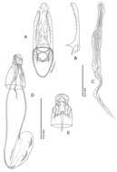
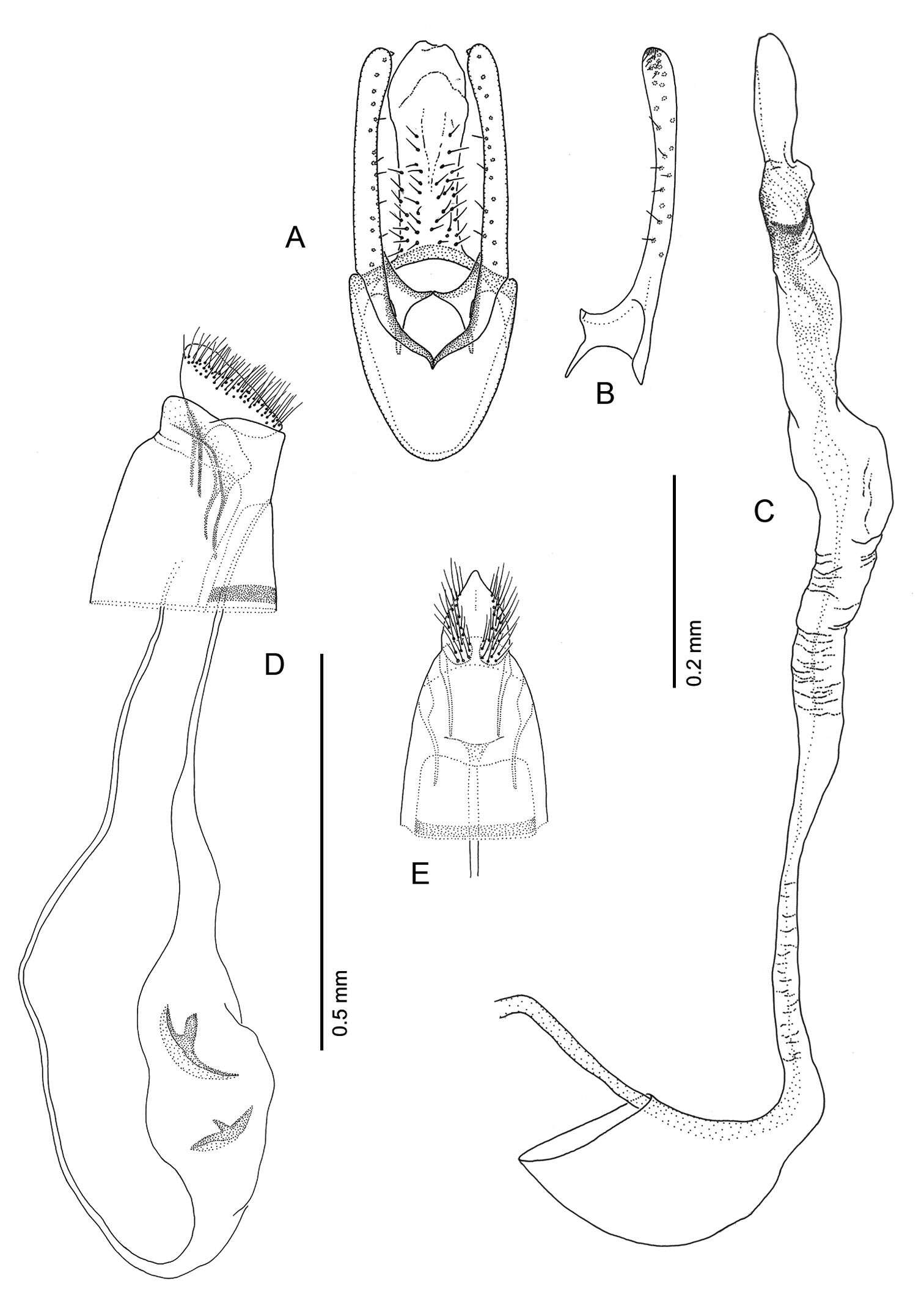
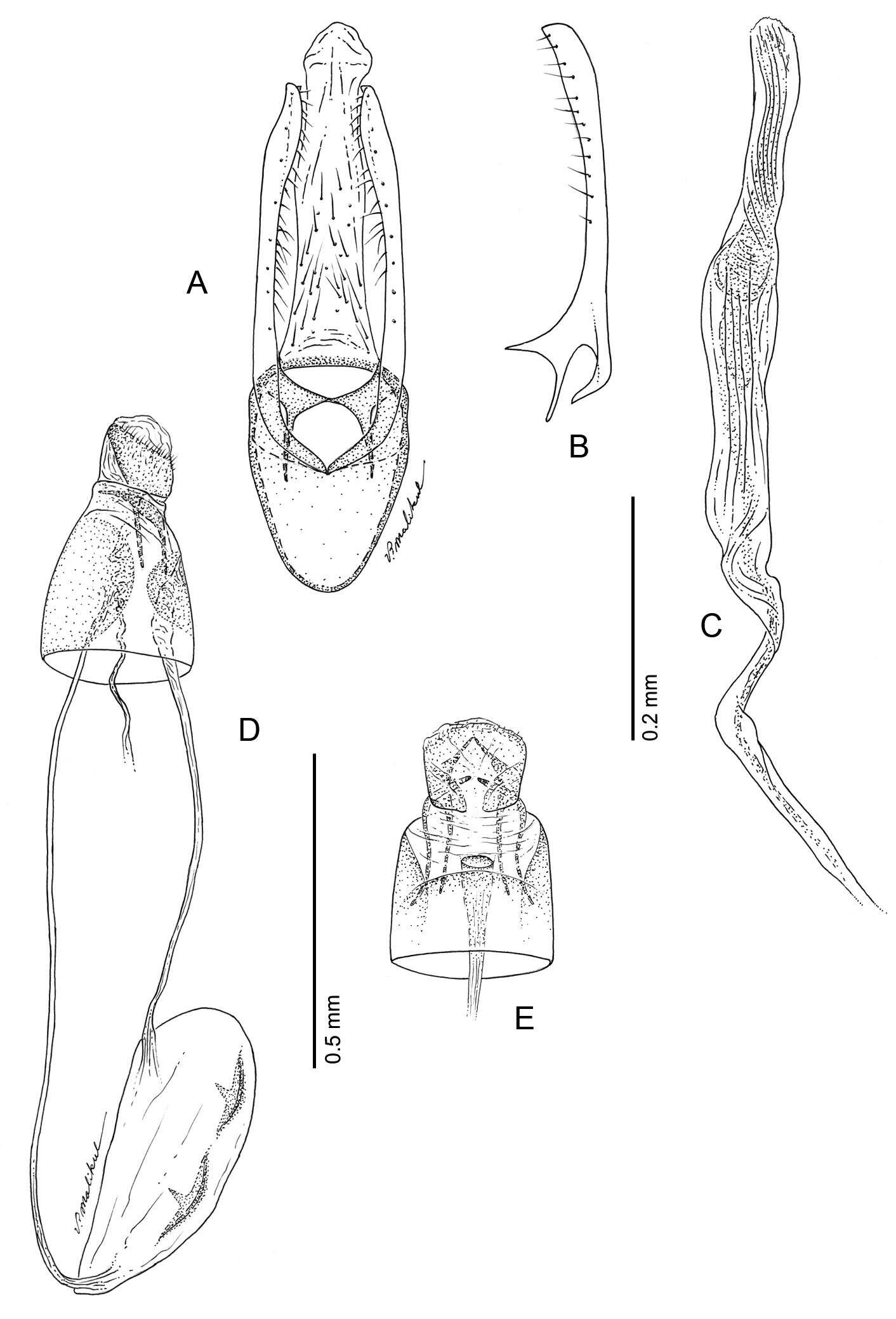
Figure 17.Phyllocnistis subpersea sp. n. genitalia. A Male, ventral view B Mesal view of valva C Aedeagus D Female, lateral view E Ventral view of D segments 7-10.
-
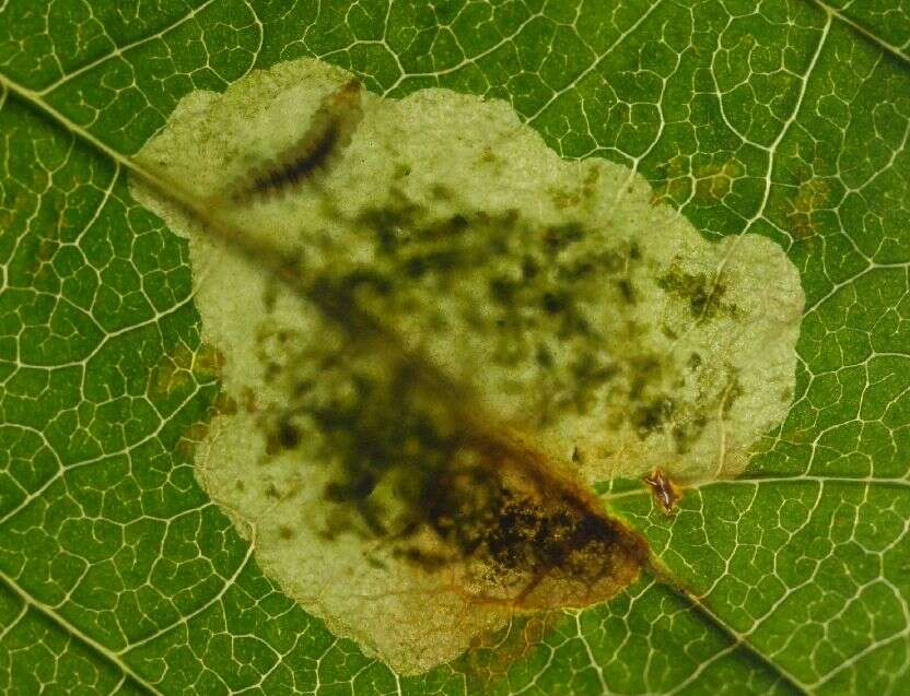
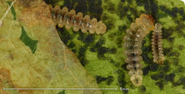
Fig. 2. Tissue feeding instar in mine
-
Donald R. Davis, David L. Wagner
Zookeys
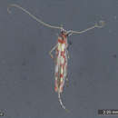
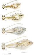
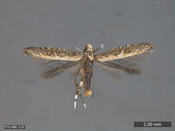
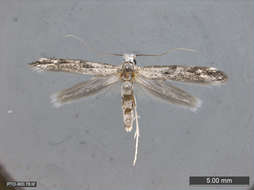
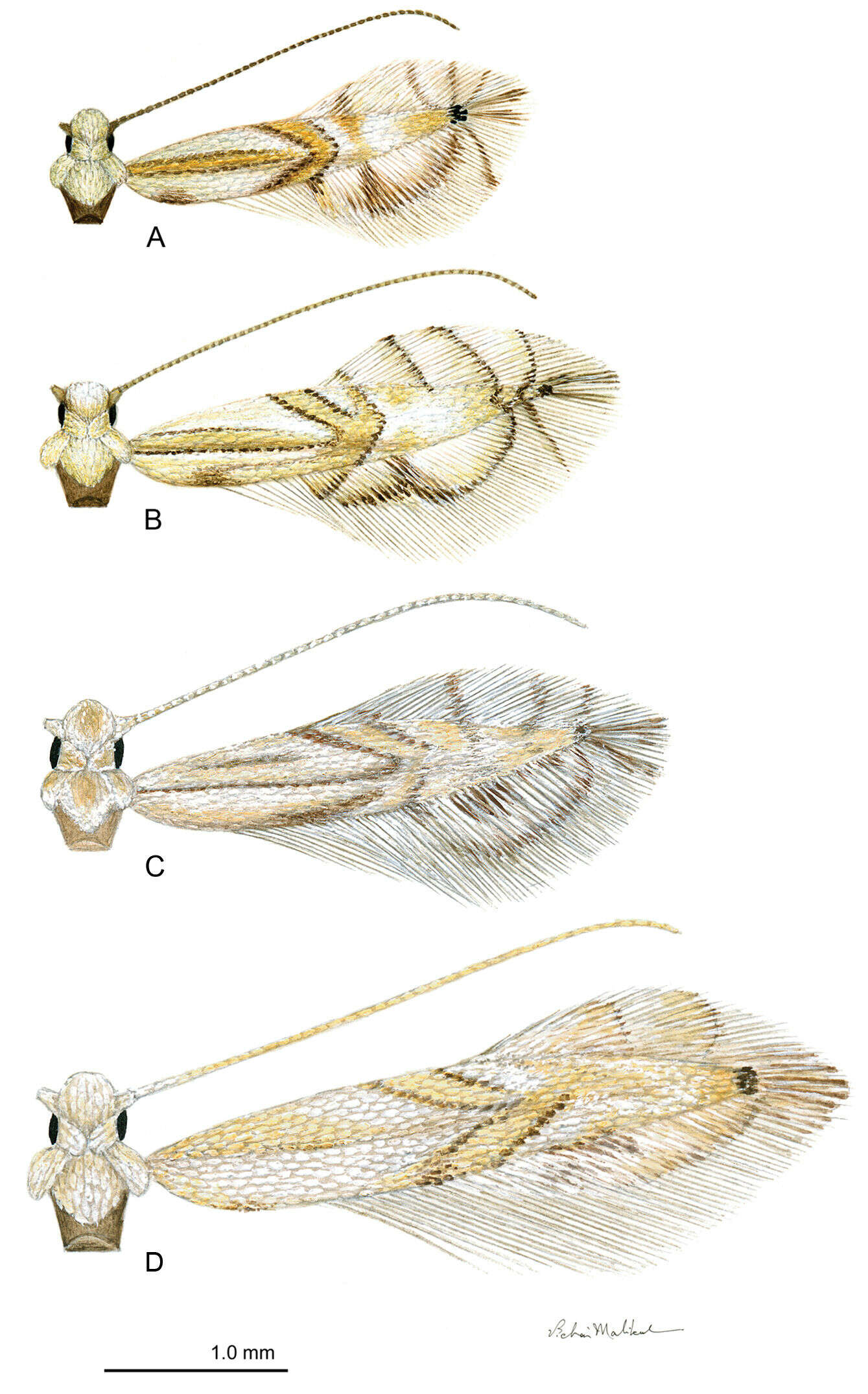
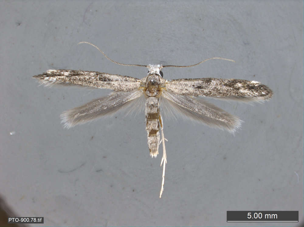
Figure 2.Phyllocnistis adults. A Phyllocnistis hyperpersea sp. n. (2.1 mm) B Phyllocnistis subpersea sp. n. (2.5 mm) C Phyllocnistis longipalpa sp. n. (2.6 mm) D Phyllocnistis perseafolia sp. n. (3.0 mm). (Drawn approximately to scale; forewing length in parentheses.)
-
Fig. 9. Cocoon
-
Donald R. Davis, David L. Wagner
Zookeys
Figure 18.Phyllocnistis longipalpa sp. n. genitalia. A Male, ventral view B Mesal view of valva C Aedeagus D Female, lateral view E Ventral view of D segments 7-10.
-
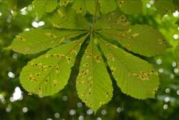
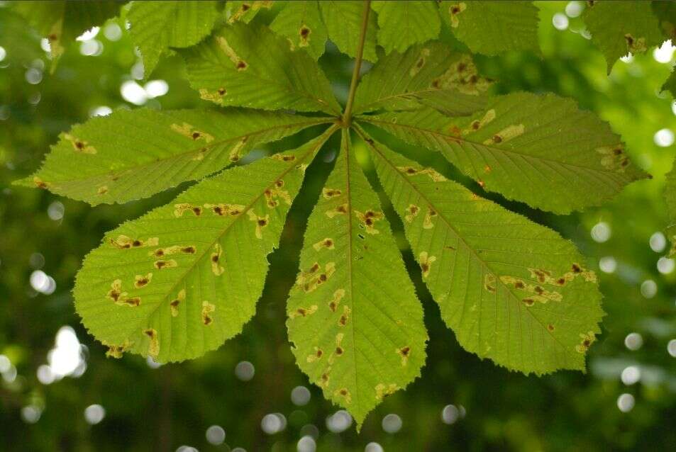
Fig. 1. Mines on horse chestnut leaf
-
Donald R. Davis, David L. Wagner
Zookeys
Figure 2.Phyllocnistis adults. A Phyllocnistis hyperpersea sp. n. (2.1 mm) B Phyllocnistis subpersea sp. n. (2.5 mm) C Phyllocnistis longipalpa sp. n. (2.6 mm) D Phyllocnistis perseafolia sp. n. (3.0 mm). (Drawn approximately to scale; forewing length in parentheses.)
-
Donald R. Davis, David L. Wagner
Zookeys
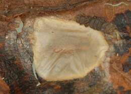
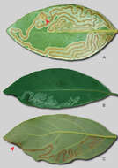
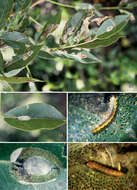
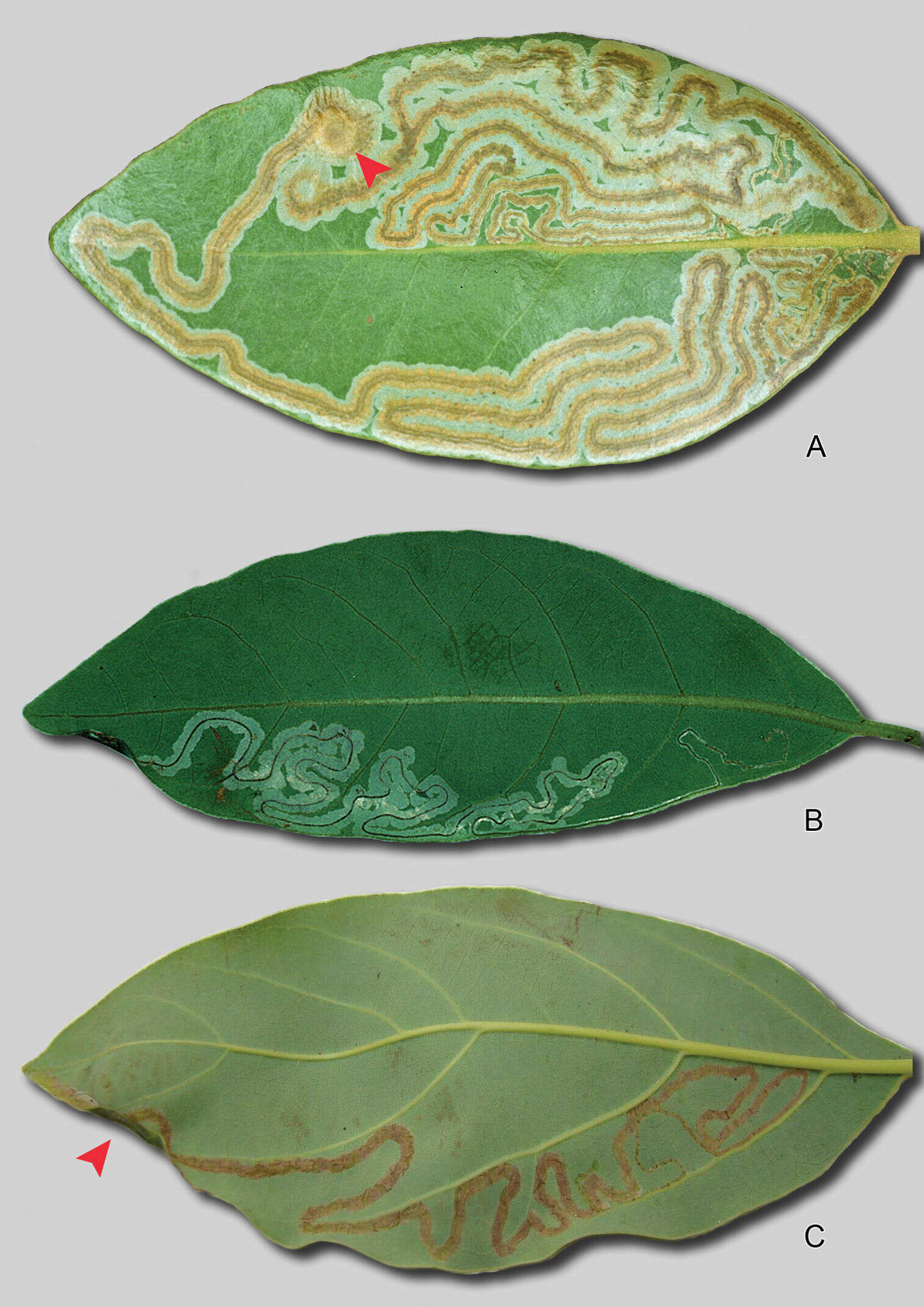
Figure 3.Phyllocnistis leafmines. A Phyllocnistis hyperpersea sp. n., upper-side mine on Persea borbonia B Phyllocnistis subpersea sp. n., lower side mine on Persea borbonia. Pupal crypts indicated by arrows in A and C. C Phyllocnistis perseafolia sp. n., lower-side mine on Persea americana (~ 15 cm).
-
Donald R. Davis, David L. Wagner
Zookeys
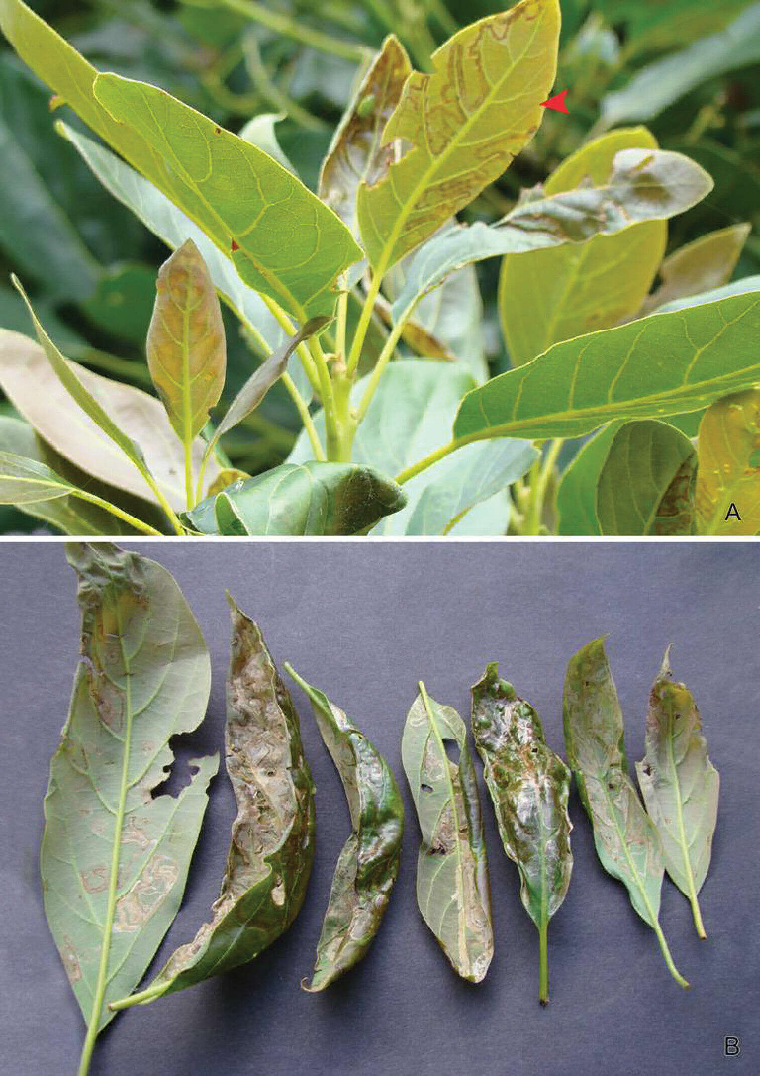
Figure 4.Leafmines of Phyllocnistis perseafolia sp. n. on Persea americana. A General habitus, note lower side mine (arrow) B Leaf damage caused by upper and lower side larval mining.
-
Donald R. Davis, David L. Wagner
Zookeys
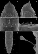
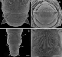
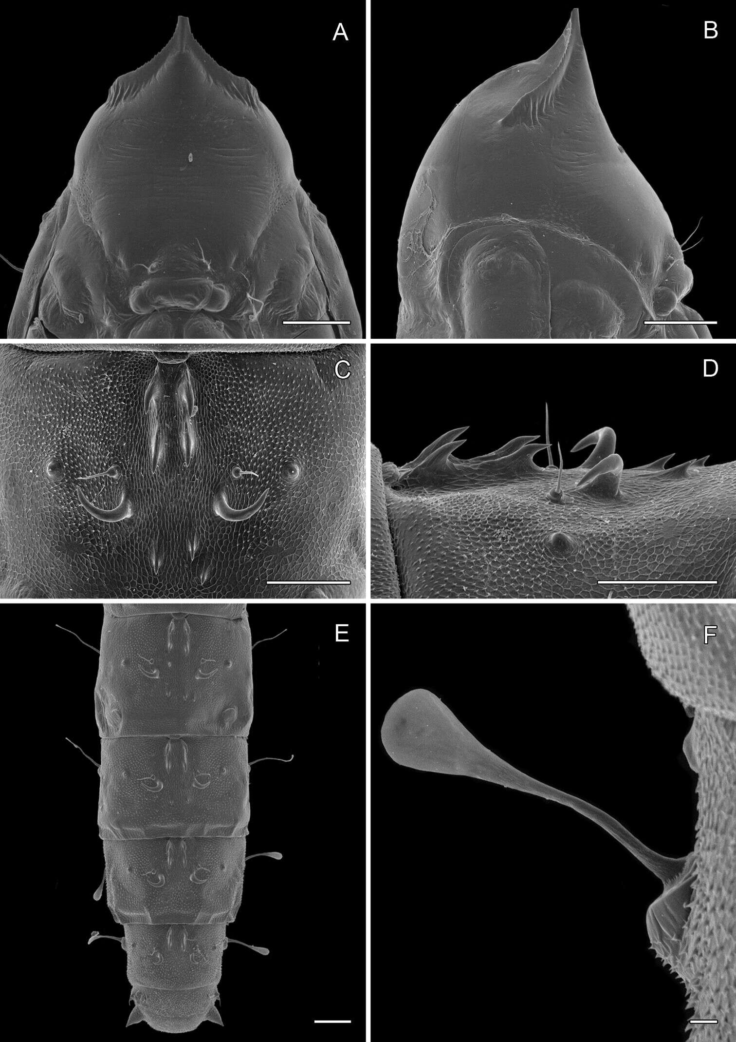
Figure 14.Phyllocnistis perseafolia sp. n. pupa. A Head, ventral view (176 µm) B Head, lateral view (200 µm) C Dorsal spines of abdominal tergum 5 (76 µm) D Lateral view of C (60 µm) E Abdominal terga 4-10 (100 µm) F Lateral seta of abdominal segment 6 (10 µm). (Length of bar scales shown in parentheses.)
-
Donald R. Davis, David L. Wagner
Zookeys
Figure 15.Phyllocnistis perseafolia sp. n. pupa. A Abdominal terga 7-10 (100 µm) B Caudal end of abdomen (100 µm) C Abdominal sterna 6-10 (100 µm) D Spinules of sternum 6 in longitudinal rows (100 µm). (Length of bar scales shown in parentheses.)
-
Donald R. Davis, David L. Wagner
Zookeys
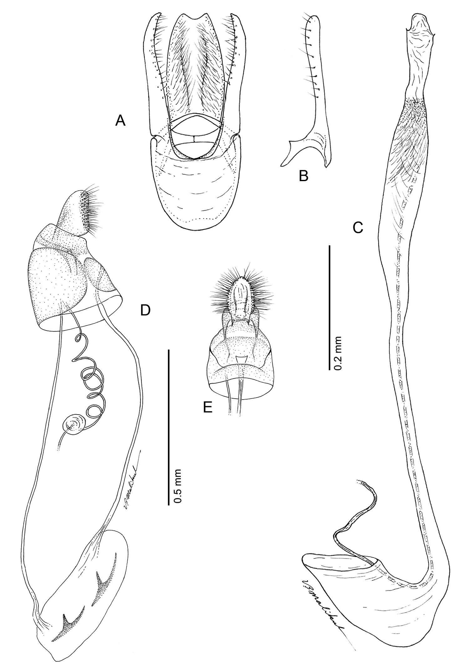
Figure 19. Phyllocnistis perseafolia sp. n. genitalia. A Male, ventral view B Mesal view of valva C Aedeagus D Female, lateral view E Ventral view of D segments 7-10.
-
Donald R. Davis, Jurate De Prins
Zookeys
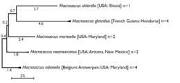
Figure 1.Compressed subtree sequenced data for cytochrome c oxidase I (COI) of Macrosaccus, derived from 13 samples among 5 species based upon neighbor-joining analysis with Kimura 2-parameter model. Numbers above branches indicate branch length. Sequence lengths obtained for all samples were 658bp each.
-
Donald R. Davis, Jurate De Prins
Zookeys
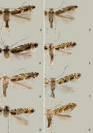
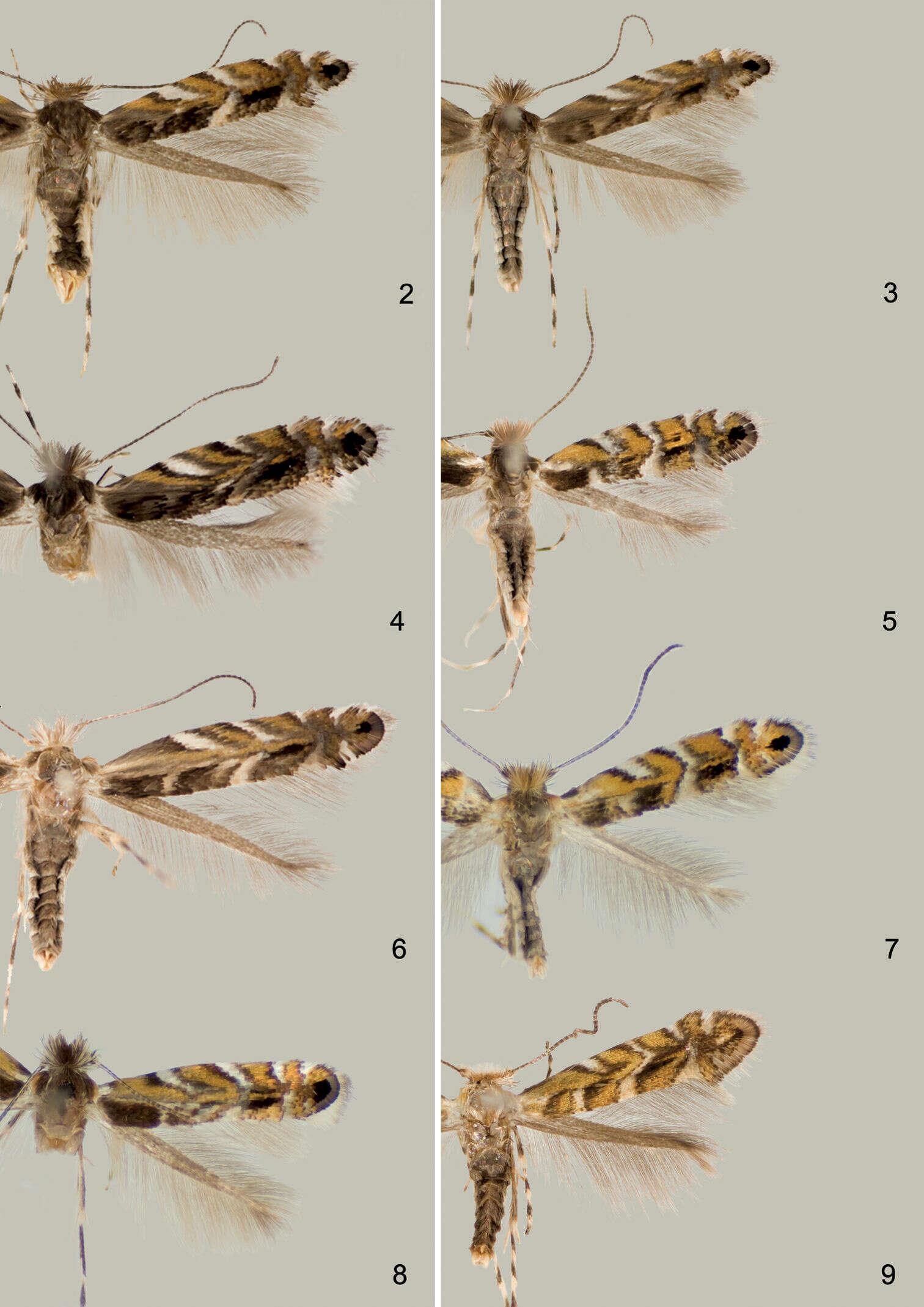
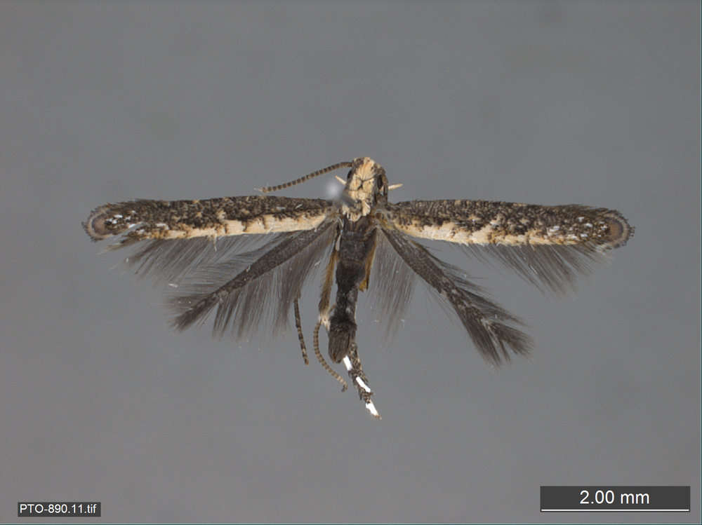
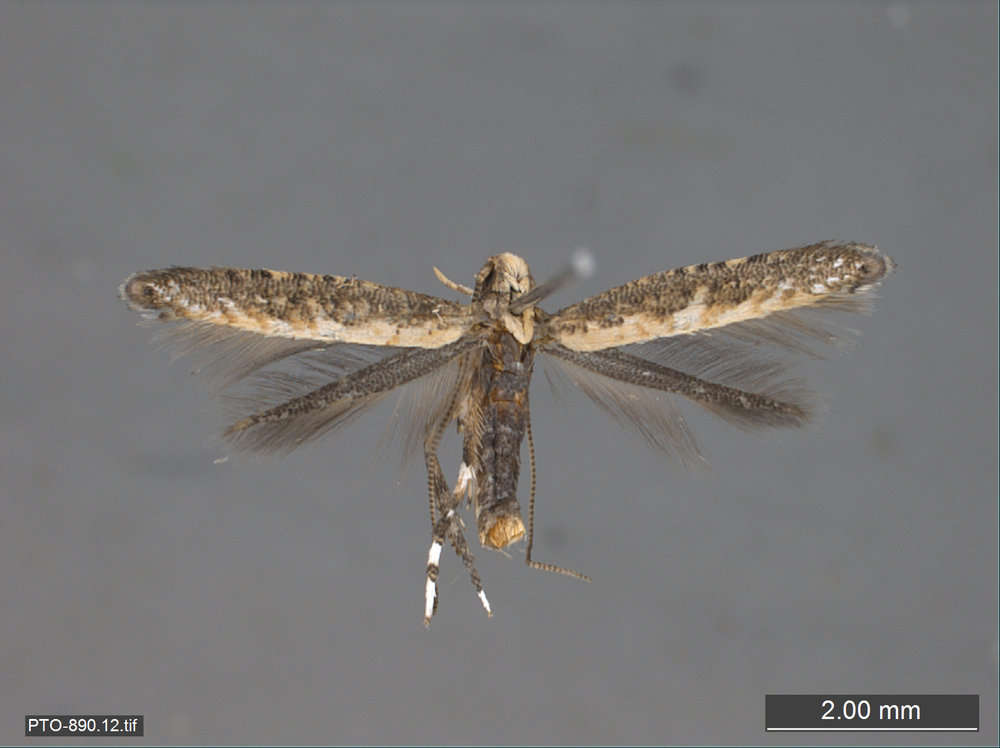
Figures 2–9.Adults 2–4. Macrosaccus robiniella. 2♂, USA: Maryland, (2.8 mm) 3 ♂, USA: Maryland, (3.0 mm) 4 BELGIUM: Antwerp, (3.0 mm) 5 Macrosaccus morrisella, ♂, USA: Maryland, (2.5 mm) 6 Macrosaccus neomexicanus, USA: Arizona, (3.2 mm) 7 Macrosaccus uhlerella, USA: Illinois, (2.5 mm) 8 Macrosaccus uhlerella, USA: Illinois, (3.0 mm) 9 Macrosaccus gliricidius, ♂, HONDURAS: Morazán, (2.4 mm). (Forewing lengths in parentheses).
-
Donald R. Davis, Jurate De Prins
Zookeys
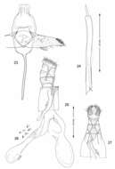
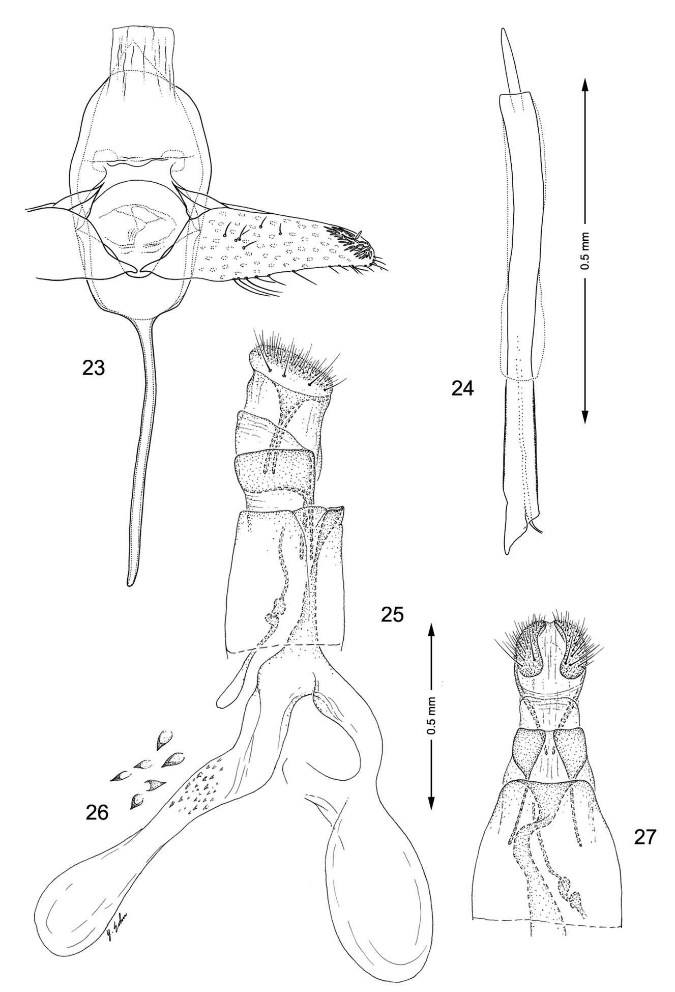
Figures 23–27.Genitalia, Macrosaccus neomexicanus. 23–24 Male. 23 Genital capsule, ventral view 24 Aedeagus 25–27 Female. 25 Lateral view 26 Detail of signa within corpus bursae 27 Segments 7–10, ventral view.
-
Donald R. Davis, Jurate De Prins
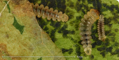
Zookeys
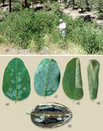
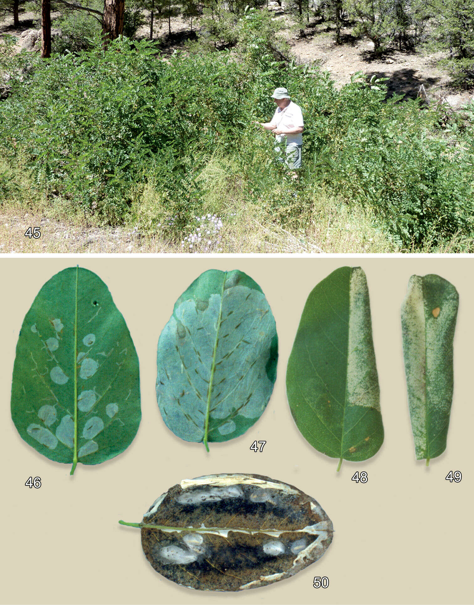
Figures 45–50.Habitat andleafmines of Macrosaccus neomexicanus on Robinia neomexicana. 45 Mixed pine-juniper habitat of Robinia neomexicana, Kaibab National Forest, Coconino Co., Arizona, ~2130 m 46 Multiple early instar serpentine and blotch mines on abaxial leaf surface 47 Later stage abaxial mines after multiple blotch mines begin to coalesce 48 Late stage tentiform blotch mine, adaxial view 49 Completely folded leaf resulting from double tentiform mines, adaxial view 50 Opened (with ventral leaf epidermis removed) aggregate blotch mines with 8 pupal cocoons, abaxial view.
-
Donald R. Davis, Jurate De Prins
Zookeys
Figure 1.Compressed subtree sequenced data for cytochrome c oxidase I (COI) of Macrosaccus, derived from 13 samples among 5 species based upon neighbor-joining analysis with Kimura 2-parameter model. Numbers above branches indicate branch length. Sequence lengths obtained for all samples were 658bp each.
-
Donald R. Davis, Jurate De Prins
Zookeys
Figures 2–9.Adults 2–4. Macrosaccus robiniella. 2♂, USA: Maryland, (2.8 mm) 3 ♂, USA: Maryland, (3.0 mm) 4 BELGIUM: Antwerp, (3.0 mm) 5 Macrosaccus morrisella, ♂, USA: Maryland, (2.5 mm) 6 Macrosaccus neomexicanus, USA: Arizona, (3.2 mm) 7 Macrosaccus uhlerella, USA: Illinois, (2.5 mm) 8 Macrosaccus uhlerella, USA: Illinois, (3.0 mm) 9 Macrosaccus gliricidius, ♂, HONDURAS: Morazán, (2.4 mm). (Forewing lengths in parentheses).
-
Donald R. Davis, Jurate De Prins
Zookeys
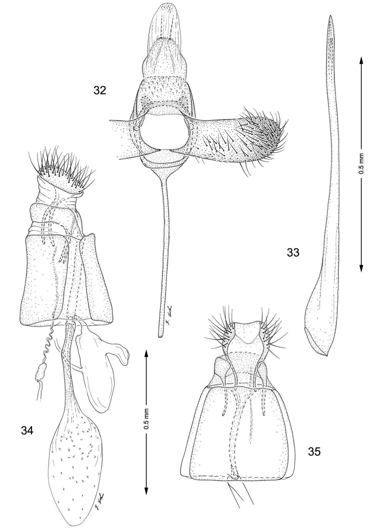
Figures 32–35.Genitalia, Macrosaccus gliricidius. 32–33 Male. 32 Genital capsule, ventral view 33 Aedeagus 34–35 Female. 34 Lateral view 35 Segments 7–10, ventral view.
-
Donald R. Davis, Jurate De Prins
Zookeys
Figures 54–58.Leafmines of Macrosaccus gliricidius on Gliricidia sepium. 54 General damage to host 55 Adaxial blotch mine 56 Late instar tissue feeding larva 57 Open blotch mine with single cocoon 58 Pupa with cocoon removed. Photographs by R. Cave.
-
Cameraria ohridella larva
-
All Biocode files are based on field identifications to the best of the researcher’s ability at the time.
-
All Biocode files are based on field identifications to the best of the researcher’s ability at the time.
-
All Biocode files are based on field identifications to the best of the researcher’s ability at the time.